
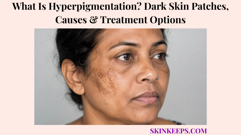
Hyperpigmentation is darkening of part of the skin compared with the person’s surrounding natural skin colour. It is a visible skin change, not one disease, and may appear as one spot, several flat marks, symmetrical facial patches or widespread darkening.
Common causes include acne, eczema, skin injury, ultraviolet exposure, hormonal influences and medicines. Treatment begins by identifying the pattern and controlling the cause, because harsh exfoliation, unsafe lightening products or poorly selected procedures can trigger more pigment.
How Can You Recognize Hyperpigmentation on the Skin?
Hyperpigmentation is recognized when part of the skin becomes darker than the person’s surrounding baseline colour.
- Flat brown, tan or dark-brown spots.
- Grey-brown or blue-grey patches.
- Black or nearly black areas.
- Red-brown marks after inflammation.
- Mottled or uneven pigmentation.
- Symmetrical facial patches.
- Marks matching previous acne, eczema or injury.
- Freckle-like sun spots.
- Dark velvety thickening in folds.
- Diffuse darkening over broad areas.
A raised lump, scale, thickening, ulcer or changing border means another process may accompany the colour change.
Figure 1. Hyperpigmentation is recognized by colour change relative to baseline, but body location, symmetry, texture, depth and evolution determine whether the pattern suggests PIH, melasma, acanthosis, sun damage or a suspicious lesion.
Where Can Dark Skin Patches Develop?
Dark patches can develop anywhere, but location often points toward the most likely cause.
| Location | Pattern Clue |
|---|---|
| Cheeks, forehead and upper lip | Melasma or facial PIH |
| Previous acne or eczema sites | Post-inflammatory hyperpigmentation |
| Face, hands, shoulders and forearms | Freckles or solar lentigines |
| Neck, armpits or groin | Acanthosis or friction |
| Chest, back or neck with scale | Tinea versicolor |
| Palms, soles, scars or mouth | Possible systemic pigmentation |
| Beneath a nail | Individual nail-lesion assessment |
How Does the Skin Produce Excess Pigment?
Hyperpigmentation often begins when melanocytes produce more melanin or distribute pigment differently.
Melanocytes make melanin inside melanosomes and transfer it to surrounding keratinocytes. Inflammation, hormones, ultraviolet radiation and visible light can increase this activity.
When injury disrupts the epidermal–dermal boundary, melanin can drop into the dermis and become trapped inside macrophages, creating slower-fading grey-blue colour.
Figure 2. Melanocyte activation increases melanin transfer, and inflammation can leave epidermal or dermal pigment; picking, burning products and repeated procedures can restart this pathway.
How Do Epidermal and Dermal Hyperpigmentation Differ?
What Does Epidermal Hyperpigmentation Look Like?
Epidermal pigment is usually tan, brown or dark brown, often with clearer borders and a better response to topical treatment.
What Does Dermal Hyperpigmentation Look Like?
Dermal pigment is often grey, blue-grey or slate-grey, with less distinct borders and slower or incomplete fading.
What Is Mixed-Depth Hyperpigmentation?
Mixed pigmentation contains both epidermal and dermal pigment, so the brown component may improve while deeper grey colour persists.
| Depth | Typical Colour | Treatment Response |
|---|---|---|
| Epidermal | Tan to dark brown | More responsive |
| Dermal | Grey to blue-grey | Slower and less responsive |
| Mixed | Brown plus grey | Variable |
Which Main Types of Hyperpigmentation Can Occur?
Hyperpigmentation can be grouped by trigger, distribution and whether it represents a diffuse pattern or an individual lesion.
| Type | Examples | Main Direction |
|---|---|---|
| Post-inflammatory | After acne, eczema, injury or procedures | Control inflammation |
| Hormonal/light-associated | Melasma | Photoprotection and maintenance |
| Sun-related | Freckles and solar lentigines | Sun protection and lesion care |
| Friction/metabolic | Friction pigment and acanthosis | Remove driver and assess metabolism |
| Medicine/chemical | Drug pigment or ochronosis | Review exposure |
| Systemic | Addison disease or iron/metabolic disorders | Medical testing |
| Melanocytic lesion | Mole, lentigo or melanoma | Lesion assessment |
| Congenital | Café-au-lait or patterned marks | Age/pattern-specific evaluation |
What Is Post-Inflammatory Hyperpigmentation?
Post-inflammatory hyperpigmentation is a flat dark mark that appears after inflammation, irritation or injury begins to heal.
Flat dark marks after acne are post-inflammatory colour changes, not automatically permanent acne scars.
- Matches the shape and location of the prior skin problem.
- Is flat rather than raised or indented.
- May be brown, dark brown, grey-brown or blue-grey.
- Can remain after the active rash or bump resolves.
- May darken with ultraviolet or visible-light exposure.
- Can occur in every skin tone and persist longer in darker skin.
Which Skin Problems Commonly Leave Post-Inflammatory Dark Marks?
Many rashes, bumps, injuries and procedures can leave PIH after the active problem heals.
Uncontrolled dermatitis / eczema can keep creating inflammation, so fading creams alone may fail while scratching and barrier damage continue.
Repeated inflamed bumps from folliculitis can leave follicle-centred marks after the bumps flatten.
- Acne, eczema, contact dermatitis and psoriasis.
- Lichen planus, folliculitis and ingrown hairs.
- Impetigo, fungal infection and viral rashes.
- Insect bites, burns, cuts and abrasions.
- Scratching, cosmetic procedures and chemical irritation.
- Shaving, waxing and hair-removal injury.
How Is Acne Hyperpigmentation Different From an Acne Scar?
Acne hyperpigmentation is a flat colour change, while a scar changes skin texture or contour.
| Feature | Post-Acne Pigment | Acne Scar |
|---|---|---|
| Surface | Flat | Indented or raised |
| Main change | Colour | Structure |
| Natural course | May fade | Usually persists |
| Treatment focus | Cause control and pigment care | Scar procedure or remodelling |
PIH and true scars can occur together.
Why Does Picking or Scrubbing Make Dark Marks Worse?
Picking or scrubbing can restart inflammation and stimulate more pigment production.
A product that repeatedly stings, burns or causes rash may trigger allergic contact dermatitis or irritation that leaves more pigment behind.
- Picking ruptures healing skin.
- Scrubbing creates micro-injury.
- Scab removal can bleed and scar.
- Harsh acids can trigger irritant dermatitis.
- Injury can push pigment into the dermis.
- Burning is not proof that treatment is working.
What Is Melasma—and How Does Its Pattern Differ?
Melasma is a recurrent pigment disorder that usually causes symmetrical brown or grey-brown facial patches.
- Both cheeks are commonly involved.
- The forehead, upper lip, nose, chin or jawline may be affected.
- The skin is usually flat and unscaled.
- Development is gradual.
- Sunlight and visible light worsen the pattern.
- Recurrence is common.
- It can occur during or outside pregnancy and can affect men.
What Causes or Worsens Melasma?
Melasma develops when genetic susceptibility, hormones and light exposure combine to overactivate pigment production.
- Ultraviolet radiation.
- Visible light.
- Pregnancy.
- Estrogen- and progesterone-related influences.
- Oral contraceptives or hormone-replacement therapy.
- Family susceptibility.
- Heat in some people.
- Irritating skin-care products.
- Selected photosensitizing medicines.
Melasma is not caused by poor hygiene and is not contagious.
How Are Sun Spots and Freckles Different From Melasma?
What Are Freckles?
Freckles are small flat brown spots that often begin in childhood, darken after sun exposure and may fade during lower-exposure seasons.
What Are Solar Lentigines?
Solar lentigines are persistent sun or age spots that develop after accumulated ultraviolet exposure, commonly on the face, hands, shoulders and forearms.
How Does Melasma Differ?
Melasma forms broader, usually symmetrical facial patches with strong hormonal and light associations and a greater recurrence tendency.
| Feature | Freckles | Solar Lentigines | Melasma |
|---|---|---|---|
| Pattern | Small spots | Persistent localized spots | Broad symmetrical patches |
| Onset | Often childhood | Accumulated UV exposure | Hormonal/light influenced |
| Seasonal change | May fade | Usually persistent | Relapses with light |
| Depth | Usually epidermal | Usually epidermal | May be mixed |
How Does Friction Cause Dark Skin Patches?
Friction can darken skin by creating repeated low-grade inflammation that stimulates melanin production.
- Tight clothing can irritate folds.
- Shaving and waxing can inflame follicles.
- Eczema scratching can thicken and darken skin.
- Inner thighs, neck, elbows, knees, groin and underarms may be affected.
- Pigment may remain after rubbing stops.
- Strong exfoliation can worsen the injury.
What Is Acanthosis Nigricans?
Acanthosis nigricans causes dark, velvety, thickened skin in folds and may signal insulin resistance or another medical driver.
Dark velvety fold thickening may be acanthosis nigricans, which should not be treated as ordinary surface pigment or dirt.
- Common sites are the back of the neck, armpits, groin and knuckles.
- Skin tags may coexist.
- Associations include insulin resistance, prediabetes, type 2 diabetes and PCOS.
- Selected medicines and genetic syndromes can contribute.
- Rapid extensive adult-onset disease needs broader assessment.
Which Medicines Can Cause Hyperpigmentation?
Medicines can cause pigmentation through drug deposition, inflammation, photosensitivity or direct stimulation of melanin.
A medicine may cause immediate inflammation, a fixed reaction, or a later drug rash that leaves residual pigmentation.
- Antimalarial medicines.
- Minocycline and selected antibiotics.
- Amiodarone.
- Chemotherapy or targeted cancer medicines.
- Antipsychotic medicines and anticonvulsants.
- Hormonal medicines.
- Clofazimine.
- Fixed drug eruptions or photosensitivity reactions.
Essential medicine should be reviewed with the prescriber rather than stopped independently.
Which Skin-Lightening Products Can Paradoxically Cause Darkening?
Some lightening products worsen pigmentation through irritation, burns, steroid damage, mercury exposure or prolonged hydroquinone misuse.
- Long-term high-strength hydroquinone.
- Unregulated combination creams.
- Undeclared potent corticosteroids.
- Mercury-containing products.
- Harsh acid mixtures.
- Repeated chemical burns.
- Poor sun protection during treatment.
Exogenous ochronosis appears as irregular blue-black or grey-blue pigmentation after prolonged hydroquinone misuse and can be difficult to reverse.
Which Internal Medical Conditions Can Cause Widespread Darkening?
Widespread or unusual pigmentation can reflect an internal condition rather than a cosmetic skin problem.
| Possible Driver | Supporting Clues |
|---|---|
| Addison disease | Mouth/scar darkening, fatigue, dizziness, low blood pressure or salt craving |
| Haemochromatosis | Diffuse colour change with iron or organ clues |
| Thyroid/hormonal disorder | Systemic endocrine symptoms |
| Nutritional deficiency | Dietary, blood or neurologic clues |
| Liver/kidney disease | Known organ disease or systemic illness |
| Rare paraneoplastic pattern | Rapid unusual progression |
How Does Pregnancy Affect Hyperpigmentation?
Pregnancy can increase pigmentation through hormonal change, but it does not make every changing dark lesion harmless.
- Melasma may develop or worsen.
- Nipples and areolae may darken.
- Linea nigra can appear.
- Freckles, scars and genital skin may darken.
- Melasma may persist after delivery.
- A new asymmetrical or evolving lesion still needs examination.
Why Is Post-Inflammatory Hyperpigmentation More Persistent in Darker Skin?
PIH can be more noticeable and persistent in darker skin because inflammatory signals can trigger a strong pigment response.
- More melanin may be transferred into epidermal cells.
- Pigment can drop into the dermis after deeper inflammation.
- Dermal pigment fades slowly.
- Acne, eczema and procedures may leave long-lasting marks.
- Treatment irritation can create additional PIH.
- Visible light can worsen selected pigment disorders.
- Procedure settings must account for baseline melanin.
How Is Hyperpigmentation Different From Hypopigmentation?
Hyperpigmentation means darker skin, hypopigmentation means lighter skin and depigmentation means pigment is almost or completely absent.
| Change | Meaning | Examples |
|---|---|---|
| Hyperpigmentation | Darker than baseline | PIH or melasma |
| Hypopigmentation | Lighter than baseline | After inflammation or infection |
| Depigmentation | Near-complete pigment loss | Vitiligo pattern |
How Is Hyperpigmentation Different From a Bruise?
A bruise is blood colour beneath the skin, while hyperpigmentation usually reflects pigment that persists after inflammation or injury.
| Feature | Hyperpigmentation | Bruise |
|---|---|---|
| Cause | Melanin or deposited pigment | Leaked blood |
| Short-term colour | Relatively stable | Changes through several colours |
| Tenderness | Often absent | May be tender |
| Course | Weeks, months or longer | Usually days to weeks |
How Can Tinea Versicolor Resemble Hyperpigmentation?
Tinea versicolor can create brown, pink or lighter patches with fine scale, often on the chest, back, shoulders or neck.
- Fine powdery scale.
- Mild itch in some people.
- Greater visibility after tanning.
- Recurrence in warm or humid conditions.
- Clinical examination, scraping or Wood lamp may help.
- Normal colour may take weeks or months to return after yeast treatment.
How Is a Benign Dark Patch Different From Melanoma?
A benign patch is usually stable and patterned, while melanoma is more concerning when a spot is new, changing, irregular or different from other marks.
| ABCDE | Warning Sign |
|---|---|
| A — Asymmetry | One half differs from the other |
| B — Border | Irregular, scalloped or poorly defined |
| C — Colour | Several colours in one lesion |
| D — Diameter | Often over about 6 mm, but can be smaller |
| E — Evolving | Change in size, shape, colour or symptoms |
- A spot that looks different from all others.
- Bleeding, pain, itching or ulceration.
- A non-healing sore.
- A dark line beneath a nail.
- Pigment spreading onto nail-fold skin.
- A rapidly growing raised lesion.
How Do Clinicians Diagnose the Cause of Hyperpigmentation?
Clinicians diagnose the cause by matching distribution, symmetry, depth, texture and evolution with inflammation, hormones, light, medicines and systemic clues.
- Age and speed of onset.
- Previous acne, eczema, injury or procedure.
- Sun and visible-light exposure.
- Pregnancy or hormonal medicine.
- Medicine and product timeline.
- Friction, shaving or waxing.
- Systemic symptoms.
- Nail, oral or palm/sole involvement.
- Change in one individual lesion.
How Do Wood-Lamp Examination and Dermoscopy Help?
What Can a Wood Lamp Show?
A Wood lamp can make some epidermal pigment more obvious and may show fluorescence from selected infections such as tinea versicolor.
What Can Dermoscopy Show?
Dermoscopy magnifies pigment structures and can support assessment of melanocytic lesions, solar lentigines, melasma, PIH and nail bands.
Neither tool replaces biopsy when a lesion remains suspicious.
Which Blood Tests Are Used for Widespread or Atypical Hyperpigmentation?
Blood tests are used when the distribution or symptoms suggest an endocrine, metabolic, nutritional or organ-related cause.
| Clinical Clue | Possible Testing |
|---|---|
| Mouth/scar darkening, dizziness or low pressure | Morning cortisol, ACTH and electrolytes |
| Velvety folds or metabolic risk | Glucose and HbA1c |
| Thyroid symptoms | Thyroid-function tests |
| Bronze diffuse pigment or organ clues | Ferritin and iron studies |
| Liver or kidney disease clues | Liver and kidney tests |
| Fatigue or nutritional concern | CBC and selected vitamin testing |
When Is a Skin Biopsy Needed for Dark Pigmentation?
Biopsy may be needed for an individual lesion that is changing, irregular, bleeding, ulcerated, nail-related or diagnostically uncertain.
- Asymmetry or irregular border.
- Several colours.
- Rapid growth or evolution.
- Bleeding, ulceration or non-healing.
- A spot different from all others.
- A widening nail band or nail-fold pigment.
- A raised or firm lesion.
- Atypical widespread pigmentation when tissue diagnosis is needed.
Does Every Dark Skin Patch Require Treatment?
Not every benign patch needs treatment, but suspicious, systemic, infectious, inflammatory or distressing pigmentation needs diagnosis or care.
- Typical stable PIH can be observed or treated.
- Melasma treatment is optional but often requires maintenance.
- Acanthosis should trigger cause assessment.
- Tinea versicolor needs antifungal treatment.
- Changing individual lesions need assessment before fading treatment.
- Diffuse systemic pigmentation needs medical evaluation.
Why Is Treating the Underlying Skin Condition the First Step?
Pigment continues to form while acne, eczema, folliculitis, infection, friction or irritation remains active.
- Control active acne.
- Repair eczema and reduce scratching.
- Treat folliculitis or ingrown-hair inflammation.
- Treat infection.
- Reduce rubbing and hair-removal injury.
- Stop irritating products.
- Review medicine-related causes safely.
How Does Sunscreen Help Prevent and Fade Hyperpigmentation?
Sunscreen reduces ultraviolet stimulation of melanocytes and helps prevent existing pigment from becoming darker.
- Choose broad-spectrum SPF 30 or higher.
- Apply to exposed skin every morning.
- Use enough product for complete coverage.
- Reapply during prolonged outdoor exposure, swimming or sweating.
- Add shade, hats and protective clothing.
- Use a tolerable formula so daily use is sustainable.
Why Can Tinted Sunscreen Be Better for Melasma and Dark Spots?
Tinted sunscreen containing iron oxides adds protection from visible light, which can worsen melasma and hyperpigmentation.
- Look for broad-spectrum SPF 30 or higher.
- Check that the product is tinted and lists iron oxides.
- Choose a shade that encourages adequate application.
- Use hats and shade as additional protection.
- Avoid a formula that stings or causes rash.
How Should a Pigment-Safe Skin-Care Routine Be Built?
A pigment-safe routine uses gentle products, one active at a time and daily photoprotection.
| Time | Routine |
|---|---|
| Morning | Gentle cleanser, moisturizer, tinted broad-spectrum sunscreen |
| Evening | Gentle cleanser, one pigment-active treatment, moisturizer |
| Weekly review | Check irritation, new marks and trigger control |
| Avoid | Picking, scrubs, bleach, lemon juice and multiple acids |
How Does Hydroquinone Treat Hyperpigmentation?
Hydroquinone reduces melanin production by inhibiting tyrosinase and can improve selected epidermal pigmentation.
- Use only on diagnosed pigment rather than changing lesions.
- Apply to affected areas according to a clinical plan.
- Monitor irritation and uneven lightening.
- Use daily photoprotection.
- Avoid continuous unsupervised use for years.
- Stop and seek assessment for blue-black or grey-blue darkening.
- Pregnancy safety is not established.
What Is Triple-Combination Treatment for Melasma?
Prescription triple-combination treatment joins hydroquinone, a retinoid and a topical corticosteroid to target several melasma pathways.
| Component | Role |
|---|---|
| Hydroquinone | Reduces pigment production |
| Retinoid | Improves turnover and treatment penetration |
| Corticosteroid | Reduces inflammation and irritation |
It is a time-limited prescription strategy, not an indefinite bleaching cream.
How Does Azelaic Acid Help Dark Marks?
Azelaic acid can reduce inflammation, help acne control and gradually reduce selected PIH and melasma.
- Useful when acne and PIH coexist.
- Can be considered in melasma plans.
- May sting or irritate initially.
- Introduce gradually.
- Continue sunscreen.
- Pregnancy use should still be discussed when treatment is prescription-strength or combined.
How Do Topical Retinoids Treat Hyperpigmentation?
Topical retinoids increase epidermal turnover, support acne control and help disperse selected epidermal pigment.
- Tretinoin, adapalene or tazarotene may be used depending on diagnosis.
- Begin slowly to limit irritation.
- Use moisturizer and daily sunscreen.
- Do not combine several irritating products immediately.
- Worsening redness or peeling can create more PIH.
- Topical retinoids are avoided during pregnancy.
Which Other Topical Ingredients May Help Hyperpigmentation?
Several topical ingredients may support gradual fading, but evidence and irritation risk vary.
| Ingredient | Possible Role | Main Caution |
|---|---|---|
| Vitamin C | Antioxidant and pigment support | Formula stability and irritation |
| Kojic acid | Tyrosinase inhibition | Contact irritation |
| Glycolic/lactic acid | Exfoliation and hydration | PIH if overused |
| Niacinamide | Pigment-transfer support | Usually mild but not universal |
| Cysteamine | Pigment reduction | Odour and irritation |
| Arbutin/licorice | Pigment-pathway support | Variable product quality |
| Topical tranexamic acid | Investigational melasma option | Evidence and formulation vary |
Can Oral Tranexamic Acid Treat Melasma?
Oral tranexamic acid can improve selected refractory melasma, but its cosmetic use is off-label and requires clot-risk review.
- Used only after diagnosis and specialist selection.
- Review personal or family clot history.
- Review thrombophilia, smoking and estrogen exposure.
- Assess kidney and medicine factors.
- Stop and seek urgent care for chest pain, breathing difficulty, one-sided swelling or neurologic symptoms.
- Avoid self-prescribing or buying cosmetic oral treatment online.
When Can Chemical Peels Help Hyperpigmentation?
Superficial chemical peels can help selected epidermal pigmentation, but injury from an overly strong peel can create more PIH.
- Confirm the diagnosis first.
- Control active inflammation.
- Choose peel depth and agent for skin tone.
- Prepare skin when clinically appropriate.
- Use strict aftercare and photoprotection.
- Avoid deep or unsupervised home peels.
- Stop when blistering, raw skin or severe pain develops.
When Can Laser or Light Treatment Help Dark Patches?
Laser or light treatment can help selected solar lentigines, epidermal pigment or resistant lesions, but results depend on diagnosis, depth, device and skin tone.
| Pattern | Possible Role | Main Risk |
|---|---|---|
| Solar lentigo | Lesion-directed device treatment | Burn or pigment change |
| PIH | Highly selective use | Worsening PIH |
| Melasma | Limited specialist role | Relapse and rebound pigment |
| Drug pigment | Selected cases | Hyper- or hypopigmentation |
| Suspicious lesion | Not before diagnosis | Delayed cancer diagnosis |
Why Can Laser Treatment Be Riskier for Darker Skin?
Laser treatment can be riskier in darker skin because background melanin can absorb energy intended for the target pigment.
- Heat injury can cause burns or blisters.
- Inflammation can trigger PIH.
- Excess energy can cause hypopigmentation.
- Device wavelength and pulse settings matter.
- Cooling and test spots may reduce risk.
- Experience treating the person’s skin tone is essential.
How Are Sun Spots Treated Differently From Melasma or PIH?
Solar lentigines are isolated UV-related lesions, melasma is a recurrent facial field disorder and PIH follows inflammation, so treatment goals differ.
| Pattern | First Priority | Possible Treatment |
|---|---|---|
| Solar lentigo | Confirm benign lesion | Cryotherapy, laser or selected topical care |
| Melasma | Light protection and maintenance | Topical combination and selected specialist options |
| PIH | Control active inflammation | Gentle pigment treatment and time |
How Is Acanthosis Nigricans Treated?
Acanthosis treatment focuses on the metabolic, endocrine or medicine-related driver rather than bleaching alone.
- Assess insulin resistance and diabetes risk.
- Review weight, metabolic health and medicines.
- Evaluate PCOS when relevant.
- Treat the underlying driver.
- Reduce friction and moisture.
- Use selected retinoids, urea or lactic acid for texture.
- Investigate rapid or atypical adult-onset disease.
How Is Drug-Induced Pigmentation Managed?
Drug-induced pigmentation is managed by confirming the timeline and reviewing the medicine safely with the prescriber.
- Document the pattern with photographs.
- Assess whether the distribution fits the drug.
- Review necessity and alternatives.
- Do not stop essential therapy independently.
- Reduce sun exposure when photosensitivity contributes.
- Treat associated inflammation.
- Recognize that deposited pigment may persist.
How Is Hyperpigmentation Treated During Pregnancy?
Pregnancy care prioritizes photoprotection, gentle skin care and pregnancy-compatible control of active inflammation.
- Use tinted broad-spectrum sunscreen, hats and shade.
- Control acne or eczema with pregnancy-compatible treatment.
- Consider azelaic acid when clinically appropriate.
- Avoid topical and oral retinoids.
- Avoid unsupervised hydroquinone.
- Avoid cosmetic oral tranexamic acid.
- Postpone elective high-risk peels and lasers.
- Reassess melasma after delivery.
How Is Hyperpigmentation Managed in Children?
Childhood pigmentation should be classified as post-inflammatory, congenital, infectious or changing before treatment.
Multiple or patterned café-au-lait spots should be separated from ordinary PIH because some patterns require syndromic assessment.
- Ask about eczema, bites, infection or injury.
- Treat the active inflammatory condition.
- Use gentle moisturizer and sunscreen.
- Avoid strong combination lightening products.
- Use retinoids or acids only with age-appropriate guidance.
- Assess changing moles.
- Consider fungal infection when scale is present.
- Refer widespread, patterned or unexplained pigmentation.
Which Hyperpigmentation Treatment Mistakes Should Be Avoided?
Unsafe treatment can cause burns, skin thinning, more PIH, hypopigmentation, mercury toxicity or delayed melanoma diagnosis.
| Mistake | Possible Harm | Safer Direction |
|---|---|---|
| Household bleach or lemon juice | Burn and PIH | Use regulated treatment |
| Unlabelled lightening cream | Mercury or steroid exposure | Check ingredients and source |
| Hydroquinone for years | Ochronosis | Time-limited supervised use |
| Several acids at once | Irritation and darker marks | Introduce one active |
| Steroid solely for lightening | Skin thinning and acne | Use only for indicated inflammation |
| Laser without diagnosis | Burn or delayed melanoma diagnosis | Assess first |
| Fading cream on changing mole | Delayed cancer diagnosis | Urgent lesion review |
| No sunscreen | Continued melanocyte activation | Daily photoprotection |
How Long Does Hyperpigmentation Take to Fade?
Hyperpigmentation usually fades slowly, and timing depends on depth, skin tone, trigger control and treatment tolerance.
| Pattern | Typical Course |
|---|---|
| Epidermal PIH | May improve over several months |
| Deep PIH | May take months to years |
| Melasma | Usually needs months and maintenance |
| Drug pigmentation | May persist after medicine change |
| Solar lentigo | Often remains without lesion treatment |
| Irritation-related pigment | Timeline resets if irritation continues |
Can Hyperpigmentation Return After Successful Treatment?
Pigmentation can return when light exposure, inflammation, friction, hormonal influence or another trigger continues.
- Melasma frequently recurs.
- New acne or eczema creates new PIH.
- Friction can recreate dark fold areas.
- New solar lentigines can form.
- Medicine or hormone triggers may continue.
- Maintenance sunscreen and low-irritation care remain necessary.
Can Hyperpigmentation Be Permanently Cured?
Whether pigment can fully clear depends on cause, depth and whether the trigger can be controlled.
- PIH may clear after inflammation stops.
- Individual sun spots can be removed, but new ones may form.
- Melasma often behaves as a chronic relapsing condition.
- Friction pigment may improve when rubbing stops.
- Medicine pigment may fade slowly after safe substitution.
- Dermal pigment can persist.
- The goal is not to lighten unaffected natural skin.
How Can Hyperpigmentation Affect Mental Health and Quality of Life?
Hyperpigmentation can affect confidence, social comfort and treatment choices even when the pigment is medically benign.
- Avoidance of photographs or social events.
- Distress after acne has healed.
- Financial burden from multiple products.
- Pressure to lighten natural skin colour.
- Risk of unsafe bleaching.
- Frustration with slow results.
- Distrust after failed procedures.
- Need for cosmetic camouflage while treatment works.
When Should Dark Skin Patches Be Checked by a Clinician?
Dark patches should be checked when the cause is unclear, the pattern changes, systemic clues appear or treatment could be risky.
- Sudden, rapidly spreading or widespread darkening.
- Mouth, palms, scars or pressure points darken.
- Systemic symptoms occur.
- The area is thick or velvety.
- Scale or itch suggests infection or dermatitis.
- Pigment follows a medicine change.
- Gentle treatment gives no improvement after several months.
- Pregnancy affects treatment choice.
- A child has several congenital marks.
- Pigment lies beneath a nail.
- A procedure is being considered.
Which Pigmented Changes Require Prompt or Urgent Assessment?
Prompt assessment is needed when pigment suggests melanoma, nail melanoma, systemic disease, rapidly progressive acanthosis or medication-related illness.
- A new spot unlike the others.
- Asymmetry or irregular border.
- Several colours in one lesion.
- Rapid growth or evolution.
- Bleeding, ulceration or non-healing.
- Persistent pain or itching in one spot.
- A widening nail band or pigment on the nail fold.
- Sudden widespread darkening.
- Mouth pigmentation with fatigue, dizziness or weight loss.
- Rapid velvety fold thickening without an expected metabolic pattern.
Prompt route: do not use fading cream, acid, peel or laser to test whether an evolving, bleeding, ulcerated or nail-related lesion is harmless.
Figure 3. Safe treatment starts with pattern recognition and cancer screening, followed by cause control, UV and visible-light protection, gradual topical care and carefully selected specialist procedures.
What Should You Remember About Hyperpigmentation?
Hyperpigmentation means skin darker than its surrounding baseline colour and is a finding rather than one diagnosis.
- Epidermal pigment is usually brown and more treatment-responsive.
- Dermal pigment often appears blue-grey and fades more slowly.
- PIH develops after inflammation or injury.
- Melasma creates recurrent symmetrical facial patches.
- Acanthosis produces dark velvety fold thickening.
- Medicines and systemic disease can create localized or diffuse pigment.
- New or changing spots must be assessed for melanoma.
- Control the underlying cause before fading pigment.
- Daily broad-spectrum sunscreen is essential.
- Tinted sunscreen with iron oxides adds visible-light protection.
- Irritation can worsen pigmentation.
- Peels and lasers require skin-tone-appropriate selection.
- The goal is an even tone, not bleaching unaffected skin.
Frequently Asked Questions About Hyperpigmentation?
Is hyperpigmentation a disease or a skin symptom?
Hyperpigmentation is a skin finding, not one disease. It means an area is darker than the surrounding natural skin colour.
What do hyperpigmented patches look like?
They may be flat brown, tan, grey-brown, blue-grey, black, mottled or symmetrical facial patches, including marks after acne, eczema or injury.
What is the difference between epidermal and dermal pigmentation?
Epidermal pigment is usually brown and more responsive to topical treatment, while dermal pigment often looks grey-blue and fades more slowly.
Why does inflammation leave dark marks?
Inflammation activates melanocytes and can deposit pigment in the epidermis or deeper dermis after acne, eczema, injury or procedures.
Is post-inflammatory hyperpigmentation an acne scar?
No. PIH is a flat colour change; an acne scar changes texture by creating indentation or raised tissue.
Why is PIH more persistent in darker skin tones?
A strong pigment response, greater visible melanin transfer and dermal pigment can make PIH more noticeable and slower to fade.
Can eczema or psoriasis cause hyperpigmentation?
Yes. Chronic inflammation, scratching and healing can leave dark marks after eczema, psoriasis or other rashes.
How is melasma different from ordinary sun spots?
Melasma usually causes symmetrical facial patches influenced by hormones, ultraviolet light and visible light, while solar lentigines are localized spots from accumulated UV exposure.
Can pregnancy cause facial hyperpigmentation?
Yes. Pregnancy can trigger melasma and other darkening, but changing individual lesions still need assessment.
Does friction cause dark skin patches?
Yes. Repeated rubbing, shaving, waxing or tight clothing can trigger low-grade inflammation and persistent pigment.
What causes dark velvety skin on the neck?
Dark velvety neck or fold thickening may be acanthosis nigricans, which is often linked to insulin resistance and needs metabolic assessment.
Which medicines can cause skin pigmentation?
Possible causes include antimalarials, minocycline, amiodarone, chemotherapy, targeted therapies, clofazimine and medicines causing photosensitivity or fixed drug eruptions.
Can hydroquinone make the skin darker?
Yes. Prolonged high-strength or uncontrolled use can cause exogenous ochronosis, a difficult blue-black or grey-blue pigmentation.
How is hyperpigmentation different from a bruise?
A bruise is blood beneath the skin and changes colour over days or weeks, while hyperpigmentation is a pigment change that is usually more stable.
Can fungal infection cause dark or light patches?
Yes. Tinea versicolor can produce brown, pink or lighter finely scaly patches, often on the trunk, and colour may take time to normalize after treatment.
When can a dark patch be melanoma?
Concern rises with asymmetry, irregular border, several colours, evolution, bleeding, ulceration, a non-healing sore, an unusual nail band or a spot different from the others.
Which tests identify the cause of widespread pigmentation?
Testing can include glucose, thyroid, adrenal, iron, liver, kidney, blood-count, vitamin or hormonal studies when the history and examination support them.
Does every dark spot require a biopsy?
No. Typical PIH or melasma is often diagnosed clinically, while an isolated changing, irregular, bleeding, nail-related or uncertain lesion may require biopsy.
Why is sunscreen essential for fading dark marks?
Ultraviolet exposure stimulates melanocytes, darkens existing pigment and can trigger new pigmentation, so exposed-skin treatment is less reliable without daily protection.
Is tinted sunscreen better for melasma?
Tinted broad-spectrum sunscreen containing iron oxides adds visible-light protection, which can be helpful for melasma and dark spots, particularly in darker skin tones.
How does hydroquinone fade hyperpigmentation?
Hydroquinone reduces pigment production by inhibiting tyrosinase and is used for selected epidermal pigmentation under an appropriate treatment plan.
Can azelaic acid treat acne marks and melasma?
Yes. Azelaic acid can reduce inflammation, help acne control and gradually reduce selected PIH and melasma.
How long do retinoids take to fade dark spots?
Retinoids work gradually through cell turnover and cause control; visible improvement usually takes months and irritation can slow progress.
Can oral tranexamic acid treat melasma safely?
It can help selected refractory melasma but is off-label, requires clot-risk review and should be prescribed by an experienced clinician.
Can chemical peels worsen hyperpigmentation?
Yes. A peel that is too deep, strong or poorly matched to skin tone can cause burns, PIH, hypopigmentation or scarring.
Is laser treatment safe for darker skin?
It can be used selectively by experienced clinicians, but background melanin increases heat-injury and pigment-change risk, so diagnosis, device, settings and test spots matter.
Which hyperpigmentation treatments should be avoided during pregnancy?
Avoid topical and oral retinoids, unsupervised hydroquinone, cosmetic oral tranexamic acid and elective high-risk peels or lasers unless specialist guidance supports treatment.
How many months does hyperpigmentation take to fade?
Superficial pigment may improve over months, while deep grey-blue pigment and persistent PIH can take months to years.
Can dark patches return after treatment?
Yes. Melasma, acne-related PIH, friction pigmentation and sun spots can recur when triggers continue.
Can hyperpigmentation be permanently cured?
It depends on the cause. PIH may clear when inflammation stops, but melasma often relapses and dermal pigment may persist.
Which pigmentation changes require urgent medical assessment?
Prompt assessment is needed for a new different-looking spot, ABCDE changes, bleeding, ulceration, a non-healing sore, spreading nail pigment, sudden widespread darkening, mouth pigmentation with systemic symptoms or rapid velvety thickening.
Which Sources Support This Hyperpigmentation Guidance?
DermNet — Pigmentation Disorders — Pigment terminology, broad classification, sunscreen and treatment limits for epidermal versus dermal pigment.
DermNet — Postinflammatory Hyperpigmentation — PIH after injury or inflammation, epidermal and dermal melanin deposition, darker-skin persistence and procedure risk.
NCBI Bookshelf / StatPearls — Postinflammatory Hyperpigmentation — PIH mechanism, brown epidermal versus blue-grey dermal pigment, months-to-years persistence and cause control.
DermNet — Melasma — Symmetrical facial distribution, hormonal and light triggers, treatment options, recurrence and procedure caution.
American Academy of Dermatology — Melasma Self-Care — SPF 30 or higher, tinted sunscreen with iron oxides, visible-light protection and gentle skin care.
American Academy of Dermatology — Melanoma Signs — ABCDE warning signs, ugly-duckling change, bleeding, non-healing sores and nail melanoma clues.
DermNet — Hydroquinone — Tyrosinase inhibition, irritation, exogenous ochronosis and pregnancy uncertainty.
U.S. FDA — Skin-Lightening Product Safety — Warnings about harmful or unapproved skin-lightening products containing hydroquinone or mercury.
This SkinKeeps article is educational and does not diagnose or replace medical care. Seek assessment for a new, changing, irregular, bleeding, ulcerated or non-healing dark spot; a widening nail band; sudden widespread darkening; mouth pigmentation with fatigue, dizziness or weight loss; or rapid velvety fold thickening. Do not use fading creams, acids, peels or lasers on suspicious lesions, household bleach, mercury or unlabelled products, chronic unsupervised hydroquinone, steroid creams solely for lightening, lemon juice or several exfoliants together. During pregnancy, avoid retinoids and obtain guidance before prescription pigment treatment.



