The main receptors that respond to deep pressure in the dermis are Pacinian corpuscles, which detect strong mechanical compression and high-frequency vibration rather than delicate surface touch.
A vibrating mobile phone or firm compression from a massage feels distinctly different from a soft brush against the skin because the body uses entirely different biological hardware to process them. Gentle brushing activates superficial receptors, while a heavy, rapid thump bypasses the surface to trigger massive, deep-set sensory organs embedded closer to the muscle.
Understanding which receptors respond to deep pressure in the dermis requires looking at their anatomical depth, their complex lamellated structure, their rapid adaptation pattern, their precise mechanical transduction, how they compare to surface receptors, and why systemic impairment compromises their function.
What is the biological baseline for how Pacinian corpuscles respond to deep pressure?
Pacinian corpuscles respond to deep pressure because they are large, rapidly adapting mechanoreceptors positioned in deep cutaneous or subcutaneous tissue, where stronger force and vibration can reach them while weak superficial contact is filtered out.
These receptors operate as specialized deep mechanosensory corpuscles rather than generic free nerve endings. Their biology is explicitly tuned to deep pressure and vibration, functioning as the body’s primary alarm system for massive, rapid mechanical shifts rather than for sensing fine surface texture.
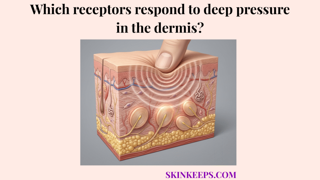
This baseline depends on a massive biological footprint. A 2021 review describes Pacinian corpuscles as large ovoid formations up to 5 × 3 mm distributed in the deep dermis and hypodermis, while the NCBI Neuroscience chapter places them directly in subcutaneous tissue and notes that they account for 10–15% of the cutaneous receptors in the hand (Cobo et al. — 2021 — Journal of Clinical Medicine / PMC) (Purves et al. — 2001 — Neuroscience / NCBI Bookshelf).
How does anatomical depth help Pacinian corpuscles respond to deep pressure?
Anatomical depth helps Pacinian corpuscles respond to deep pressure because receptors located in the deep dermis and hypodermis are exposed mainly to stronger transmitted force, not to weak superficial contact.
Light surface contact produces exceptionally shallow deformation that dissipates before reaching the deep connective tissue. By anchoring deeply, Pacinian corpuscles ensure they are only triggered when the applied mechanical force is strong enough or resonant enough to travel completely through the epidermal and dermal layers.
This deep placement directly connects to their low spatial sensitivity and broad-field deep-force detection. The 2015 PLOS Computational Biology model found that deep dermal placement gives the Pacinian corpuscle a larger receptive field and lower spatial sensitivity, because force location matters less when the receptor is embedded deeply. In the model, the dermal corpuscle center sat about 2.375 mm beneath the surface, meaning deep embedding actively reduces dependence on the exact point of skin indentation (Quindlen, Lai, and Barocas — 2015 — PLOS Computational Biology).
How does the structure of Pacinian corpuscles help them respond to deep pressure?
The structure of Pacinian corpuscles helps them respond to deep pressure because their onion-like lamellated capsule mechanically filters sustained compression and emphasizes transient deep-force changes.
This receptor is an engineering marvel comprised of an inner core housing the nerve ending, surrounded by an outer core of concentric lamellae (layers), enclosed by an outer fibrous capsule. Crucially, these concentric lamellar layers are separated by a fluid-filled space.
When steady, static pressure is applied to the skin, this fluid simply shifts sideways, effectively filtering out the constant load. The central nerve only fires when the pressure is dynamic or vibrating fast enough to overcome this liquid cushion.
The NCBI Neuroscience chapter describes the Pacinian corpuscle as having an onion-like capsule with an inner core separated from the outer lamellae by a fluid-filled space, while the PLOS model describes 30 or more collagenous lamellae around a central myelinated fiber. That layered arrangement is thought to function as a high-pass mechanical filter that shields the axon from low-frequency, high-amplitude sustained force (Purves et al. — 2001 — Neuroscience / NCBI Bookshelf) (Quindlen, Lai, and Barocas — 2015 — PLOS Computational Biology).
How does rapid adaptation explain how Pacinian corpuscles respond to deep pressure?
Rapid adaptation explains how Pacinian corpuscles respond to deep pressure because these receptors fire most strongly when force begins, changes rapidly, vibrates, or ends, not when heavy pressure simply remains constant.
In clinical neurobiology, a rapidly adapting receptor is a sensor that stops sending signals if a stimulus remains perfectly still. It generates an intense burst of signaling at the exact moment of impact (onset) and another burst when the pressure is removed (offset).
This biological design makes the corpuscle hyper-sensitive to continuous, rapid changes in compression—the exact definition of vibration. A steady, unmoving hand resting on a desk will silence the receptor within fractions of a second.
The NCBI Neuroscience chapter reports that Pacinian corpuscles respond to transient disturbances at 250–350 Hz, while the 2021 sensory-corpuscle review gives a wider operational vibration range of 20–1500 Hz with maximum sensitivity at 200–400 Hz. These ranges definitively support the idea that Pacinian corpuscles are tuned to rapidly changing deep-force events, not steady static pressure (Purves et al. — 2001 — Neuroscience / NCBI Bookshelf) (Cobo et al. — 2021 — Journal of Clinical Medicine / PMC).
How does mechanical transduction facilitate how Pacinian corpuscles respond to deep pressure?
Mechanical transduction facilitates how Pacinian corpuscles respond to deep pressure by converting deep tissue deformation into neural strain at the receptor’s central afferent axon.
Mechanotransduction is the direct physical translation of a mechanical bump into a biological electrical spark. The sequence operates precisely: deep force or high-frequency vibration reaches the corpuscle, the outer lamellae shift, the fluid transfers the kinetic energy, and the central axon is physically strained.
This mechanical strain rips open stretch-gated ion channels within the nerve, flooding the axon with electrical charge that fires the action potential straight to the spinal cord.
The 2015 PLOS model states that the working mechanism is stretch along the long axis of the receptor and central nerve fiber, which opens stretch-gated cation channels and initiates the response. In the same model, 10 μm surface indentations were sufficient to generate measurable strain within embedded Pacinian corpuscles, showing how small external displacement can be transformed into deep neural strain when the receptor architecture is properly engaged (Quindlen, Lai, and Barocas — 2015 — PLOS Computational Biology).
How do Pacinian corpuscles compare to other receptors in how they respond to deep pressure?
Pacinian corpuscles differ from other cutaneous receptors because they are the principal deep, rapidly adapting receptors for high-frequency vibration and deep pressure, not for fine surface touch, sustained pressure, or skin stretch.
While Meissner’s corpuscles catch flutter and light slip at the top of the dermis, Pacinian corpuscles handle the heavy, booming resonance deep below. Ruffini endings detect sustained, slow tissue stretching, while Merkel cells manage continuous, static fine-edge detection.
Because they are buried deep within the tissue, a single Pacinian corpuscle governs a massive receptive field, covering entirely broad areas of skin compared to the microscopic precision of surface receptors.
Master Comparison Table
| Receptor type | Primary stimulus | Main location | Adaptation | Main role |
|---|---|---|---|---|
| Pacinian corpuscles | Deep pressure, high-frequency vibration | Deep dermis / hypodermis | Rapidly adapting | Deep-force and vibration detection |
| Meissner’s corpuscles | Light touch, flutter | Superficial dermis | Rapidly adapting | Dynamic fine touch |
| Ruffini endings | Stretch, sustained force | Deeper dermis | Slowly adapting | Skin tension awareness |
| Merkel-cell system | Fine detail, sustained light pressure | Epidermis | Slowly adapting | Static form / edge detection |
The NCBI Neuroscience chapter reports that Meissner afferents account for about 40% of sensory innervation in the human hand, Merkel receptors about 25%, Ruffini receptors about 20%, and Pacinian corpuscles about 10–15%. It also distinguishes Pacinian corpuscles by their 250–350 Hz sensitivity, which is much higher than the 30–50 Hz low-frequency range emphasized for Meissner corpuscles (Purves et al. — 2001 — Neuroscience / NCBI Bookshelf).
What conditions impair the ability of Pacinian corpuscles to respond to deep pressure?
Peripheral neuropathic disease can impair the ability of Pacinian corpuscles to respond to deep pressure by reducing corpuscle number, distorting corpuscle structure, and weakening mechanosensory signaling.
While cosmetic conversations often focus on collagen, functional sensory loss stems from nerve and receptor degradation. It is vital to separate the proven destruction caused by neuropathy from the more subtle, systemic changes associated with normal chronological aging.
How does peripheral neuropathy impair how Pacinian corpuscles respond to deep pressure?
Peripheral neuropathy impairs how Pacinian corpuscles respond to deep pressure because diabetic neuropathic disease can reduce Pacinian corpuscle density, damage the inner core, and disrupt the corpuscle’s axonal and Schwann-like components.
In diabetic distal symmetric polyneuropathy, the metabolic toxicity fundamentally attacks the neural architecture inside the lamellated capsule, destroying the biological wire required to fire the mechanotransduction signal.
A 2021 Journal of Clinical Medicine paper reports that the number of Pacinian corpuscles was severely decreased in non-painful diabetic neuropathy and almost disappeared in painful diabetic neuropathy, with additional loss of axonal and Schwann-like cell marker expression and major inner-core destruction (García-Mesa et al. — 2021 — Journal of Clinical Medicine (MDPI)).
How should aging be framed when discussing Pacinian corpuscle function?
Aging should be framed cautiously when discussing Pacinian corpuscle function because current reviews report little or no major age-related structural alteration in Pacinian corpuscles themselves, even though broader tactile performance may still decline with age.
While the overall somatosensory system may exhibit slower processing speeds as decades pass, the Pacinian corpuscle structure largely resists the severe degeneration seen in superficial touch receptors.
Both the 2021 sensory-corpuscle review and the 2019 aging review indicate that Pacinian corpuscles generally show no relevant age-related alterations, meaning the aging subsection should be written carefully without exaggerated claims of Pacinian-specific degeneration (Cobo et al. — 2021 — Journal of Clinical Medicine / PMC) (García-Piqueras et al. — 2019 — Ageing of the Somatosensory System at the Periphery).
What are the key summary facts for how Pacinian corpuscles respond to deep pressure?
The key facts are that Pacinian corpuscles are deep rapidly adapting mechanoreceptors, their lamellated capsule filters steady force, and their central axon is specialized for deep pressure and high-frequency vibration signaling.
What steps can you take to support how Pacinian corpuscles respond to deep pressure?
Pacinian corpuscles respond to deep pressure best when the deep tissue environment and peripheral nerve pathway remain structurally intact.
Sensory Preservation Checklist
Quick Answers About How Pacinian Corpuscles Respond to Deep Pressure
What receptors respond to deep pressure in the dermis?
Pacinian corpuscles are the main receptors that respond to deep pressure in the dermis and nearby subcutaneous tissue. They are specialized for dynamic deep-force events and high-frequency vibration rather than delicate surface touch.
Where are Pacinian corpuscles located?
Pacinian corpuscles are located in the deep dermis, hypodermis, and even deeper structures such as interosseous membranes. Their deep position helps them respond to strong transmitted force rather than weak superficial contact.
Why do Pacinian corpuscles detect vibration so well?
Pacinian corpuscles detect vibration well because their lamellated capsule and rapid adaptation make them highly sensitive to fast-changing mechanical events, especially high-frequency vibration in the hundreds-of-hertz range.
How are Pacinian corpuscles different from Meissner’s corpuscles?
Pacinian corpuscles are deeper and more vibration-focused, while Meissner’s corpuscles are superficial and specialized for dynamic light touch and flutter. Pacinian sensitivity peaks in a much higher frequency band.
Can neuropathy damage Pacinian corpuscles?
Yes. Diabetic neuropathy can severely reduce Pacinian corpuscle number and distort their structure, especially in more advanced or painful forms of distal diabetic sensorimotor polyneuropathy.
Conclusion
In conclusion, Pacinian corpuscles respond to deep pressure because their deep placement, lamellated mechanical filter, and rapid adaptation pattern are all tuned to dynamic deep-force and vibration signaling.
Anchored far below the surface in the deep reticular dermis and hypodermis, their unique onion-like structure effectively dissipates sustained, static force, ensuring that the central nerve only fires during abrupt mechanical changes. This explicit mechanotransduction ensures that the skin’s warning system can flawlessly distinguish a high-frequency vibration from a gentle surface brush, though diseases like diabetic neuropathy threaten to sever this crucial biological wiring permanently.