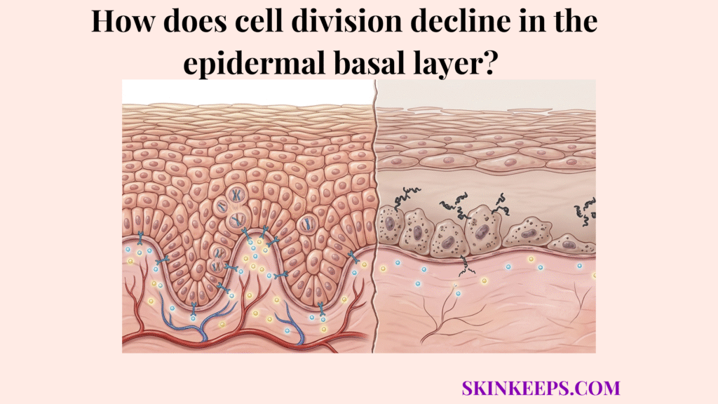
Cell division declines in the epidermal basal layer when the skin’s renewal cells become less efficient at entering, sustaining, and completing mitosis over time. As basal-cell signaling weakens, metabolic support falls, and the renewal reserve becomes less robust, fewer fresh keratinocytes are produced and the epidermis renews itself more slowly.
This decline does not only affect glow or smoothness. When basal-layer division slows, the skin often becomes less full, slower to recover, more fragile, and less efficient at maintaining a strong outer barrier. That is why basal-layer slowdown is closely tied to dullness, thinning, and reduced resilience. Reviews of epidermal aging describe this broader decline as involving lower proliferative activity, reduced growth-factor support, and diminished tissue renewal over time.
Understanding epidermal basal-layer cell division decline—the gradual loss of efficiency in basal-cell mitotic renewal—makes it easier to explain what fails first, how the slowdown progresses, what signs reveal weaker renewal output, and what actually helps support aging or stressed skin.
What begins first in epidermal basal-layer cell division decline?
Epidermal basal-layer cell division decline often begins as a loss of renewal efficiency before it becomes obvious as visible skin aging. The basal layer is the deepest epidermal layer where new keratinocytes are generated through mitosis, the process of cell division that produces replacement cells.
The earliest change is often reduced efficiency in the basal renewal pool rather than total absence of dividing cells. Initial biological fatigue occurs long before the surface barrier feels thin or reactive, creating a gap between deep cellular health and surface appearance.
Basal cells may still be present, but they divide less consistently, less productively, or with weaker regenerative persistence. In Rübe et al., 2021, the proportion of Ki67-positive epidermal keratinocytes fell from 8.5 ± 0.3% in young skin to 6.3 ± 0.3% in middle-aged skin and 4.2 ± 0.2% in aged skin, which strongly supports the idea that decline begins as weaker proliferative activity rather than sudden failure.
How does reduced stem-cell reserve drive epidermal basal-layer cell division decline?
Reduced stem-cell reserve—the pool of renewal-capable cells that supports ongoing epidermal replacement—drives epidermal basal-layer cell division decline because the skin loses part of the cellular depth it depends on for consistent renewal.
The basal layer depends on a stable pool of renewal-capable cells, and over time that pool becomes less robust and less reliable. This depletion limits the “emergency backup” cells required during times of barrier injury or environmental stress, making the epidermis increasingly vulnerable.
The problem is not always total absence of basal cells; it is often reduced regenerative strength within the cells that remain. Rübe et al., 2021 also found that H2A.J-positive basal-layer cells rose from about 5% in young epidermis to about 20% in aged epidermis, while integrin-α6–positive basal stem/progenitor compartments showed evidence of age-related decline, supporting the idea that reserve quality worsens as proliferative aging advances.
How does weaker signaling drive epidermal basal-layer cell division decline?
Weaker signaling drives epidermal basal-layer cell division decline by reducing the cell-cycle signaling—molecular instructions that help cells enter and complete division—that normally keep renewal active and well coordinated.
Basal cells rely on molecular cues that regulate entry into the cell cycle, persistence in division, and orderly replacement output. Without strong signals, cells may remain in a resting phase for longer periods, resulting in a delayed renewal rhythm and a surface that looks less fresh.
With age or chronic stress these signals become less effective, so the rate and quality of division decline. Wang et al., 2020 specifically describe reduced epidermal growth-factor signaling alongside lower keratinocyte proliferation in aging skin, which fits this signaling-loss model.
How do growth and differentiation signals weaken during epidermal basal-layer cell division decline?
Growth and differentiation signals weaken during epidermal basal-layer cell division decline when the molecular cues that regulate cell-cycle entry and coordination become less effective over time. This breakdown in communication ensures that the basement membrane provides less “instruction” to the cells anchored upon it, leading to a stalling of turnover.
As these cues fade, the orderly progression of cells from the base to the surface becomes erratic. This contributes to the uneven skin texture and persistent dullness that characterizes aging tissue.
Maintaining strong intracellular communication is vital; without it, the basal layer cannot sustain the high-volume production required to keep the stratum corneum durable and reflective.
How does hormonal change contribute to epidermal basal-layer cell division decline?
Hormonal change contributes to epidermal basal-layer cell division decline by altering the tissue environment that normally helps support steady renewal. This does not create basal decline alone, but it can lower the quality of the environment in which renewal must occur.
Hormonal shifts, such as those occurring during menopause or the late Luteal phase where androgen dominance can shift tissue behavior, directly influence the metabolic speed of keratinocyte factories.
Lower levels of estrogen, in particular, are associated with thinner epidermis and reduced mitotic activity, reinforcing the fact that systemic health is mirrored in basal cellular speed.
How does lower cellular energy worsen epidermal basal-layer cell division decline?
Lower cellular energy worsens epidermal basal-layer cell division decline because dividing cells need strong metabolic support to sustain renewal output. Mitosis is inherently energy-demanding, requiring significant ATP stores to manage chromosome replication and cellular split.
Aging and chronic stress reduce how efficiently cells maintain and use the resources needed for productive cycling. This metabolic fatigue makes division slower, less consistent, and less resilient under external environmental pressure, leading to a visibly thinner surface.
Declining basal-layer function should be framed as a support failure as well as a signaling failure. Wang et al., 2020 describe epidermal aging as involving metabolic and functional deterioration rather than simple chronological slowdown alone, highlighting the importance of cellular fuel.
How does epidermal basal-layer cell division decline reduce epidermal output over time?
Epidermal basal-layer cell division decline reduces keratinocyte output—the supply of new epidermal cells produced from below—by shrinking the stream of new cells that normally replenishes the skin from below.
When fewer basal cells divide successfully, fewer keratinocytes move upward through the epidermal layers. This “supply shortage” results in a thinner tissue architecture that is less capable of maintaining its protective density and youthful volume.
Over time this reduces thickness, slows turnover, and weakens visible renewal. Abd El-Aal et al., 2012 reported mean epidermal thickness of 39.84 ± 7.4 μm in younger skin versus 21.81 ± 5.4 μm in older skin, providing direct evidence that lower long-term epidermal output is reflected in thinner aged tissue.
How do aging and chronic stress accelerate epidermal basal-layer cell division decline?
Aging and chronic stress accelerate epidermal basal-layer cell division decline by increasing damage pressure while reducing the skin’s ability to renew itself efficiently.
Aging weakens renewal reserve and recovery speed, while chronic stress adds UV burden, inflammation, and pollution. The result is a faster drop in productive basal-layer division because the skin is being asked to repair more while renewing less efficiently, creating a “deficit” in cellularity.
Wang et al., 2020 characterize epidermal aging as a compounded decline in proliferative and functional capacity, which aligns with this amplified-stress model. Chronic barrier irritation further worsens this by forcing the epidermal barrier into a disruption cycle.
How does aging accelerate epidermal basal-layer cell division decline?
Aging accelerates epidermal basal-layer cell division decline by weakening renewal reserve, signaling quality, and recovery speed over time. This makes basal division slower and less resilient as the epidermis loses renewal momentum, resulting in a surface that stays rough for longer periods.
This intrinsic decline becomes more visible as the skin produces fewer fresh cells with the same consistency. Over decades, the “factory” simply produces a lower volume of units, leading to the hallmark thinning seen in mature skin.
Biological markers of aging, such as senescent cell accumulation, act as background noise that distracts the remaining healthy cells from their primary task of division.
How does chronic stress accelerate epidermal basal-layer cell division decline?
Chronic stress accelerates epidermal basal-layer cell division decline by adding cumulative repair demand to a renewal system that is already becoming less efficient. UV exposure, inflammation, pollution, and over-exfoliation all increase the stress load, weakening renewal efficiency.
Repetitive stress forces the basal layer to spend its limited energy on survival and emergency patching rather than robust, orderly replacement. This trade-off ensures that visible slowdown appears earlier than it would in protected skin.
Long-term environmental load acts as an extrinsic accelerator, pushing the basal compartment into premature fatigue and reducing the overall resilience of the skin barrier.
Which signs suggest epidermal basal-layer cell division decline is becoming more visible?
Epidermal basal-layer cell division decline may be becoming more visible when the skin looks slower, less radiant, less smooth, and less responsive to recovery. Recognizing these shifts allows for early intervention using a Barrier Health Checker.
Warning signs can include duller skin tone, slower fading of post-inflammatory marks, and skin that looks older even without obvious inflammation. These visible signs reflect weaker output from below rather than only surface dryness or roughness.
Skin-aging reviews consistently link declining basal renewal with reduced epidermal freshness and slower recovery characteristics. This corresponds to the structural reality that surface texture is anchored by deep regenerative health and consistent mitotic flux.
How does healthy basal renewal compare with epidermal basal-layer cell division decline?
Healthy basal renewal keeps the epidermis well supplied and responsive, while epidermal basal-layer cell division decline reduces output and slows visible renewal.
| Renewal State | Basal Mitosis | Recovery Speed | Visible Quality |
|---|---|---|---|
| Healthy Renewal | Strong and efficient | Faster | Resilient & Smooth |
| Decline State | Slower and less efficient | Slower | Dull & Less Responsive |
This comparison confirms that the defining difference is not just surface texture, but the speed of the underlying renewal engine. Healthy skin refreshes itself effortlessly, while skin in decline struggles to clear older cells and replace them with fresh ones.
What factors make epidermal basal-layer cell division decline worse?
Epidermal basal-layer cell division decline worsens when cumulative damage and chronic stress reduce renewal capacity faster than the skin can compensate. Chronic UV exposure and repeated barrier damage are the primary external accelerators.
Ignoring early signs of slowdown and pushing the skin with harsh, frequent treatments often backfires. Aging-skin literature consistently supports this cumulative-burden model, where stress keeps accumulating while reserve, signaling, and recovery quality all keep weakening simultaneously.
One major but hidden driver is why hot water strips protective oils from the epidermis, as thermal stress adds more repair demand to an already fatigued renewal system.
What habits and ingredients help support skin affected by epidermal basal-layer cell division decline?
The best support for skin affected by epidermal basal-layer cell division decline combines careful renewal support, lower barrier stress, and consistent UV protection. The goal is to protect remaining renewal capacity and support productive turnover carefully.
Routine consistency is vital; sporadic high-intensity treatments often cause more harm than good by triggering inflammation. A long-term maintenance approach ensures the basal layer has the stability it needs to function at its best.
Targeting the biological source requires highly specific, clinically proven ingredients that reach the deeper layers of the epidermis without causing a collapse in surface tolerance.
How do retinoids help support skin affected by epidermal basal-layer cell division decline?
Retinoids help support skin affected by epidermal basal-layer cell division decline by improving renewal signaling over time when they are introduced carefully. They bind to nuclear receptors that directly stimulate cellular proliferation and normalize the entire turnover cycle from the deepest layers upward.
Retinoids are infinitely more relevant here than physical scrubs because they influence the deep biology of cellular turnover. In Mukherjee et al., 2006, histologic studies of topical tretinoin showed increased epidermal thickness within the first 3 to 6 months, while some compaction changes were reported as early as 15 days in certain studies.
The core clinical goal is to achieve sustainable renewal support. Using a tool like the Routine Stability Index (RSI) can help you ensure that your retinoid usage does not exceed your skin’s capacity for recovery.
How does barrier support help skin affected by epidermal basal-layer cell division decline?
Barrier support helps skin affected by epidermal basal-layer cell division decline because a stable surface reduces the stress burden on already slower renewal systems. Lower irritation gives aging skin more capacity for productive recovery instead of spending that capacity on repeated repair.
Simple, non-stinging care is mandatory. Lueangarun et al., 2019 found that a ceramide-containing moisturizer improved hydration, TEWL, and pH for up to 24 hours after a single application, supporting barrier care as a practical support strategy for slower-renewing skin.
Stable hydration ensures the enzymatic environment in the granular layer remains functional, allowing cells to mature correctly as they are pushed upward from the basal compartment.
How does sun protection help slow epidermal basal-layer cell division decline?
Sun protection helps slow epidermal basal-layer cell division decline by reducing cumulative UV injury to skin that is already renewing more slowly. Daily SPF lowers one of the most important external accelerators of structural decline and DNA mutation.
Protection helps preserve renewal quality over time rather than allowing cumulative damage to deepen the slowdown. Berry, 2023 and Quan, 2023 both frame photoprotection and topical retinoids as core preventive supports in skin aging rather than optional add-ons.
By cutting off the UV trigger, the basal layer can dedicate its energy to maintenance and renewal rather than constant, high-intensity DNA repair protocols.
Problem: epidermal basal-layer cell division decline is increasing
Implication: fewer fresh cells are being produced and recovery is slowing
Solution: support renewal carefully, protect the barrier, and reduce cumulative stress
What are the key takeaways about epidermal basal-layer cell division decline?
The key takeaways about epidermal basal-layer cell division decline are that renewal reserve, signaling, and energy support become less efficient over time, which lowers keratinocyte output and visible renewal.
Summary Points
- Epidermal basal-layer cell division decline happens when renewal reserve, signaling, and energy support become less efficient over time
- The result is lower keratinocyte output, slower upward replacement, and weaker visible renewal
- Aging and chronic stress accelerate the decline by increasing damage pressure and reducing recovery quality
- The best support strategy focuses on renewal support, barrier stability, and daily protection rather than aggressive forcing
Frequently Asked Questions
Does epidermal basal-layer cell division decline mean the skin stops renewing?
The skin does not usually stop renewing completely. The problem is reduced efficiency, weaker output, and less resilient recovery over time, resulting in a thinner and more fragile tissue architecture that looks duller.
Is epidermal basal-layer cell division decline only an aging issue?
Aging is a major driver, but chronic UV exposure, inflammation, harsh routines, and repeated barrier stress can accelerate the decline significantly even in younger adults, making early prevention vital.
Can faster exfoliation fix epidermal basal-layer cell division decline?
No. Forcing the surface through aggressive exfoliation does not directly restore basal mitotic efficiency and may worsen barrier stress if overdone, potentially accelerating the deep cellular decline.
What helps most when epidermal basal-layer cell division decline is already visible?
The most useful strategy is careful renewal support plus barrier protection plus daily UV defense, rather than aggressive overcorrection that might inflame the already sensitive surface and slow recovery further.
What daily steps help reduce the impact of epidermal basal-layer cell division decline?
Daily steps help reduce the impact of epidermal basal-layer cell division decline by lowering cumulative stress and supporting the skin’s reduced renewal capacity.
Long-term resilience matters more than fast surface correction when renewal systems are already becoming less efficient. Shifting from “stripping” to “supporting” is the key behavioral change required for healthy aging skin.
Daily Renewal Support Checklist
Basal-layer division does not usually stop suddenly. It becomes less efficient, less productive, and less resilient over time.
Build your routine around protection, lower stress, and careful renewal support if your goal is stronger long-term epidermal resilience.