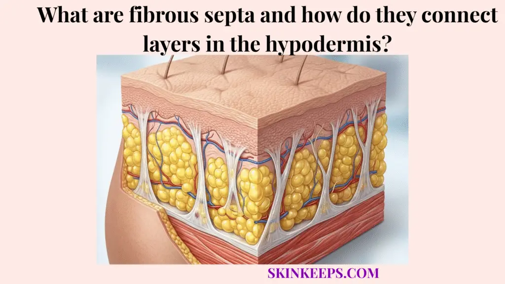
Fibrous septa are collagen-rich connective tissue bands in the hypodermis that divide adipose lobules, carry vessels and nerves, and connect the dermis to deeper fascial support layers. These structures ensure the subcutaneous tissue functions as an integrated structural network rather than a loose layer of free-floating fat.
This guide covers the biological composition and mechanical importance of this deep connective tissue web. We explain how dermis-to-fascia bridging supports skin shape, how structural breakdown occurs, and how to approach clinical options and daily preservation realistically.
Why is the deep connective tissue web important for skin architecture?
The deep connective tissue web is important for skin architecture because fibrous septa create internal support that keeps the dermis connected to fat compartments, vessels, nerves, and deeper fascial planes. This internal linkage ensures that the skin maintains its predictable position over underlying structures.
By distributing force and tension across a broad area, the hypodermis supports adipose storage, insulation, and structural anchoring simultaneously.
How do fibrous septa support skin firmness and contour?
Fibrous septa support skin firmness and contour by creating tension-bearing connective pathways between the dermis, adipose compartments, and fascia. These internal scaffolding bands prevent tissue from simply collapsing under its own weight.
This organized tension provides the necessary resistance to maintain a stable, predictable surface contour over soft fat layers.
How do fibrous septa contribute to biomechanical shock absorption?
Fibrous septa contribute to biomechanical shock absorption by distributing pressure through connective tissue pathways while adipose lobules compress under mechanical load. The compressible fat acts as soft padding, while the tensile bands catch and spread the kinetic energy across the network.
This coordinated force distribution reduces concentrated stress that could otherwise rupture delicate local blood vessels.
What is the biological composition of fibrous septa within subcutaneous tissue?
The biological composition of fibrous septa within subcutaneous tissue is mainly connective tissue matrix containing collagen, elastin, fibroblasts, vessels, nerves, and supporting extracellular material. These structures consist primarily of type I collagen and type III collagen interwoven with elastic fibers to create a resilient mesh.
This living tissue actively adapts and remodels to maintain the structural continuity of the hypodermis.
How do collagen and elastin form the septal matrix?
Collagen and elastin form the septal matrix by giving fibrous septa tensile strength, flexibility, and recoil inside the hypodermal connective network. Collagen fibers provide the rigid, tension-bearing strength needed to hold layers together without tearing.
Elastin complements this rigidity by allowing the bands to stretch slightly and return to their original shape after mechanical loading.
How do fibroblasts maintain the fibrous septal web?
Fibroblasts maintain the fibrous septal web by producing and remodeling extracellular matrix proteins that help repair ordinary connective tissue wear. These specialized cells constantly monitor tissue tension and synthesize new collagen to replace degraded fibers.
This ongoing cellular turnover is essential for keeping the septal architecture strong and functional over time.
How does this fascial web physically bridge the dermis to the underlying muscle fascia?
This fascial web physically bridges the dermis to underlying muscle fascia by extending connective septa from the reticular dermis through subcutaneous fat toward superficial fascia and deeper fascial support planes. This vertical and oblique network acts as a structural tie-down system.
By spanning the entire thickness of the fat layer, the hypodermis anchors the skin to deeper structures effectively.
How do fibrous septa attach to the reticular dermis?
Fibrous septa attach to the reticular dermis by blending into the deeper collagen network of the dermis and creating an upper anchoring zone for subcutaneous connective tissue. This continuous integration ensures that upward pressure and downward tension are transferred smoothly between the skin and fat.
Without this seamless junction, the layers would easily shear apart during routine body movement.
How do fibrous septa attach to superficial fascia?
Fibrous septa attach to superficial fascia by traveling downward through adipose lobules and linking subcutaneous compartments to deeper connective tissue planes. In facial anatomy, this connection often interacts with the superficial musculoaponeurotic system (SMAS) to support complex expressive movements.
This basal attachment provides the necessary foundation to keep the entire tissue block grounded.
| Anatomical Layer | Type of Attachment | Biomechanical Function |
|---|---|---|
| Reticular dermis | Upper collagen integration | Anchors septa to the dermal support layer |
| Hypodermal fat lobules | Compartmental separation | Organizes adipose tissue into stable regions |
| Fibrous septa | Vertical/oblique connective bridges | Carry tension and guide neurovascular pathways |
| Superficial fascia | Lower connective support plane | Links subcutaneous tissue to deeper structures |
| SMAS / regional fascia | Region-specific facial support | Coordinates facial soft tissue support |
Why is compartmentalization of adipose lobules essential for structural stability?
Compartmentalization of adipose lobules is essential for structural stability because fibrous septa divide soft fat into organized units that resist uncontrolled shifting while allowing vessels and nerves to pass through the tissue. This division ensures that fat remains properly positioned rather than sliding freely under the skin.
Because hypodermal anchoring affects skin mobility, these structured pockets maintain body shape during active gliding.
How do fibrous septa limit excessive adipose shifting?
Fibrous septa limit excessive adipose shifting by creating connective tissue partitions that hold adipose lobules in organized compartments. These walls act like a honeycomb, encasing small groups of adipocytes to preserve localized volume.
This containment prevents the soft lipid tissue from pooling or migrating unevenly under gravitational stress.
How do fibrous septa act as pathways for vessels, lymphatics, and nerves?
Fibrous septa act as pathways for vessels, lymphatics, and nerves because connective tissue partitions provide structured routes through the subcutaneous fat layer. These neurovascular bundles travel safely within the collagen-rich walls rather than being crushed by shifting fat.
This architectural design ensures steady tissue perfusion and sensory continuity across the hypodermal bridge.
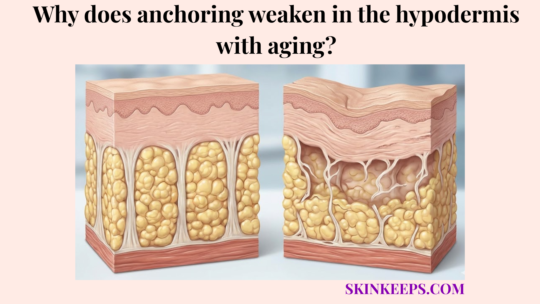
Which metabolic factors can weaken inter-layer connective bridges?
Metabolic factors can weaken inter-layer connective bridges when inflammation, UV-induced enzymes, hormonal change, or collagen aging disrupt the extracellular matrix that supports fibrous septa. As these biochemical stressors accumulate, anchoring weakens in the hypodermis with aging.
This gradual loss of structural integrity leads to reduced tissue resilience and diminished contour stability.
How do matrix metalloproteinases degrade septal collagen?
Matrix metalloproteinases degrade septal collagen by breaking down extracellular matrix proteins when inflammatory or UV-related signaling increases enzyme activity. These specialized MMPs cleave existing collagen fibers, which may compromise the tensile strength of the local septal network over time.
This continuous enzymatic degradation steadily weakens the mechanical connections holding the tissue layers together.
How can menopausal estrogen decline affect collagen maintenance?
Menopausal estrogen decline can affect collagen maintenance by reducing hormonal support for skin thickness, fibroblast activity, and extracellular matrix renewal. As estrogen levels drop during menopause, fibroblasts often decrease their overall production of new structural proteins.
Some menopause skin reviews cite up to 30% collagen loss in the first 5 years after menopause, representing a general skin collagen context that may also affect broader subcutaneous support. [PMC]
What misconceptions should be avoided when repairing a compromised subdermal connective network?
Misconceptions should be avoided when repairing a compromised subdermal connective network because deep connective tissue support cannot be restored by surface products or forceful manual techniques alone. Many aesthetic claims confuse superficial skin smoothing with actual hypodermal bridging reconstruction.
Understanding tissue depth helps patients avoid spending time on methods incapable of remodeling deep septa.
Why can’t topical collagen rebuild fibrous septa?
Topical collagen cannot rebuild fibrous septa because topical collagen products mainly act at or near the skin surface and cannot mechanically reconstruct deep hypodermal connective pathways. The molecules are typically too large to reach the subcutaneous tissue, and surface hydration does not trigger deep fascial repair.
While helpful for epidermal texture, they do not reinforce the internal architectural web.
Why can’t deep massage reconnect degraded fibrous bands?
Deep massage cannot reconnect degraded fibrous bands because manual pressure may mobilize tissue temporarily, but it cannot rebuild collagen architecture or restore lost attachment points. Pressing forcefully on the skin does not stimulate fibroblasts to bridge micro-tears in the septal matrix.
In fact, excessively aggressive manipulation may cause unnecessary inflammation in already fragile connective tissue.
Which clinical modalities may reinforce degraded hypodermal retaining bands?
Clinical modalities may reinforce degraded hypodermal retaining bands by stimulating collagen remodeling, heating selected tissue layers, or supporting extracellular matrix renewal in clinician-selected patients. Since energy-based devices tighten dermal tissue, these targeted therapies aim to provoke a localized biological healing response.
Results vary, but they offer a mechanical approach to supporting septal resilience.
How do radiofrequency microneedling and ultrasound support collagen remodeling?
Radiofrequency microneedling and ultrasound may support collagen remodeling by delivering controlled energy into selected skin or subcutaneous layers and triggering wound-healing or thermal remodeling responses. Radiofrequency microneedling drives heat directly into the tissue, while microfocused ultrasound (HIFU or MFU-V) targets deeper fascial planes.
MFU-V protocols can use treatment depths such as 1.5 mm, 3.0 mm, and 4.5 mm, with some systems providing visualization up to 8.0 mm for precise layer targeting. [JCAD]
How do PLLA and CaHA support injectable biostimulation?
PLLA and CaHA support injectable biostimulation by acting as collagen-stimulating materials that may improve skin quality or connective tissue support when placed appropriately by trained clinicians. Poly-L-lactic acid (PLLA) and calcium hydroxylapatite (CaHA) incite a mild inflammatory response that encourages local fibroblasts to lay down new collagen.
This controlled biostimulation helps maintain tissue architecture over several months, rather than providing instant structural lifting.
| Procedure | Depth / Target Concept | Main Target | Collagen Stimulation Type |
|---|---|---|---|
| Radiofrequency microneedling | Dermal or subdermal energy delivery | Dermal matrix and selected support planes | Wound-healing + thermal remodeling |
| Microfocused ultrasound / HIFU | Layer-targeted focused ultrasound | Dermis/subdermis/deeper support layers | Thermal coagulation + remodeling |
| PLLA | Injectable collagen stimulation | Dermal/subdermal tissue quality | Gradual collagen biostimulation |
| CaHA | Injectable collagen stimulation | Dermal/subdermal support | Immediate scaffold + collagen stimulation |
| Retinoids / topical actives | Epidermal/dermal biology | Surface-to-dermal support | Dermal remodeling support, not septal repair |
| Topical collagen | Skin surface | Hydration/surface texture | Does not rebuild deep septal architecture |
What daily checklist helps protect deep structural fascial connections?
A daily checklist helps protect deep structural fascial connections by supporting collagen maintenance, reducing avoidable mechanical strain, and protecting connective tissue from inflammation, UV damage, and extreme tissue stress. Preserving the existing septal network is far more reliable than attempting to rebuild it later.
Simple, consistent habits offer the safest path to long-term structural preservation.
How can nutrition support septal collagen maintenance?
Nutrition can support septal collagen maintenance by providing adequate amino acids, vitamin C, copper, zinc, and overall dietary sufficiency for normal extracellular matrix turnover. These essential nutrients support ongoing fibroblast activity without requiring extreme dietary restrictions or high-dose supplementation.
A consistently balanced diet ensures the tissue has the resources to execute normal micro-repairs.
How can biomechanics protect the hypodermal connective web?
Biomechanics can protect the hypodermal connective web by reducing repeated extreme stretching, high-shear stress, and avoidable trauma to the subcutaneous support network. Gradual weight management prevents rapid expansion from overloading the fibrous septa.
During high-impact movement, proper supportive garments can also improve comfort and reduce localized strain on the connective bridging.
Daily Septal Architecture Preservation Checklist
FAQs About Fibrous Septa and Hypodermal Layer Connections
Are fibrous septa the same as retinacula cutis?
Fibrous septa and retinacula cutis are closely related terms used to describe connective tissue bands that travel through subcutaneous fat. They help link the dermis to deeper fascia while organizing adipose lobules and neurovascular pathways.
Do fibrous septa only hold fat in place?
No. Fibrous septa do more than hold fat in place. They connect layers, distribute force, support tissue contour, guide vessels and nerves, and help the hypodermis function as an architectural bridge between skin and deeper support planes.
How do fibrous septa connect the dermis to fascia?
Fibrous septa connect the dermis to fascia by extending from the deep dermal collagen network through adipose lobules toward superficial fascia or regional fascial planes. This creates continuity between skin, subcutaneous tissue, and deeper support structures.
Can topical collagen rebuild fibrous septa?
No. Topical collagen cannot rebuild deep fibrous septa. It may support surface hydration or texture, but septal repair requires deeper extracellular matrix remodeling than topical collagen can mechanically provide.
Can massage reconnect weak fibrous septa?
Massage cannot reconnect weak fibrous septa or rebuild lost collagen architecture. Gentle massage may support comfort or tissue mobility, but forceful pressure should not be presented as a way to reconstruct deep connective bridges.
Which treatments may support septal collagen remodeling?
Energy-based treatments and injectable biostimulators may support collagen remodeling in selected patients. MFU-V protocols can use treatment depths such as 1.5 mm, 3.0 mm, and 4.5 mm when targeting skin support layers. [JCAD]
How can daily habits protect fibrous septa?
Daily habits may protect fibrous septa by reducing avoidable mechanical strain and supporting collagen maintenance. Gradual weight change, regular movement, UV protection, adequate nutrition, and avoiding painful mechanical tools all support a healthier tissue environment.
Conclusion
Fibrous septa connect layers in the hypodermis by organizing adipose lobules, linking the dermis to fascia, and providing structured pathways for vessels, lymphatics, and nerves. This connective web helps the hypodermis act as a stable architectural layer rather than a loose fat compartment.
At SkinKeeps, we explain skin structure through evidence-based dermatology and anatomy. Understanding fibrous septa helps readers see how deep connective tissue supports skin shape, mobility, and long-term resilience.