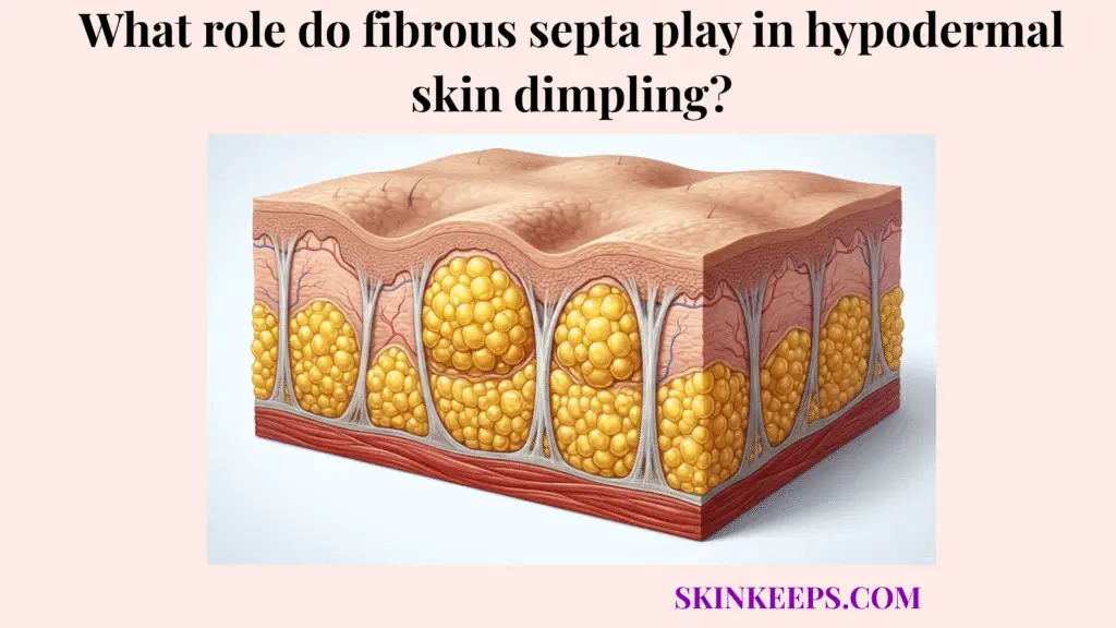
Fibrous septa play a central role in hypodermal skin dimpling because these collagen-rich connective tissue bands anchor the skin to deeper structures while dividing subcutaneous fat into compartments. This anchoring creates septal tension across the dermal-fat interface, which fundamentally shapes the visible skin surface.
This guide covers how connective tissue architecture drives cellulite-related dimpling, why certain biological factors increase tissue tension, and which clinical treatments target these specific bands. The focus remains strictly on the structural mechanics of the connective network rather than treating dimpling as a simple issue of excess fat.
What exactly are fibrous septa and how does their anatomical anchoring set the stage for skin dimpling?
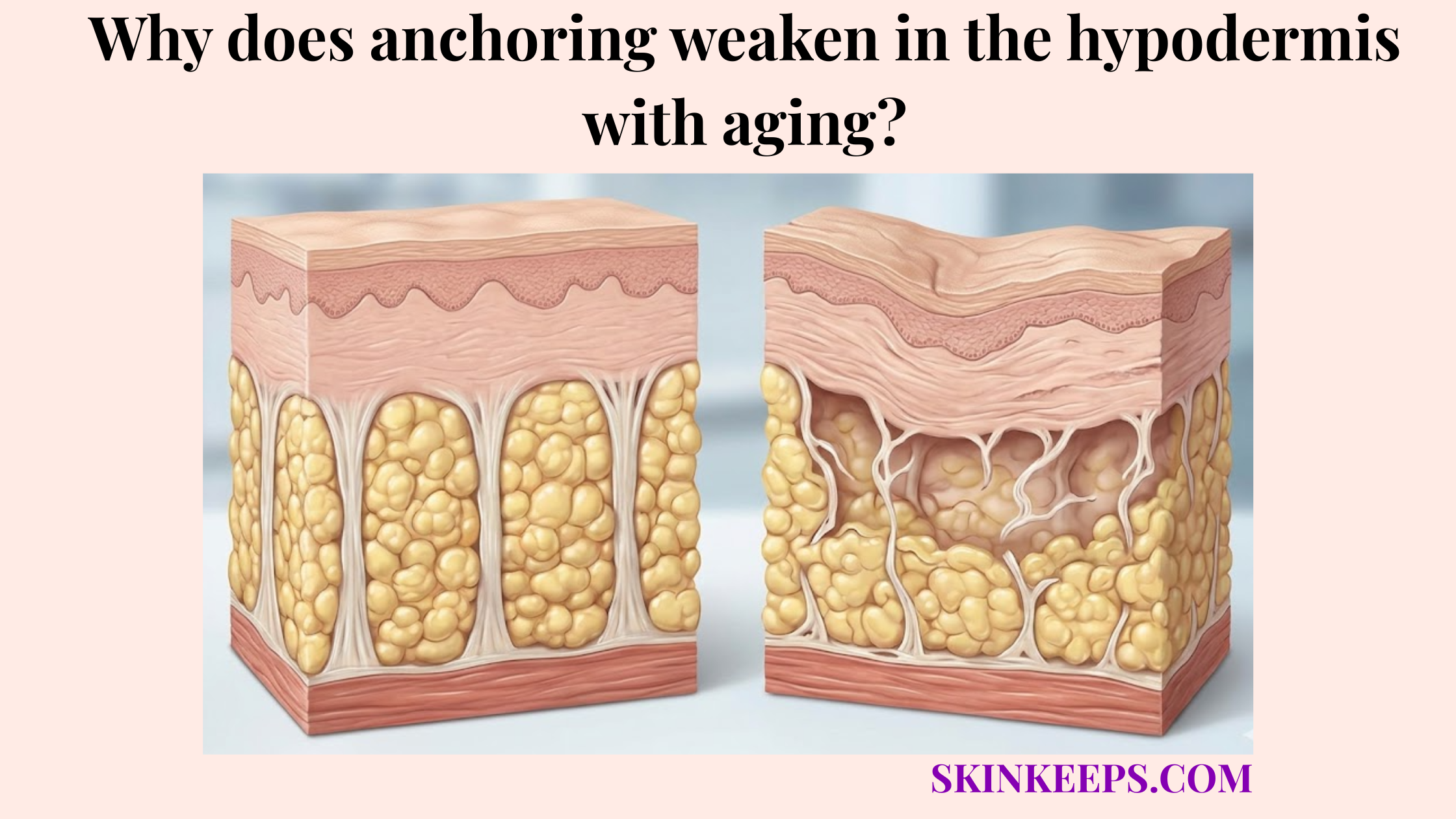
Fibrous septa set the stage for skin dimpling because they anchor the dermis to deeper fascia while separating subcutaneous fat into compartments that can bulge unevenly when tissue tension changes. These connective tissue bands actively organize the hypodermis, preventing it from functioning as a free-floating liquid layer.
When these structural anchors tighten, shorten, or encounter expanding fat volume, they pull the skin surface inward. This dynamic creates the visible surface depressions known as cellulite.
How do fibrous septa act as biological retaining walls?
Fibrous septa act as biological retaining walls by compartmentalizing subcutaneous fat and creating collagen-based connections between the dermis, hypodermis, and deeper fascia. This fat compartmentalization forms a necessary structural support network beneath the skin surface.
Cellulite affects approximately 80–90% of postpubertal women, which supports explaining these septal mechanics as a common skin architecture pattern rather than a rare disorder. [PubMed]
How does the dermal-fascial connection influence skin surface smoothness?
The dermal-fascial connection influences skin surface smoothness because elastic septa allow the skin to move over deeper tissue, while shortened or rigid septa restrict that movement at fixed anchor points. Normal, flexible bands permit a smooth tissue glide when the body moves.
Conversely, stiffened anchor points restrict this movement, pulling the surface inward. This restriction forms the biological foundation of a visible dimple.
Why do sclerotic connective tissue bands cause visible surface dimpling?
Sclerotic connective tissue bands cause visible surface dimpling because stiffened septa pull selected points of the skin downward while nearby fat compartments push upward. This sclerosis involves a distinct loss of flexibility within the fibrous septa, turning normal anchors into rigid cords.
The opposing forces of downward pull and upward pressure create the classic uneven topography of hypodermal skin dimpling. Addressing this condition requires targeting the rigid bands rather than focusing solely on adipocyte volume.
How does the tethering effect create the downward pull of a dimple?
The tethering effect creates the downward pull of a dimple when a shortened or stiffened septum holds one skin point lower than the surrounding surface. This localized tethering concentrates tension at fixed anchor points along the dermal-fat interface.
Aging, inflammation, or glycation may contribute to this septal stiffness, turning flexible bands into unyielding tethers. The Nürnberger–Müller scale grades cellulite from 0 to 3, with higher grades reflecting more visible dimpling at rest or with standing. [PMC]
How does adipose herniation amplify the visible dimple?
Adipose herniation amplifies the visible dimple because fat lobules press upward around tethered septa, making the pulled-down skin point look deeper by comparison. This upward pressure originates from adipocytes expanding within their confined subcutaneous compartments.
This upward protrusion severely contrasts with the downward tethering, maximizing the visible depth of the surface depression. Cleveland Clinic reports that 80–90% of women after puberty have cellulite, while less than 10% of men have cellulite. [Cleveland Clinic]
| Anatomical Component | Normal State | Sclerotic / Dimpled State | Visible Surface Result |
|---|---|---|---|
| Fibrous septa | Flexible connective bands organize fat compartments | Shortened or stiffened bands pull downward | Localized dimples |
| Fat lobules | Sit evenly within subcutaneous compartments | Press upward around tethered septa | Raised areas beside depressions |
| Dermis | Smooth surface layer over fat compartments | Shows uneven tension from below | Dimpled surface pattern |
| Fascia | Deep anchoring layer | Acts as lower anchor for tense septa | Fixed pull lines |
| Collagen network | Provides strength and flexibility | May stiffen or lose elasticity | Reduced tissue glide |
How does tissue architecture differ between genders regarding hypodermal tethering?
Tissue architecture differs between genders regarding hypodermal tethering because septal orientation and fat-compartment structure can make upward fat protrusion more visible in many female patterns than in many male patterns. The female tissue architecture often features bands that run perpendicularly to the skin surface.
By contrast, the male connective tissue network generally features oblique, crisscrossing bands that distribute tension more evenly. This anatomical variance heavily dictates regional vulnerability to visible dimpling.
Why does the female vertical matrix make dimpling more visible?
The female vertical matrix can make dimpling more visible because many female septa run more perpendicular to the skin, allowing fat lobules to protrude upward between vertical septa. This direct vertical alignment offers little structural resistance to expanding subcutaneous fat compartments.
When these fat lobules press outward, the parallel vertical bands pull straight down, rendering the protrusion highly apparent. A 2023 review describes female fibrous septa as oriented vertically to the dermis, while male septa are oriented at approximately 45° and crisscross through subcutaneous tissue. [PMC]
Why does the male crisscross network reduce visible protrusion?
The male crisscross network can reduce visible protrusion because oblique septa distribute tension in multiple directions and limit the upward bulging pattern that creates dimples. This crisscross septal network forms a tighter structural mesh that contains fat lobules more securely.
This mechanical containment helps explain why hypodermal skin dimpling is significantly less prominent in male tissue. Male fibrous septa are described as approximately 45° to the dermis and crisscrossing through subcutaneous tissue, which helps explain why cellulite is less common in men. [PMC]
| Architecture Pattern | Septal Orientation | Mechanical Effect | Visible Result |
|---|---|---|---|
| Many female patterns | More vertical/perpendicular septa | Fat can protrude between bands | Dimpling becomes more visible |
| Many male patterns | More oblique/crisscrossed septa | Tension spreads in multiple directions | Protrusion is often less visible |
| Aging tissue | Septa may stiffen or shorten | Tethering increases | Existing dimples may deepen |
| Higher fat volume | Fat lobules press more strongly upward | Push effect increases | Dimples may appear more prominent |
Which biological stressors accelerate stiffening of subcutaneous fibrous walls?
Biological stressors may accelerate stiffening of subcutaneous fibrous walls by affecting collagen flexibility, tissue oxygenation, microcirculation, inflammation, and fluid balance within the hypodermal connective network. These localized stressors alter the physiological environment supporting the collagen-rich septa.
Over time, compromised tissue conditions may accelerate connective tissue aging and transform pliable bands into rigid tethers. Addressing these stressors may gently support connective tissue flexibility.
How can advanced glycation end-products stiffen collagen-rich septa?
Advanced glycation end-products can stiffen collagen-rich septa by increasing abnormal cross-linking within collagen fibers, which may reduce septal flexibility over time. This glycation process occurs naturally as sugars bind to structural proteins within the extracellular matrix.
As these bonds accumulate, they may contribute to collagen stiffness, exacerbating the downward tethering effect on the skin. This biochemical mechanism demonstrates why connective tissue flexibility requires more than surface hydration.
How can hypoxia and lymphatic stagnation influence septal stiffness?
Hypoxia and lymphatic stagnation may influence septal stiffness by reducing tissue oxygenation and fluid movement, which can support low-grade inflammation and connective tissue rigidity in cellulite-prone areas. Lymphatic drainage relies on consistent muscle activity to clear fluids from the subdermal compartments.
When this microcirculatory support slows, the resulting stagnation may support gradual fibrosis within the surrounding septal network.
What are the biggest myths about releasing dermal-fat interfacial tension?
The biggest myths about releasing dermal-fat interfacial tension come from assuming that surface-level treatments can permanently stretch, cut, or remodel deep fibrous septa anchored within the hypodermis. Many consumers mistakenly believe that vigorous surface exfoliation or fat-reduction alone will cure structural tethering.
In reality, addressing hypodermal skin dimpling requires acknowledging the mechanical limitations of superficial treatments. Reliable improvement demands septa-targeted interventions rather than overpromising cosmetic creams.
Why can’t dry brushing physically stretch or break fibrotic septa?
Dry brushing cannot physically stretch or break fibrotic septa because surface friction does not apply controlled release force to the deep connective tissue bands that create tethered dimples. While this surface exfoliation may temporarily increase skin redness or surface circulation, it cannot remodel deep tissue architecture.
Aggressive brushing can irritate sensitive skin without offering any genuine structural release.
Why does the fat-melting misconception miss the septa problem?
The fat-melting misconception misses the septa problem because reducing fat volume alone does not necessarily release the fibrous bands that pull the skin downward. While reducing adipocyte size may diminish upward fat protrusion, the underlying sclerotic septa often remain fully intact.
Consequently, significant dimpling may persist even when body fat decreases. AAD states that treatments such as creams cannot remove cellulite permanently, while procedures that target tough bands beneath the skin work through different mechanisms. [AAD]
Which clinical procedures successfully sever or remodel fibrotic hypodermal tethers?
Clinical procedures may sever or remodel fibrotic hypodermal tethers by mechanically releasing selected septa, stimulating tissue remodeling, or chemically targeting collagen-based bands under clinician supervision. These septa-targeted treatments bypass the surface to directly interact with the connective tissue network.
Each intervention carries varying levels of evidence regarding appearance improvement and treatment longevity. Proper clinician guidance helps match the mechanical release strategy to the patient’s specific tissue architecture.
How does mechanical release through subcision target fibrous septa?
Mechanical release through subcision targets fibrous septa by using a controlled instrument to release selected bands that pull the skin downward into visible dimples. This clinician-guided procedure severs the rigid anchors, allowing the depressed dermal-fat interface to lift.
Side effects can include bruising, soreness, swelling, and bleeding under the skin. AAD reports that a 232-patient Cellfina study found 99% patient satisfaction and results lasting 2 years or possibly longer, which supports subcision as a septa-targeting option for selected patients. [AAD]
How do acoustic and enzymatic approaches remodel septal tension?
Acoustic and enzymatic approaches remodel septal tension through different mechanisms: acoustic treatments use mechanical wave stimulation, while collagenase injections historically targeted collagen-based septa. Acoustic wave therapy offers indirect appearance improvement but generally requires ongoing maintenance to sustain results.
Enzymatic collagenase treatments, such as Qwo, represented a biological approach to dissolving collagen bands. The FDA label for Qwo reported injection-site bruising in 84% of treated subjects, and Endo announced in 2022 that it would cease production and sale of Qwo, so collagenase should be discussed historically rather than promoted as a current routine option. [FDA] and [Endo]
| Procedure | Mechanism of Action | Degree of Tension Release | Clinical Longevity Language |
|---|---|---|---|
| Manual subcision | Releases selected fibrous bands | Direct mechanical release | Can improve tethered dimples in selected patients |
| Tissue-stabilized guided subcision | Controlled septal release | Direct, targeted release | Evidence includes multi-year follow-up |
| Cellfina | Device-guided septal release | Septa-focused | AAD cites high satisfaction in one 232-patient study |
| Acoustic wave therapy | Mechanical wave stimulation | Indirect remodeling | May improve appearance; maintenance may be needed |
| Collagenase / Qwo | Enzymatic collagen targeting | Biological septal modification | Historical option; production and sale ceased |
| Topical creams | Surface hydration/texture change | No septal release | Temporary appearance changes only |
What daily checklist helps preserve elasticity of the fascial and septal network?
A daily checklist helps preserve elasticity of the fascial and septal network by supporting collagen maintenance, circulation, movement, and gentle tissue handling without claiming to permanently remove dimpling. Consistent daily management fosters a healthier hypodermal environment and supports overall connective tissue flexibility.
These supportive habits aim for realistic management rather than impossible cures.
How can nutrition support collagen-rich septa?
Nutrition can support collagen-rich septa by providing adequate amino acids, vitamin C, and overall dietary quality needed for normal connective tissue turnover. These nutrients assist the connective matrix in maintaining baseline tensile strength and flexibility.
Avoiding highly refined, high-sugar diets may also help mitigate glycation-related stiffness over the long term.
How can movement and circulation habits support septal flexibility?
Movement and circulation habits can support septal flexibility by helping muscle activity, blood flow, and lymphatic movement maintain a healthier tissue environment. Regular walking and resistance training naturally assist microcirculatory support and fluid balance beneath the skin.
Gentle massage may temporarily support surface circulation, but it does not permanently stretch subcutaneous bands.
Daily Fascial Elasticity & Tension Management Checklist
FAQs About Fibrous Septa and Hypodermal Skin Dimpling
Are fibrous septa normal structures?
Yes. Fibrous septa are normal connective tissue bands that organize subcutaneous fat and help anchor skin to deeper structures. Dimpling becomes visible when septal tension, fat protrusion, and dermal support create uneven surface mechanics.
Do fibrous septa cause cellulite by themselves?
No. Fibrous septa contribute to dimpling, but they do not act alone. Cellulite-related texture usually involves septal tension, fat lobule protrusion, dermal support, tissue architecture, hormones, and genetic patterns.
Why is cellulite more common in women?
Cellulite is more common in women partly because septal architecture and fat distribution can make protrusion more visible. Reviews describe cellulite as affecting about 80–90% of postpubertal women, so it should be framed as common rather than abnormal.
Can dry brushing break fibrous septa?
No. Dry brushing may temporarily increase surface redness or circulation, but it does not apply controlled release force to deep fibrous septa. Aggressive brushing can also irritate sensitive skin, so any brushing should be gentle.
Does fat loss remove septal dimpling?
Fat loss may reduce fullness around dimples for some people, but it does not necessarily release tethered septa. This is why cellulite-related dimpling can persist even when body fat decreases.
Which treatment targets fibrous septa most directly?
Subcision targets fibrous septa most directly because it mechanically releases selected tethering bands beneath dimples. AAD reports that a 232-patient Cellfina study found 99% satisfaction and results lasting 2 years or possibly longer.
Is Qwo still a routine cellulite treatment option?
Qwo should be discussed cautiously. Its FDA label reported injection-site bruising in 84% of treated subjects, and Endo announced in 2022 that it would cease production and sale of Qwo.
Conclusion
Fibrous septa shape hypodermal skin dimpling by anchoring the dermis to deeper tissue, dividing subcutaneous fat into compartments, and creating localized tension when those bands stiffen or shorten. This complex biomechanical relationship demonstrates that cellulite-related texture relies heavily on structural tethering, not solely on adipocyte volume.
While superficial treatments cannot sever deep fascial connections, targeted clinical interventions may offer realistic appearance improvement for selected individuals. At SkinKeeps, we explain skin structure through evidence-based dermatology so readers can understand visible skin changes without shame, exaggeration, or false treatment promises.
Understanding fibrous septa as a structural tension system helps readers approach skin dimpling with clarity, caution, and realistic expectations.