
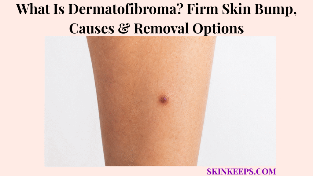
A dermatofibroma is a usually benign firm skin growth made of fibrous tissue that can feel like a small button, pebble, or scar-like knot in the skin.
Dermatofibroma is usually harmless when classic and stable, but changing or atypical bumps need medical review. This page covers appearance, dimple sign, locations, causes, similar bumps, diagnosis, biopsy, observation, removal options, scar expectations, recurrence, mistakes, and doctor-warning signs.
What Is Dermatofibroma and Why Does It Feel Firm?
A dermatofibroma is a usually benign firm skin growth made of fibrous tissue that can feel like a small button, pebble, or scar-like knot in the skin.
It is also called benign fibrous histiocytoma, histiocytoma, fibrous skin nodule, or benign dermal nodule.
The firm feel comes from fibrous tissue within the dermis, which is why it usually cannot be squeezed out like a pimple.
Why Is Dermatofibroma Considered Benign?
Dermatofibroma is considered benign because it is a non-cancerous fibrous skin lesion that usually stays localized in the skin.
A classic stable dermatofibroma often remains unchanged for months or years and may not need treatment when diagnosis is clear.
Benign does not mean every firm bump should be self-diagnosed, because biopsy may be needed when the lesion is uncertain or atypical.
Why May It Feel Like a Scar, Pebble, or Hard Knot?
A dermatofibroma may feel like a scar, pebble, or hard knot because fibrous tissue forms within the dermis and can feel fixed under the skin surface.
It may feel like a small button beneath the skin and can be slightly raised, flat, or slightly depressed.
It is not an active pus-filled bump, and repeated squeezing or picking usually adds irritation instead of removing the lesion.
Practical rule: A classic stable dermatofibroma can often be observed, but a changing, bleeding, ulcerated, painful, irregular, or uncertain firm bump should be checked.
What Does Dermatofibroma Look and Feel Like?
Dermatofibroma usually appears as a small firm bump that may be brown, pink, red-brown, tan, purple, gray, darker than surrounding skin, or skin-colored.
It may look scar-like, button-like, pebble-like, round, oval, flat, raised, slightly depressed, smooth, shiny, rough, or slightly scaly.
What Are Common Dermatofibroma Features?
Common dermatofibroma features include a firm papule or nodule, round or oval shape, stable course, and scar-like or button-like feel.
The color can vary across skin tones and may look brown, gray, purple, reddish-brown, tan, pink, darker, or skin-colored.
Many are solitary, but some people can have more than one.
What Symptoms Can Happen?
Many dermatofibromas cause no symptoms, but some can itch, feel tender, become irritated by shaving or clothing, or cause cosmetic concern.
A bump may hurt if it is bumped, rubbed, scratched, or caught by clothing.
Bleeding usually follows trauma; spontaneous or frequent bleeding needs review.
What Signs Are Less Typical and Need Caution?
Rapid growth, frequent bleeding, ulceration, irregular color, irregular border, persistent pain, large size, sudden multiple lesions, or a bump that looks very different from other skin spots needs caution.
These features should be checked before calling the lesion dermatofibroma.
Atypical changes can overlap with melanoma, DFSP, cysts, scars, and other tumors that need different care.
| Feature | Typical Dermatofibroma Clue | When to Check |
|---|---|---|
| Feel | Firm, button-like, scar-like. | Soft, cystic, rapidly enlarging, or very painful. |
| Color | Brown, pink, tan, purple, gray, darker, or skin-colored. | Multiple colors or irregular pigment. |
| Course | Stable for months or years. | Recent rapid change. |
| Surface | Smooth, shiny, rough, or slightly scaly. | Ulceration, bleeding, persistent crusting. |
| Symptoms | Usually none or mild itch/tenderness. | Persistent pain or spontaneous bleeding. |
| Number | Usually solitary. | Multiple sudden new firm bumps. |
What Is the Dermatofibroma Dimple Sign?
The dimple sign means a dermatofibroma may sink inward when the skin around it is gently pinched from the sides.
This is also called the pinch sign. It can support recognition, but it should not be used as a home cancer-excluding test.
Why Does the Bump Dimple When Pinched?
The bump may dimple when pinched because dermatofibroma can be fixed to deeper dermal tissue, so side pressure pulls the surface inward.
This creates a central dimple or indentation.
The sign can be useful during examination, especially when the bump otherwise has a classic stable pattern.
Why Is the Dimple Sign Not Enough by Itself?
The dimple sign is not enough by itself because not every dermatofibroma dimples clearly and other lesions can sometimes look similar.
Skin cancers and dermatofibrosarcoma protuberans can mimic dermatofibroma.
Dermoscopy or biopsy may be needed if the bump is changing, bleeding, ulcerated, painful, irregular, enlarging, recurrent, or uncertain.
Dimple-sign caution:
- Dimple sign is a helpful clue, not a final diagnosis.
- It does not rule out every serious mimic.
- A changing, bleeding, ulcerated, painful, irregular, or rapidly growing bump still needs medical review.
- Do not rely on a home pinch test for suspicious lesions.
Where Does Dermatofibroma Usually Appear?
Dermatofibromas most often appear on the legs, especially the lower legs, but they can also occur on the thighs, arms, shoulders, trunk, upper back, or other skin areas.
They may appear at sites of prior bites, shaving nicks, ingrown hairs, splinters, thorn pricks, or minor injuries.
Location helps recognition, but it does not diagnose the bump by itself.
| Site | Common Trigger Clue | Mimic to Consider |
|---|---|---|
| Lower leg | Prior bite, shaving nick, small injury. | Mole, scar, DFSP, melanoma. |
| Thigh | Firm stable bump. | Ingrown hair, cyst, scar. |
| Arm | Bite or trauma history. | Wart, scar, mole. |
| Shoulder / upper back | Firm scar-like bump. | Keloid, cyst, DFSP. |
| Trunk | Less classic but possible. | Mole, cyst, skin cancer mimic. |
| Shaving area | Repeated irritation. | Ingrown hair, folliculitis, scar. |
What Causes Dermatofibroma?
The exact cause of dermatofibroma is not always clear, but many are thought to form after a local skin reaction such as an insect bite, shaving nick, ingrown hair, splinter, thorn prick, or minor trauma.
Not every dermatofibroma has a remembered trigger. Some appear without an obvious bite or injury.
Can an Insect Bite or Small Injury Cause Dermatofibroma?
An insect bite or small injury may precede dermatofibroma because the skin can respond to local inflammation by forming a firm fibrous nodule.
The bump can remain long after the original irritation is gone.
A painful bump after a splinter or thorn injury should still be checked if a retained foreign body, infection, or another diagnosis is possible.
Is Dermatofibroma Caused by Infection?
Dermatofibroma is not usually an active infection and is not caused by poor hygiene.
It is not contagious and cannot be squeezed like pus.
If there is pus, warmth, spreading redness, worsening pain, or fever, another diagnosis or secondary infection should be considered.
Can Multiple Dermatofibromas Happen?
Most people have one dermatofibroma, but some people can have several, and sudden multiple new firm bumps should be medically reviewed.
Multiple sudden lesions are less typical than a single stable bump.
Medical review is safer when new firm bumps appear quickly, especially with immune suppression or diagnostic uncertainty.
| Possible Trigger | How It May Relate | Care Implication |
|---|---|---|
| Insect bite | Local inflammatory reaction. | History clue. |
| Shaving nick | Minor trauma. | Avoid repeated irritation. |
| Ingrown hair | Local inflammation. | Mimic or trigger clue. |
| Splinter / thorn | Skin injury. | Check retained foreign body if painful. |
| Unknown | No clear trigger. | Still common. |
| Multiple sudden bumps | Unusual pattern. | Medical review. |
Who Is More Likely to Get Dermatofibroma?
Dermatofibromas are common in adults and often appear on the limbs, especially the legs, sometimes after minor skin trauma.
Anyone can develop one, and clinical descriptions often note them more often in adults than children.
Multiple, unusual, very large, rapidly changing, or atypical lesions need review rather than routine reassurance.
- Adult.
- Firm lower-leg or limb bump.
- Stable for months or years.
- Possible prior bite, shaving nick, splinter, or minor trauma.
- Button-like or scar-like feel.
- Possible dimple sign.
- No rapid growth, bleeding, ulceration, or irregular color.
How Is Dermatofibroma Different From Other Skin Bumps?
Dermatofibroma can resemble a mole, cyst, wart, scar, keloid, ingrown hair bump, melanoma, or dermatofibrosarcoma protuberans, so stability, firmness, color, dermoscopy, and biopsy matter.
This comparison prevents two mistakes: over-reassuring a suspicious lesion and using destructive treatment before the diagnosis is secure.
How Is Dermatofibroma Different From a Mole?
A mole is a melanocytic lesion, while dermatofibroma is a fibrous lesion that may be pigmented and firm.
Some dermatofibromas are brown or dark and can resemble pigmented lesions.
Changing pigment, irregular border, asymmetry, multiple colors, or a lesion that looks different from other spots should be checked before reassurance.
How Is Dermatofibroma Different From a Cyst?
A cyst is often a deeper, round, mobile lump that may have a central punctum, while dermatofibroma usually feels firmer and more fixed in the skin.
Cysts can become inflamed or drain keratin-like material if they rupture.
A painful enlarging lump, pus-filled bump, or warm swollen lesion needs evaluation.
How Is Dermatofibroma Different From a Wart?
A wart often has a rough viral surface, while dermatofibroma is usually a firm, scar-like, non-contagious dermal nodule.
Warts may show a verrucous surface or black dots.
Surface freezing or wart acid may not remove dermatofibroma and should not be used before diagnosis is clear.
How Is Dermatofibroma Different From a Keloid or Hypertrophic Scar?
A keloid or hypertrophic scar forms from a known wound or procedure, while dermatofibroma may follow minor injury but becomes a discrete firm dermal nodule.
Keloids grow beyond the original wound boundary, while hypertrophic scars usually stay within it.
Keloid-prone patients should discuss scar risk before removing a benign dermatofibroma for cosmetic reasons.
How Is Dermatofibroma Different From DFSP?
Dermatofibrosarcoma protuberans is a rare locally aggressive skin cancer that can begin as a firm scar-like patch or nodule and may slowly enlarge over time.
DFSP is not the same as dermatofibroma, but early firm scar-like lesions can create diagnostic confusion.
A deep, plaque-like, enlarging, recurrent, atypical, or uncertain firm lesion may need biopsy or excision planning.
| Condition | Main Clue | Why Confusion Happens | Safer Next Step |
|---|---|---|---|
| Dermatofibroma | Firm stable dimpled nodule. | Pigmented or scar-like bump. | Observe if classic and confirmed. |
| Mole | Pigmented melanocytic lesion. | Brown bump overlap. | Check ABCDE-type changes. |
| Cyst | Mobile lump, punctum, drainage possible. | Firm bump overlap. | Evaluate if inflamed or enlarging. |
| Wart | Rough viral surface. | Raised bump overlap. | Diagnose before freezing. |
| Keloid | Raised scar beyond wound. | Firm scar-like overlap. | Scar-directed care. |
| Melanoma | Changing irregular pigment. | Pigmented dermatofibroma mimic. | Prompt dermoscopy/biopsy. |
| DFSP | Slowly enlarging firm plaque or nodule. | Can mimic dermatofibroma. | Deep biopsy/excision planning. |
A pigmented firm bump that changes color, grows, bleeds, or looks asymmetric should not be dismissed as dermatofibroma when melanoma is a possible mimic.
Blue-gray or dark firm bumps can overlap with blue nevus or other pigmented lesions, so new or changing color deserves assessment.
A shiny, bleeding, or non-healing bump may raise concern for basal cell carcinoma, while a crusted, growing, ulcerated, or tender lesion may need review for squamous cell carcinoma.
A waxy or stuck-on growth can resemble benign keratosis, but firm dermal nodules and surface keratoses have different exam clues.
How Is Dermatofibroma Diagnosed?
Dermatofibroma is often diagnosed by skin examination and dermoscopy, but biopsy is used when the bump has atypical features or the diagnosis is uncertain.
Diagnosis depends on the whole picture: feel, color, stability, symptoms, location, dimple sign, dermoscopy, and whether the lesion resembles a mimic.
What Does a Dermatologist Check?
A dermatologist checks the bump’s size, color, border, firmness, dimple sign, stability, symptoms, location, dermoscopic pattern, and history of injury or irritation.
The exam may also consider whether it looks like a mole, melanoma, DFSP, cyst, wart, scar, keloid, ingrown hair bump, or other skin tumor.
Photos over time can help show whether the bump is stable or changing.
What Dermoscopy Features May Support Dermatofibroma?
Dermoscopy may support dermatofibroma when a central white scar-like area and peripheral pigment network are seen, but patterns can vary.
Dermoscopy supports diagnosis, but it does not replace biopsy when the lesion is atypical.
Asymmetric colors, asymmetric structures, unusual pigment, ulceration, or rapid change should shift the plan toward diagnostic confirmation.
When Is Biopsy or Excision Needed?
Biopsy or excision is needed when a firm bump is changing, bleeding, ulcerated, painful, irregularly colored, atypical on dermoscopy, recurrent, deep, rapidly growing, or uncertain.
Biopsy may also be chosen when there is concern for DFSP, melanoma, cyst, wart, scar, or another tumor.
Pathology helps confirm what the lesion is instead of relying on appearance alone.
- When the bump first appeared.
- Whether it has changed in size, color, shape, or symptoms.
- Photos over time.
- Any bleeding, crusting, ulceration, pain, or itching.
- Whether it catches on shaving or clothing.
- Past insect bite, shaving nick, splinter, thorn prick, or injury.
- Whether the bump dimples when gently pinched.
- Personal or family history of skin cancer.
- Prior removal, freezing, shaving, or biopsy.
- Whether the lesion came back after removal.
- Any new multiple similar bumps.
Does Dermatofibroma Need Treatment?
Dermatofibroma does not usually need treatment when it is classic, stable, asymptomatic, and confidently diagnosed.
Observation is common because a benign stable bump may remain unchanged for years.
Treatment may be considered for itching, tenderness, repeated shaving or clothing irritation, cosmetic concern, anxiety, or diagnostic uncertainty.
| Situation | Best Direction | Why |
|---|---|---|
| Classic and stable | Observe. | Usually benign. |
| Itchy or tender | Discuss treatment. | Symptom relief. |
| Shaving or clothing irritation | Discuss removal options. | Practical comfort. |
| Cosmetic concern | Discuss scar tradeoff first. | Scar may be more noticeable. |
| Changing, bleeding, or ulcerated | Biopsy or excision. | Rule out mimics. |
| Unclear diagnosis | Dermatology evaluation. | Avoid missed cancer or wrong removal. |
What Removal Options Are Used for Dermatofibroma?
Dermatofibroma removal options depend on whether the goal is diagnosis, symptom relief, cosmetic improvement, or complete removal.
The key tradeoff is that removal can leave a scar, while partial methods may flatten the bump without removing the deeper fibrous component.
When Is Surgical Excision Used?
Surgical excision is often used when complete removal or pathology confirmation is the goal.
It may be preferred when the diagnosis is uncertain, the bump is changing, or complete removal is desired.
Excision may require stitches and leaves a scar, so the reason for removal should be clear before the procedure.
When Are Shave Removal, Punch Removal, or Surface Flattening Used?
Shave removal, punch removal, or surface flattening may be used when the goal is to reduce a raised or irritating bump, but they may not remove the deeper component.
These options may help when the bump catches on shaving or clothing.
Because deeper fibrous tissue can remain, the lesion can persist, remain palpable, or recur.
When Are Cryotherapy or Laser Considered?
Cryotherapy or laser may be considered for selected raised or cosmetically bothersome dermatofibromas, but diagnosis should be secure before destructive treatment.
These methods may flatten, lighten, or reduce the appearance of selected lesions.
Pigment change, scarring, incomplete response, or recurrence can occur, so they should not be used as home treatment or before diagnosis is clear.
Why Do Removal Scars Matter?
Removal scars matter because dermatofibroma is benign and the scar from treatment can sometimes be more noticeable than the original bump.
Legs can scar noticeably or heal slowly in some people, and keloid-prone skin needs extra caution.
Cosmetic expectations should be discussed before removal, especially when the lesion is stable and not medically concerning.
| Option | Best-Fit Goal | Limitation |
|---|---|---|
| Observe | Classic stable lesion. | Bump remains. |
| Excision | Complete removal + pathology. | Scar and stitches. |
| Punch removal | Small lesion or tissue sample. | Scar; may not fit all lesions. |
| Shave flattening | Raised irritating bump. | Deeper part may remain. |
| Cryotherapy | Selected protruding irritated lesion. | Pigment change or partial result. |
| Laser | Cosmetic surface change in selected cases. | Not always complete or diagnostic. |
Can Dermatofibroma Come Back After Removal?
Dermatofibroma can recur or remain palpable if the deeper fibrous part is not fully removed, especially after partial flattening methods.
Complete excision gives the best chance of removing the deeper component, but it also creates a surgical scar.
A recurrent, growing, bleeding, painful, ulcerated, or changing lesion should be reassessed and may need pathology review.
What Dermatofibroma Mistakes Should You Avoid?
The biggest dermatofibroma mistake is trying to pop, cut, freeze, burn, or dissolve a firm bump at home without knowing what it is.
Do not squeeze it like a pimple, use wart remover unless the diagnosis is confirmed, freeze or burn it at home, or repeatedly pick and scratch it.
Do not ignore a bump that is growing, bleeding, ulcerating, changing color, becoming painful, recurring after removal, or looking different from your other skin spots.
| Mistake | Why It Fails | Better Action |
|---|---|---|
| Squeezing | Nothing to drain; irritation risk. | Leave alone or evaluate. |
| Cutting at home | Bleeding, infection, and scar risk. | Dermatology removal. |
| Wart acid/freezing | Wrong diagnosis risk. | Confirm lesion first. |
| Ignoring change | Missed mimic risk. | Biopsy if atypical. |
| Dimple-sign overconfidence | Does not rule out all mimics. | Use dermoscopy/biopsy when needed. |
| Cosmetic removal without scar talk | Scar may disappoint. | Discuss expectations first. |
When Should a Dermatofibroma-Like Bump Be Checked by a Doctor?
A dermatofibroma-like bump should be checked when it is new and uncertain, changing, rapidly growing, bleeding, ulcerated, painful, irregularly colored, unusually large, deep, recurrent, or different from your other skin spots.
Prompt review is also safer when the person has a skin cancer history, immune suppression, many atypical moles, a new firm lesion in a child, or a large or unusual bump.
Which Bump Changes Need Prompt Review?
Prompt review is needed for rapid growth, new asymmetry, irregular border, multiple colors, new black or blue areas, bleeding without injury, ulceration, persistent crusting, increasing pain, or recurrence after removal.
A firm plaque that keeps enlarging, a deep bump, or a lesion larger than expected should not be managed with home treatment.
Medical review can decide whether dermoscopy, biopsy, excision, monitoring, or another workup is appropriate.
Which Patients Should Be More Cautious?
People with skin cancer history, immune suppression, many atypical moles, a new firm lesion in a child, or a large or unusual bump should be more cautious.
Keloid-prone patients considering removal should discuss scar risk before treatment.
Anyone unsure whether the lesion is a mole, cyst, wart, scar, or skin cancer should get a clinician’s assessment before trying removal.
Seek medical review if a firm bump is:
- New and uncertain.
- Rapidly growing.
- Changing in color, size, shape, or symptoms.
- Bleeding without injury.
- Ulcerated or persistently crusted.
- Painful or increasingly tender.
- Irregularly colored.
- Asymmetric or irregularly bordered.
- Very dark, blue-black, or multi-colored.
- Large, deep, or plaque-like.
- Recurrent after removal.
- Different from other skin spots.
- In a person with skin cancer history, immune suppression, many atypical moles, or diagnostic uncertainty.
What Should You Remember About Dermatofibroma?
Dermatofibroma is usually a benign firm skin bump, so the safest approach is to recognize classic features, avoid self-removal, and check any changing or atypical lesion.
Observation is often reasonable when the lesion is classic, stable, and confidently diagnosed, while biopsy or removal may be needed when symptoms, uncertainty, or atypical features are present.
- Dermatofibroma is usually benign.
- It often feels firm, scar-like, pebble-like, or button-like.
- It often appears on legs or limbs.
- The dimple sign can support recognition but does not prove diagnosis.
- Causes are not always clear, but minor trauma, shaving irritation, splinters, or insect bites may be involved.
- Classic stable lesions often need no treatment.
- Removal is optional for symptoms, irritation, cosmetic concern, or diagnosis.
- Excision gives the best chance of complete removal but leaves a scar.
- Partial removal may leave a bump behind or allow recurrence.
- Changing, bleeding, ulcerated, painful, irregular, or rapidly growing bumps need dermatologist review.
Frequently Asked Questions About Dermatofibroma
Is dermatofibroma cancer?
No. Dermatofibroma is usually a benign fibrous skin lesion, but uncertain or atypical bumps may need biopsy because dermatofibroma can resemble DFSP, melanoma, or other tumors.
What does dermatofibroma feel like?
It can feel like a firm, button-like, scar-like, pebble-like, or hard nodule in the skin, sometimes with a dimple or indentation when gently pinched.
What causes dermatofibroma?
The exact cause is often unknown, but some follow minor injury, insect bite, thorn prick, splinter, shaving irritation, ingrown hair, or localized inflammation.
Does dermatofibroma need to be removed?
Not usually. Classic, stable, asymptomatic dermatofibromas can often be left alone; removal may be considered for irritation, tenderness, cosmetic concern, or diagnostic uncertainty.
What is the best removal option for dermatofibroma?
The best option depends on the goal. Complete surgical excision gives the best chance of full removal and pathology confirmation, while shave, punch, cryotherapy, or laser may improve selected raised lesions but can leave part of the lesion behind or cause pigment change or scarring.
Can dermatofibroma come back after removal?
Yes, especially if only the surface is removed and the deeper fibrous component remains. Recurrent, growing, painful, bleeding, or changing lesions should be reassessed.
Can I pop a dermatofibroma?
No. Dermatofibroma is a firm fibrous lesion, not pus or a pimple. Squeezing can irritate the skin and will not remove the deeper fibrous bump.
When should a dermatofibroma-like bump be checked?
A firm bump should be checked for rapid growth, bleeding, ulceration, pain, irregular color, asymmetry, size increase, recurrence, diagnostic uncertainty, or history of skin cancer.
Sources & Evidence About Dermatofibroma
DermNet — Dermatofibroma was used for dermatofibroma as a benign firm papule or nodule, common limb and leg location, dimple sign, no-treatment framing, and biopsy or diagnostic excision for atypical features.
StatPearls / NCBI Bookshelf — Dermatofibroma was used for dermatofibroma as a common benign cutaneous soft-tissue lesion, the dimple-sign mechanism, dermoscopy pattern, complete excision, cryotherapy context, and recurrence or variant caution.
Cleveland Clinic — Cellular Dermatofibroma was used for firm bump presentation, dimple or indent description, and biopsy to distinguish dermatofibroma from another type of skin lesion.
American Academy of Dermatology — DFSP Diagnosis & Treatment was used for DFSP biopsy and diagnostic boundary when a firm lesion is suspicious, atypical, or enlarging.
American Academy of Dermatology — DFSP Signs & Symptoms was used for DFSP as a firm or scar-like skin change that can enlarge, supporting the mimic-safety boundary.
Cleveland Clinic — Dermatofibrosarcoma Protuberans was used for DFSP differential context and biopsy-based diagnosis when a firm skin growth is not clearly benign.
Educational Disclaimer: This SkinKeeps article is for educational purposes only and does not diagnose or replace medical care. A firm, hard, scar-like, pigmented, new, changing, rapidly growing, bleeding, ulcerated, painful, crusting, irregularly colored, asymmetric, large, deep, plaque-like, recurrent, infected-looking, pus-filled, or uncertain skin bump should be checked by a qualified healthcare professional or dermatologist. Do not squeeze, cut, freeze, burn, pick, or treat a suspected dermatofibroma with wart remover, acids, blades, or home procedures. Biopsy, dermoscopy, or excision may be needed when the diagnosis is uncertain or when melanoma, dermatofibrosarcoma protuberans, cyst, wart, scar, or another skin tumor is possible.



