
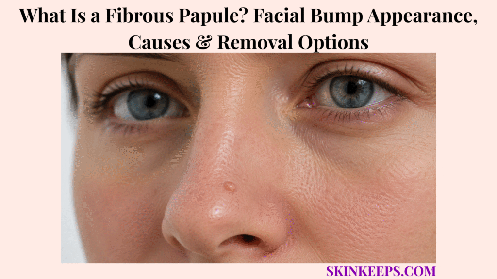
A fibrous papule is a small benign facial bump made of fibrous tissue, collagen, fibroblast cells, and small blood vessels. Also called a solitary angiofibroma, it most often appears on or around the nose and usually remains stable and painless.
This page covers appearance, location, causes, acne confusion, facial-bump lookalikes, diagnosis, biopsy, observation, removal choices, home-removal risks, recurrence, aftercare, and warning signs. A new, changing, bleeding, crusted, ulcerated, painful, itchy, pearly, or non-healing bump should be examined.
How Does a Fibrous Papule Form as a Facial Bump?
A fibrous papule forms as a small benign overgrowth of collagen-rich tissue, fibroblasts, and small blood vessels in the skin.
It is commonly described as a solitary angiofibroma because it combines fibrous connective tissue with a vascular component. The usual result is a smooth, firm, dome-shaped papule on the nose or central face.
A typical fibrous papule is noncancerous and does not transform into cancer. It is not a collection of pus, a blocked pore, or an infection, and it may remain unchanged for years.
Key distinction: the lesion itself is usually harmless, but diagnosis still matters because basal cell carcinoma, moles, sebaceous hyperplasia, and adnexal tumours can look similar.
Figure 1. A fibrous papule is a benign fibrovascular bump. A classic stable lesion may be observed, while an uncertain or changing lesion should be examined and may need biopsy.
What Does a Fibrous Papule Look and Feel Like?
A fibrous papule usually looks like a small, smooth, firm, dome-shaped bump that may be skin-coloured, white, pink, red, brown, or pigmented.
Which Appearance Clues Are Typical?
Typical clues include a shiny or smooth surface, firm texture, solitary pattern, and a stable course rather than repeated inflammation or drainage.
Many fibrous papules measure about 1–6 mm. They are usually non-tender and do not contain pus. Surface vessels may occasionally be visible, but the lesion should not be labelled from one feature or a photograph alone.
Which Symptoms Are Uncommon?
Pain, persistent itch, repeated spontaneous bleeding, crusting, ulceration, and continued growth are not typical of a stable fibrous papule.
Scratching, shaving, squeezing, or picking can temporarily cause tenderness, swelling, or bleeding. A bump that develops symptoms without clear trauma, fails to heal, or continues changing should be re-evaluated.
| Feature | Typical Fibrous Papule Clue | When to Be Cautious |
|---|---|---|
| Shape | Smooth dome-shaped papule. | Irregular, indented, or ulcerated growth. |
| Texture | Firm and often shiny. | Crusted, scaly, repeatedly bleeding, or open sore. |
| Size | Usually small, often 1–6 mm. | Continues enlarging. |
| Colour | Skin-coloured, pink, red, white, brown, or pigmented. | New colour change or several irregular colours. |
| Symptoms | Usually painless and not itchy. | Pain, persistent itch, or spontaneous bleeding. |
| Course | Stable for months or years. | New, changing, or non-healing lesion. |
Where Do Fibrous Papules Usually Appear?
Fibrous papules most often appear on the nose or central face, especially the nasal tip, nasal ala, nasal sidewall, cheeks, or chin.
The nose is the classic site, including the tissue around the nostrils. A single central-face papule fits the usual pattern more closely than numerous bumps spread across the face.
Multiple similar papules do not automatically indicate a syndrome, but they change the diagnostic context. A clinician may check for other facial, nail, oral, family-history, or systemic clues.
| Site | Fibrous Papule Relevance | Main Mimic or Context |
|---|---|---|
| Nasal tip | Classic site. | Basal cell carcinoma or nevus. |
| Nasal ala | Common facial location. | Sebaceous hyperplasia or basal cell carcinoma. |
| Nasal sidewall | Central-face pattern. | Nevus or adnexal tumour. |
| Cheek | Possible site. | Nevus or angiofibroma. |
| Chin | Less typical but possible. | Acne papule or nevus. |
| Multiple facial sites | Less typical for one sporadic lesion. | Multiple angiofibromas or syndrome context. |
What Causes a Fibrous Papule?
A fibrous papule develops from a benign local overgrowth of collagen, fibroblast cells, and small blood vessels, although the exact trigger is usually unknown.
Why Is It Called a Fibrovascular Bump?
The term fibrovascular reflects the lesion’s mixture of fibrous connective tissue and blood vessels.
A papule is simply a small raised skin lesion. Under the microscope, fibrous papules commonly show fibroblast proliferation, a fibrotic stroma, and dilated vessels, which is why they are considered a form of angiofibroma.
Is It Caused by Acne, Dirt, or Poor Hygiene?
A fibrous papule is not caused by acne, dirt, poor hygiene, pus, or a clogged pore.
It is different from acne, and salicylic acid, benzoyl peroxide, retinoids, or repeated exfoliation do not remove its fibrous tissue. These products may only irritate the surrounding facial skin.
It is also different from blackheads, which involve keratin and oil inside an open follicle rather than a stable fibrovascular papule.
| Cause Idea | Correct Explanation | Care Implication |
|---|---|---|
| Acne or clogged pore | Not the underlying cause. | Acne products do not remove it. |
| Infection | Not normally infectious. | Antibiotics are not routine treatment. |
| Poor hygiene | Not a cause. | Avoid blame or aggressive cleansing. |
| Fibrovascular tissue | Collagen, fibroblasts, and vessels. | Explains the firm stable bump. |
| Unknown trigger | The initiating reason is often unclear. | Observation is acceptable if diagnosis is secure. |
| Multiple lesions | May alter the clinical context. | Broader assessment may be appropriate. |
Who Is More Likely to Get a Fibrous Papule?
Fibrous papules can affect many people and often appear in adolescence, young adulthood, or adulthood as a solitary stable nose bump.
A single lesion is the common sporadic pattern. Multiple facial angiofibroma-like papules deserve a broader review, especially when accompanied by periungual growths, oral lesions, seizures, kidney or lung history, endocrine tumours, or a relevant family history.
Conditions associated with multiple angiofibromas can include tuberous sclerosis complex, Birt-Hogg-Dubé syndrome, and multiple endocrine neoplasia type 1. The presence of several papules alone does not establish any of these diagnoses.
- Single stable papule on the nose or central face.
- Appearance during adolescence, young adulthood, or adulthood.
- Multiple facial papules rather than one solitary lesion.
- Nail, oral, scalp, or other body lesions.
- Relevant family history or known genetic condition.
- History of skin cancer, heavy sun exposure, or indoor tanning.
- A new or changing facial bump.
How Is a Fibrous Papule Different From Acne or a Pimple?
A fibrous papule can resemble a stubborn pimple, but it is usually firm, stable, non-tender, and does not drain or cycle like acne.
Acne lesions may become inflamed, form a whitehead, drain, shrink, or change over days or weeks. A fibrous papule usually remains a similar size and shape because it is made of tissue rather than removable contents.
Squeezing a fibrous papule may rupture surface vessels and cause bleeding without removing the lesion. Repeated picking can produce a more noticeable scar than the original bump.
| Feature | Fibrous Papule | Acne or Pimple |
|---|---|---|
| Feel | Firm and usually non-tender. | May be tender, soft, or inflamed. |
| Course | Stable for months or years. | Often changes over days or weeks. |
| Contents | Fibrous tissue and vessels. | Oil, keratin, and inflammation. |
| Squeezing | Usually irritates or bleeds. | May drain, but squeezing still risks scarring. |
| Treatment | Observation or clinician removal. | Acne-directed treatment. |
| Safety | Biopsy if uncertain. | Reassess any persistent non-acne bump. |
How Is a Fibrous Papule Different From Other Facial Bumps?
Fibrous papules can resemble moles, sebaceous hyperplasia, basal cell carcinoma, warts, molluscum, dermatofibroma, and adnexal tumours, so uncertain facial bumps should be examined.
How Is It Different From Basal Cell Carcinoma?
Basal cell carcinoma can appear as a firm, shiny, pearly, slowly growing, bleeding, crusted, or non-healing facial bump, while a fibrous papule is benign and usually stable.
Clinical overlap with basal cell carcinoma is the main reason a suspicious lesion should not be destroyed cosmetically before diagnosis. Visible vessels, ulceration, recurrent bleeding, or continued growth increase the need for examination and possible biopsy.
How Is It Different From a Mole or Intradermal Nevus?
A raised mole or intradermal nevus can also be dome-shaped and skin-coloured or brown, and clinical examination may not always separate it from a fibrous papule.
A stable mole may be soft or firm, while a fibrous papule is often small and distinctly firm. New change, asymmetry, irregular pigmentation, bleeding, or uncertainty warrants dermoscopy or biopsy.
An irregularly pigmented or changing bump may require assessment for melanoma or another melanocytic lesion rather than being assumed to be a pigmented fibrous papule.
How Is It Different From Sebaceous Hyperplasia?
Sebaceous hyperplasia often appears yellowish with a central dell and may occur as several forehead, nose, or cheek papules, while a fibrous papule is usually solitary and firm.
Both are benign but can resemble basal cell carcinoma. Dermoscopy may help reveal characteristic vessels or lobules, while biopsy settles cases that remain unclear.
How Is It Different From Warts or Molluscum?
Warts often have a rough surface, while molluscum commonly has a central dimple and may appear in clusters.
A fibrous papule is usually smooth, firm, and solitary. Treating an uncertain facial papule with wart acid or an at-home freezing product risks burns, pigment change, scarring, and delayed diagnosis.
How Is It Different From Adnexal Tumours or Dermatofibroma?
Benign adnexal tumours such as trichoepithelioma, trichofolliculoma, trichodiscoma, and perifollicular fibroma can resemble a fibrous papule.
A dermatofibroma is usually found away from the central face and may dimple when compressed, but a firm benign papule can still create diagnostic overlap. Pathology is useful when clinical features do not confidently identify the lesion.
| Condition | Main Clue | Why Confusion Happens | Safer Next Step |
|---|---|---|---|
| Fibrous papule | Stable firm dome-shaped papule. | Resembles several facial growths. | Examination; biopsy if uncertain. |
| Acne | Inflames, changes, or drains. | Nose-bump overlap. | Use acne care only for an acne pattern. |
| Mole or nevus | Skin-coloured or pigmented raised lesion. | Dome-shape overlap. | Dermoscopy or biopsy if changing. |
| Sebaceous hyperplasia | Yellowish papule with a central dell. | Central-face overlap. | Dermoscopy if uncertain. |
| Basal cell carcinoma | Pearly, growing, bleeding, or non-healing. | Can mimic a benign papule. | Dermatology review and biopsy. |
| Wart or molluscum | Rough surface or central dimple. | Small-papule overlap. | Clinical diagnosis before treatment. |
| Adnexal tumour | Variable solitary or multiple facial papules. | Histological overlap. | Biopsy. |
How Is a Fibrous Papule Diagnosed?
A fibrous papule may be suspected from its stable, smooth, firm appearance, but biopsy is used when the diagnosis is uncertain or tissue confirmation is needed.
What Does a Dermatologist Check?
A dermatologist checks the lesion’s size, shape, colour, surface, firmness, visible vessels, location, growth history, symptoms, and whether it is solitary or multiple.
The assessment may include dermoscopy, personal and family skin-cancer history, sun and tanning exposure, immune status, and examination for additional facial, nail, oral, or body lesions.
When Is Biopsy Useful?
Biopsy is useful when the lesion is new, changing, growing, bleeding, crusting, ulcerated, irregularly pigmented, symptomatic, or difficult to separate from basal cell carcinoma or another tumour.
Shave biopsy can sometimes provide diagnosis and cosmetic flattening in one procedure. Excision may be selected when complete removal or deeper evaluation is preferred.
What Does Pathology Show?
Pathology typically shows fibroblast proliferation, fibrotic stroma, and dilated blood vessels with angiofibroma-like features.
Several microscopic variants exist, including pigmented, clear-cell, hypercellular, pleomorphic, epithelioid, inflammatory, and granular patterns. Recognising these variants helps avoid confusion with nevi, basal cell carcinoma, or adnexal tumours.
Figure 2. Diagnosis combines the lesion’s history, clinical pattern, dermoscopy when available, and biopsy when a benign fibrous papule cannot be confidently separated from a lookalike.
- When the bump first appeared.
- Any change in size, shape, colour, or texture.
- Bleeding, crusting, ulceration, pain, itch, or failure to heal.
- Whether the lesion is solitary or multiple.
- Exact site on the nose or face.
- Previous acne products, acids, picking, freezing, or cutting attempts.
- Skin-cancer history, heavy sun exposure, or tanning-bed use.
- Relevant family history or other nail, oral, facial, or body lesions.
- Whether the goal is reassurance, biopsy, cosmetic removal, or both.
- Concerns about scar, pigment, or texture change after treatment.
Does a Fibrous Papule Need Treatment?
A typical fibrous papule does not need treatment when the diagnosis is clear, the bump is stable, and it is not causing a cosmetic concern.
Observation is a legitimate medical choice because a classic fibrous papule is harmless and has no malignant potential. The lesion may remain unchanged for years.
Removal is usually chosen for appearance, repeated irritation, or diagnosis. A suspicious bump should be evaluated rather than labelled benign merely because it resembles a fibrous papule.
| Situation | Best Direction | Why |
|---|---|---|
| Small classic stable papule | Observe. | No medical treatment is required. |
| Cosmetic concern | Discuss removal methods. | Personal preference and scar trade-offs matter. |
| Diagnosis uncertain | Biopsy. | Rule out a lookalike. |
| Growing, bleeding, or non-healing bump | Prompt dermatology assessment. | Skin cancer or another lesion is possible. |
| Multiple facial papules | Broader evaluation. | The clinical context differs from a solitary lesion. |
| Recently picked or inflamed lesion | Allow assessment after irritation settles when safe. | Trauma can distort appearance. |
What Removal Options Are Used for a Fibrous Papule?
Fibrous papules may be removed for cosmetic or diagnostic reasons by shave biopsy, excision, electrosurgery, laser therapy, or cryotherapy.
How Does Shave Biopsy or Shave Removal Work?
Shave biopsy removes the raised part of the papule and can provide tissue for pathology at the same time.
This method may flatten a small lesion with a relatively superficial wound. A slight depression, raised scar, colour change, texture change, or residual bump can still occur.
How Does Excision Work?
Excision removes the lesion with a scalpel and may be selected when complete removal or diagnostic certainty is important.
The wound may require stitches depending on size, depth, and facial location. Scar direction and visibility matter on the nose, eyelid, lip, and central face.
How Do Electrosurgery, Laser, or Cryotherapy Fit?
Electrosurgery, laser therapy, and cryotherapy may be cosmetic options for selected, confidently diagnosed fibrous papules.
These methods may flatten or destroy the lesion but may not provide tissue for pathology unless biopsy is performed first. Risks include incomplete removal, recurrence, scarring, persistent redness, pigment change, and altered texture.
Figure 3. Observation is appropriate for a confidently diagnosed stable lesion. Biopsy-based removal is useful when pathology matters, while non-biopsy cosmetic procedures require diagnostic certainty first.
| Removal Option | Best-Fit Use | Main Caution |
|---|---|---|
| Shave biopsy or removal | Flattening plus pathology. | Scar, depression, or texture change. |
| Excision | Complete removal plus pathology. | Stitches and a linear scar may be needed. |
| Electrosurgery | Selected small cosmetic papule. | Pigment or texture change. |
| Laser therapy | Cosmetic smoothing after diagnosis. | No pathology unless tissue was sampled. |
| Cryotherapy | Selected superficial lesion. | Pigment change and incomplete removal. |
| Observation | Classic stable harmless lesion. | The bump remains visible. |
Can a Fibrous Papule Be Removed at Home?
A fibrous papule should not be cut, burned, frozen, picked, squeezed, or treated with wart remover or strong acids at home.
It contains tissue and blood vessels rather than removable pus. Squeezing can cause bleeding, while cutting creates infection and scar risk. Chemical acids or home-freezing products can injure surrounding facial skin and cause permanent pigment or texture change.
Home treatment also removes the opportunity for diagnostic examination and pathology. A lesion that was actually basal cell carcinoma, a nevus, or another tumour could be delayed or partly destroyed without a diagnosis.
| Home Attempt | Why It Is Risky | Better Action |
|---|---|---|
| Squeezing | Bleeding and inflammation. | Leave it alone and arrange assessment if uncertain. |
| Needle or cutting | Infection and scarring. | Clinician biopsy or removal. |
| Acid or acne peel | Chemical burn and pigment change. | Confirm the diagnosis first. |
| Wart remover or freezing | Scarring, pigment loss, or incomplete removal. | Use only a selected clinician-directed method. |
| Repeated picking | Irritation and a larger scar. | Protect the area from trauma. |
| Cosmetic destruction before diagnosis | A cancer mimic may be missed. | Examine or biopsy first. |
Will a Fibrous Papule Come Back After Removal?
Fibrous papules rarely recur after complete removal, but a residual or recurrent bump can occur when tissue remains or healing creates a raised contour.
A person with an underlying condition associated with multiple angiofibromas may develop additional lesions even when one papule was completely removed. A returning or newly changing bump should be checked rather than repeatedly retreated at home.
Every facial procedure trades the bump for a healing site. Temporary redness and crusting are expected after many methods, while lasting pigment change, indentation, raised scar, or texture difference is possible.
| Outcome Stage | What May Happen | When to Recheck |
|---|---|---|
| Early healing | Redness, tenderness, or a small crust. | Increasing pain, pus, warmth, or spreading redness. |
| Surface recovery | The area flattens and colour gradually settles. | Persistent open wound or worsening swelling. |
| Long-term result | Small scar or texture difference may remain. | The bump enlarges again or changes. |
| New nearby papules | May be unrelated or part of a multiple-lesion pattern. | Arrange examination if numerous or atypical. |
How Should Skin Be Cared for Before and After Removal?
Skin care before and after removal should minimise irritation, protect the healing wound, and reduce scar or pigment problems.
Before treatment, avoid squeezing, scratching, harsh exfoliation, and repeated acne acids on the bump. Ask whether the diagnosis is certain, whether tissue will be sent to pathology, and what scar is expected for the chosen method.
After treatment, follow the clinician’s wound-care instructions, keep the area clean, avoid picking the crust, and do not place makeup over an open wound unless approved. Sun protection after the surface heals can reduce contrast from post-procedure pigment change.
- Do not pick, squeeze, or scratch the papule.
- Avoid harsh acne products on the lesion.
- Use a gentle cleanser and moisturiser.
- Ask whether pathology is recommended.
- Discuss scar, pigment, and texture expectations before removal.
- Follow procedure-specific wound-care instructions.
- Keep the healing site clean and protected.
- Avoid makeup over an open wound unless approved.
- Use sun protection once the clinician says the area can be exposed.
- Seek care for pus, increasing redness, warmth, swelling, fever, or worsening pain.
What Fibrous Papule Mistakes Should You Avoid?
The biggest fibrous papule mistake is treating a persistent facial bump like acne while ignoring diagnostic uncertainty or skin-cancer lookalikes.
| Mistake | Why It Fails | Better Action |
|---|---|---|
| Squeezing it like a pimple | It is not pus-filled and may bleed. | Stop trauma and obtain an examination if uncertain. |
| Using salicylic acid repeatedly | Fibrovascular tissue does not dissolve like a clogged pore. | Confirm the lesion type. |
| Cutting or freezing at home | Infection, scarring, and pigment change. | Use clinician-directed removal. |
| Ignoring growth or bleeding | A basal cell carcinoma or other tumour may be missed. | Arrange dermatology review. |
| Choosing cosmetic laser first | Pathology is lost if the diagnosis is wrong. | Establish diagnostic confidence first. |
| Expecting scar-free removal | Every procedure alters facial skin. | Compare methods and healing trade-offs. |
| Relying only on online photos | Many facial papules overlap visually. | Use examination, dermoscopy, or biopsy. |
Do not assume every nose bump is harmless, and do not assume every pigmented papule is a fibrous papule. Multiple new facial papules, especially with other body signs or family history, should also be assessed.
When Should a Facial Bump Be Checked by a Dermatologist?
A facial bump should be checked if it is new, changing, growing, bleeding, crusting, ulcerated, painful, itchy, non-healing, pearly, visibly vascular, irregularly pigmented, multiple, or difficult to identify.
Which Bump Changes Need Medical Review?
Slow continuous growth, rapid growth, spontaneous bleeding, recurrent crusting, ulceration, a sore that heals and returns, pain, itch, colour change, irregular borders, or visible vessels need medical review.
Basal cell carcinoma commonly develops on the head and neck and can look like a firm shiny bump or non-healing sore. A lesion should not be considered safe merely because it is small or has been present for some time.
Which Patient Contexts Need Extra Caution?
Extra caution is appropriate with a personal history of skin cancer, heavy ultraviolet exposure, indoor tanning, immune suppression, organ transplant, many unusual moles, or multiple facial papules.
A family history of tuberous sclerosis complex, Birt-Hogg-Dubé syndrome, or multiple endocrine neoplasia type 1 changes the context of multiple angiofibroma-like lesions. Removal from the nose, eyelid, or lip also deserves careful scar planning.
Prompt review is warranted for a growing, bleeding, pearly, ulcerated, repeatedly crusted, visibly vascular, or non-healing facial bump—especially before laser, freezing, or other cosmetic destruction.
- New or changing facial bump.
- Slow continuous or rapid growth.
- Bleeding without clear trauma.
- Crusting, ulceration, or a sore that returns.
- Pain, itch, numbness, or persistent irritation.
- Pearly or shiny surface with visible vessels.
- Irregular colour or border.
- Multiple facial papules or clusters.
- Personal skin-cancer history, heavy sun exposure, tanning-bed use, immune suppression, or organ transplant.
- Relevant family history or other nail, oral, facial, or body signs.
- Uncertain diagnosis before cosmetic removal.
What Should You Remember About Fibrous Papules?
A fibrous papule is usually a harmless facial bump, but diagnosis matters because several benign and cancerous lesions can look similar.
- A fibrous papule is often called a solitary angiofibroma.
- It usually appears on or around the nose.
- It is commonly small, firm, smooth, shiny, and dome-shaped.
- It may be skin-coloured, pink, red, white, brown, or pigmented.
- It is benign and does not become cancer.
- It is not acne, pus, dirt, or a clogged pore.
- Observation is valid when the diagnosis is clear and the lesion is stable.
- Biopsy is useful when diagnosis is uncertain or tissue confirmation is needed.
- Removal options include shave biopsy, excision, electrosurgery, laser, and cryotherapy.
- Scar, pigment, texture, pathology, and recurrence expectations should be discussed first.
- Growing, bleeding, crusted, ulcerated, painful, itchy, pearly, or non-healing facial bumps need review.
What Questions Do People Ask About Fibrous Papules?
Is a fibrous papule cancer?
No. A typical fibrous papule is benign and has no malignant potential. A changing or uncertain bump still needs assessment because basal cell carcinoma and other lesions can resemble it.
Is a fibrous papule the same as acne?
No. It contains fibrous tissue and small blood vessels rather than pus or a clogged pore. Acne products such as salicylic acid do not remove the fibrovascular tissue.
What does a fibrous papule look like?
It is usually a small, smooth, shiny, firm, dome-shaped papule on or around the nose. It may be skin-coloured, white, pink, red, brown, or pigmented and is commonly stable and painless.
What causes a fibrous papule?
It forms from a benign overgrowth involving collagen, fibroblasts, and small blood vessels. The exact reason this local overgrowth begins is often unknown.
How is a fibrous papule diagnosed?
A dermatologist reviews the bump’s history and examines its shape, surface, colour, vessels, and firmness. Dermoscopy may help, while biopsy provides confirmation when the diagnosis is uncertain.
Does a fibrous papule need removal?
No treatment is medically required when the diagnosis is clear and the lesion is stable. Removal may be chosen for cosmetic reasons or to obtain tissue for diagnosis.
Can I remove a fibrous papule at home?
No. Cutting, squeezing, acids, wart remover, or at-home freezing can cause infection, scarring, pigment change, incomplete removal, and a missed diagnosis.
When should a nose bump be checked?
A new, growing, bleeding, crusting, ulcerated, painful, itchy, pearly, visibly vascular, irregularly pigmented, or non-healing bump should be examined before cosmetic treatment.
Sources & Evidence
DermNet — Fibrous Papule of the Nose — Names, appearance, 1–6 mm size range, benign nature, biopsy features, differential diagnosis, removal options, stability, and recurrence.
Cleveland Clinic — Fibrous Papule of the Nose — Patient-friendly definition, tissue components, unknown trigger, noncancerous nature, salicylic-acid warning, removal choices, and home-removal risks.
Primary Care Dermatology Society — Fibrous Papule — Typical nose location, young-adult context, smooth firm shiny morphology, stable course, early BCC differential, and shave excision.
StatPearls / NCBI Bookshelf — Cutaneous Angiofibroma — Solitary central-face fibrous papules, fibrovascular angiofibroma framing, and the significance of multiple angiofibromas in genetic conditions.
American Academy of Dermatology — Basal Cell Carcinoma — Firm shiny raised growths, bleeding or crusting, non-healing sores, head-and-neck location, and dermatologist assessment.
Cutaneous Lesions of the Nose — PMC Review — Nasal fibrous papule pattern and the role of biopsy when BCC or benign adnexal tumours closely resemble it.
PubMed — Fibrous Papule: A Histopathologic Review — Benign nose lesion, angiofibroma-like pathology, histological variants, and diagnostic pitfalls.
This SkinKeeps article is for educational purposes only and does not diagnose or replace medical care. A new, changing, growing, bleeding, crusting, ulcerated, painful, itchy, non-healing, pearly, visibly vascular, irregularly pigmented, or uncertain facial bump should be examined by a qualified healthcare professional, particularly before cosmetic laser, freezing, electrosurgery, acid treatment, or removal. Seek prompt care for a wound with pus, increasing redness, warmth, swelling, fever, or worsening pain after trauma or a procedure. Do not diagnose a fibrous papule from photographs, squeeze it like acne, cut or burn it, use wart remover or at-home freezing, apply strong acids, repeatedly pick it, or destroy a suspicious lesion without diagnostic confidence or pathology when needed. No removal method can guarantee a scar-free or pigment-free result.



