
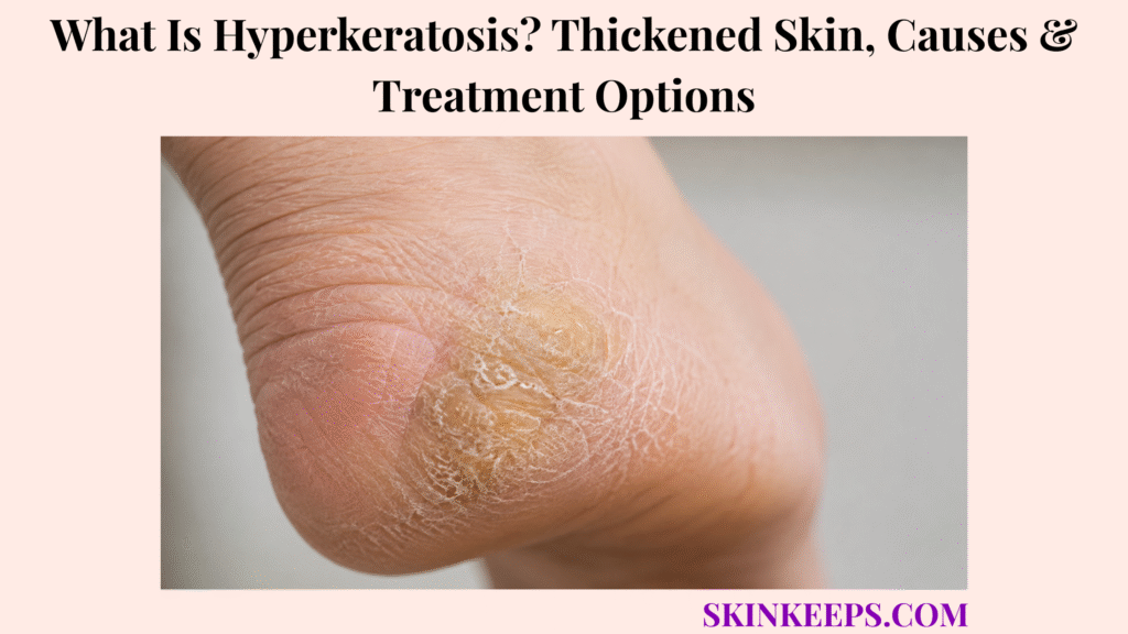
Hyperkeratosis is excessive thickening of the keratin-rich outer skin layer called the stratum corneum. It is a skin change, not one disease, and may look rough, hard, scaly, waxy, cracked, bumpy or plaque-like.
Common examples include calluses, corns, keratosis pilaris and inflammation-related thickening, but infection, inherited keratinization disorders, medicines and sun-damaged precancerous growths can produce a similar texture. Cause-specific treatment may include moisturizers, keratolytics, retinoids, pressure relief, infection treatment, procedures or biopsy.
How Can You Recognize Hyperkeratosis on the Skin?
Hyperkeratosis often feels like thick, rough, hard or scaly skin, but its cause depends on location, shape, symptoms and change over time.
- Hard, leathery or compact thickening.
- Rough or sandpaper-like texture.
- Dry scale or a waxy, ridged surface.
- Yellow, grey, white, brown or skin-coloured plaque.
- Raised follicle-centred bumps.
- Cracks or painful fissures.
- Thickening over pressure points.
- Thick material around or beneath a nail in selected disorders.
Possible symptoms include dryness, itch, tenderness, pressure pain, burning, reduced flexibility or bleeding from deep cracks.
Figure 1. Hyperkeratosis can look hard, scaly, fissured, follicular or plaque-like; pressure sites, hair follicles, inflamed areas, sun-exposed skin and oral mucosa lead to different diagnostic routes.
Where Can Hyperkeratosis Develop?
Hyperkeratosis can develop wherever the outer skin layer is repeatedly stressed, inflamed, infected, genetically altered or sun-damaged.
| Location | Common Pattern Clue |
|---|---|
| Heels, soles and toes | Pressure, footwear, callus, corn or keratoderma |
| Palms, fingers and knuckles | Tools, friction or palmoplantar disease |
| Upper arms, thighs, cheeks and buttocks | Follicular plugging |
| Elbows, knees, scalp and nails | Eczema or psoriasis |
| Face, ears, balding scalp, hands and forearms | Sun-damaged keratosis |
| Oral or genital mucosa | Separate keratotic disorder needing assessment |
What Happens Inside the Skin During Hyperkeratosis?
Hyperkeratosis develops when keratin production increases, shedding slows, or both processes occur together.
Keratinocytes mature as they move upward through the epidermis and normally form a controlled, shedding stratum corneum. Pressure, inflammation, infection, genetic change or ultraviolet damage can disturb that balance.
The result is compact keratin and a thickened outer layer.
Figure 2. The same thick texture can arise from protective pressure response, a focal corn core, HPV infection or ultraviolet-damaged keratinocytes, so surface appearance alone does not determine treatment.
Which Main Patterns of Hyperkeratosis Can Occur?
The main patterns are mechanical, follicular, inflammatory, infectious, inherited, sun-related and medicine- or systemic-disease-related.
| Pattern | Examples | Main Care Direction |
|---|---|---|
| Mechanical | Callus, corn and cracked heel | Pressure relief and safe reduction |
| Follicular | Keratosis pilaris | Moisturizer and selected keratolytic |
| Inflammatory | Eczema, psoriasis and lichenification | Control inflammation |
| Infectious | Wart or hyperkeratotic tinea | Organism-directed treatment |
| Inherited | Ichthyosis or palmoplantar keratoderma | Long-term specialist care |
| Sun-related | Actinic keratosis | Assessment and lesion/field treatment |
| Acquired/systemic | Medicine or systemic association | Identify underlying driver |
A moisturizer can improve flexibility, but it cannot eliminate a wart, cure fungus, control active psoriasis or safely treat a suspicious sun-damaged growth by itself.
How Do Friction and Pressure Cause Hyperkeratosis?
Friction and pressure stimulate extra keratin production as a protective response.
- Calluses form across broader pressure-bearing areas.
- Corns form concentrated, often painful keratin cores.
- Tight footwear and bony prominences increase focal pressure.
- Tools, sports equipment and prostheses can create hand or body lesions.
- Altered walking mechanics can concentrate weight.
- Surface reduction gives temporary relief when pressure continues.
How Are Calluses and Corns Different?
What Does a Callus Look Like?
A callus is a broad, less sharply defined area of pressure-related thickening, commonly beneath the forefoot, heel or palm.
It may look yellowish, grey or darker, and skin lines usually continue across it.
What Does a Corn Look Like?
A corn is smaller and more localized, with a dense central keratin core over a toe joint or bony prominence.
Hard corns occur on dry exposed surfaces, while soft corns develop between moist compressed toes.
Pressure-related corns and calluses commonly recur when footwear, tools, bony pressure or walking mechanics remain unchanged.
| Feature | Callus | Corn |
|---|---|---|
| Size | Broad | Small and focal |
| Border | Poorly defined | More sharply defined |
| Core | Absent | Dense central core |
| Typical site | Forefoot, heel or palm | Toe joint or bony point |
| Pain | Variable pressure tenderness | Often direct-pressure pain |
Why Do Heels Become Thick, Hard and Cracked?
Heels become thick and cracked when dry callused skin loses flexibility while walking pressure continues to stretch the heel.
The heel fat pad expands sideways during standing, while rigid callused skin resists movement. Small cracks can deepen, bleed and permit infection.
A deep heel fissure can bleed, hurt or become infected when thick skin loses flexibility under repeated pressure.
Diabetes, neuropathy or poor circulation makes heel cracks more concerning.
What Is Follicular Hyperkeratosis?
Follicular hyperkeratosis occurs when keratin collects inside hair-follicle openings and creates rough plugged bumps.
- Small rough plugs centred on follicles.
- Sandpaper-like surface.
- Redness or darker colour around follicles.
- Trapped hair shafts.
- Common involvement of upper arms, thighs, cheeks or buttocks.
How Is Keratosis Pilaris Different From Other Hyperkeratosis?
Keratosis pilaris produces many tiny follicle-centred bumps rather than broad pressure plaques or sharply inflamed scaly plaques.
| Feature | Keratosis Pilaris | Mechanical Thickening | Inflammatory Plaque |
|---|---|---|---|
| Pattern | Many follicular bumps | Broad pressure area | Defined inflamed area |
| Common site | Upper arms or thighs | Palms or soles | Eczema/psoriasis sites |
| Pain | Usually minimal | Weight-bearing pain possible | Itch or soreness |
| Course | Improves and recurs | Returns with pressure | Tracks inflammatory disease |
How Do Eczema and Psoriasis Produce Thickened Skin?
How Does Chronic Eczema Cause Hyperkeratosis?
Chronic eczema disrupts the barrier, and repeated scratching can make the skin leathery with exaggerated lines, a change called lichenification.
Repeated scratching and chronic eczema can make skin leathery and thick, so scale-softening products alone may fail while inflammation remains active.
How Does Psoriasis Cause Hyperkeratosis?
Psoriasis accelerates keratinocyte turnover so cells accumulate before they can shed normally, forming thick sharply defined plaques.
Palms, soles, elbows, knees, scalp and nails can be involved, and anti-inflammatory treatment is central.
Which Infections Can Cause Hyperkeratotic Skin?
Infections can cause hyperkeratotic skin, so thick scale should not always be treated as dryness alone.
- Human papillomavirus causing warts.
- Chronic hyperkeratotic tinea pedis.
- Selected fungal infections of palms or soles.
- Crusted scabies in an appropriate immune-risk setting.
- Secondary bacterial infection within fissures.
A moccasin-like scale on the soles may reflect athlete’s foot, which requires antifungal treatment rather than moisturizer alone.
How Is Hyperkeratosis Different From a Viral Wart?
A wart is an infectious epidermal growth caused by human papillomavirus, while a callus or corn is mainly pressure-related thickening.
| Feature | Wart | Corn or Callus |
|---|---|---|
| Cause | HPV | Pressure or friction |
| Surface | Rough or papillated | Compact or smooth-thickened |
| Skin lines | May be interrupted | Often continue |
| Dots | Tiny thrombosed capillaries may appear | Absent |
| Pain | May hurt with side squeeze | Often direct pressure |
| Spread | Can spread | Does not spread virally |
Do not cut a lesion at home to see whether it bleeds.
What Are Inherited Hyperkeratosis and Palmoplantar Keratoderma?
Inherited hyperkeratosis and palmoplantar keratoderma involve abnormal cornification that often causes persistent palm, sole or wider skin thickening.
- Symptoms may begin in infancy, childhood or adolescence.
- Palms and soles may be diffusely, focally or punctately thickened.
- Pressure areas can become painful and fissured.
- Sweating, infection or odour can coexist.
- Nails, hair, teeth or other organs may be involved in syndromic forms.
- Family members may have similar findings.
Management may include emollients, keratolytics, topical retinoids and carefully monitored oral retinoids for severe disease.
Which Medicines or Systemic Conditions Can Cause Acquired Hyperkeratosis?
New adult-onset or rapidly progressive hyperkeratosis may reflect medicine exposure, systemic disease or acquired keratoderma rather than local pressure alone.
- Selected chemotherapy or targeted cancer treatment.
- Chronic contact dermatitis or lymphoedema.
- Hypothyroid or nutritional states.
- Chronic inflammatory disease.
- Selected infection.
- Rare paraneoplastic keratoderma.
- Long-term mechanical exposure.
Rapid bilateral palm-and-sole change, mucosal involvement, systemic symptoms or unexplained weight loss needs broader assessment.
How Does Sun Damage Cause a Hyperkeratotic Lesion?
Sun damage can create hyperkeratotic lesions by producing abnormal ultraviolet-damaged keratinocytes.
A persistent rough patch on chronically sun-exposed skin may need assessment for actinic keratosis rather than repeated cosmetic exfoliation.
- Common sites include the face, ears, balding scalp, hands, forearms, neck and lips.
- The lesion may feel rough before it is easy to see.
- It may be flat, raised, scaly, crusted or horn-like.
- Some actinic keratoses progress to squamous cell carcinoma.
- Persistent or changing sun-damaged growths need assessment.
How Is Actinic Keratosis Different From a Harmless Callus?
Actinic keratosis arises from ultraviolet-damaged keratinocytes on sun-exposed skin, while a callus develops from repeated pressure or friction.
| Feature | Actinic Keratosis | Callus |
|---|---|---|
| Location | Sun-exposed skin | Pressure-bearing skin |
| Texture | Gritty, scaly or crusted | Broad hard thickening |
| Driver | Ultraviolet damage | Mechanical stress |
| Change | Can enlarge or thicken | Tracks pressure |
| Care | Clinical assessment/treatment | Pressure redistribution |
Can Hyperkeratosis Develop Inside the Mouth?
Hyperkeratosis can appear in the mouth as a white keratotic patch, but persistent oral patches need a separate diagnostic pathway.
Dental friction, biting, tobacco, betel quid, lichen planus or infection may contribute, but dysplasia and carcinoma cannot always be excluded visually.
A persistent white patch that cannot be wiped away should be assessed by oral medicine, dentistry or another appropriate specialist and may require biopsy.
How Do Clinicians Diagnose the Cause of Hyperkeratosis?
Clinicians diagnose the cause by matching texture with body site, pressure history, inflammation, infection signs, inherited clues and lesion evolution.
- Age and speed of onset.
- Family history.
- Pressure, footwear, occupation or hobby exposure.
- Itch, pain or bleeding.
- Inflammatory skin history.
- Medicine and sun exposure.
- Immune status and systemic symptoms.
- Previous treatment response.
Examination assesses border, surface, follicular distribution, skin lines, fissures, pigmentation, nails, mucosa, infection and suspicious growth.
Which Tests Can Identify the Cause of Hyperkeratosis?
Tests should answer a specific diagnostic question rather than confirm thickening alone.
| Question | Possible Test |
|---|---|
| Is fungus present? | Skin scraping or fungal culture |
| Is a fissure infected? | Bacterial culture when indicated |
| Is the lesion a wart or tumour? | Dermoscopy and possible biopsy |
| Is contact allergy contributing? | Patch testing |
| Is an inherited disorder possible? | Genetic testing |
| Is acquired keratoderma systemic? | Targeted metabolic, thyroid or broader evaluation |
When Is a Skin Biopsy Needed for Hyperkeratosis?
A skin biopsy may be needed when a thickened lesion is changing, atypical, non-healing, suspicious or not responding as expected.
- Rapid growth.
- Repeated bleeding or ulceration.
- Persistent pain.
- Irregular borders or several colours.
- A hard horn-like projection.
- A presumed callus without a pressure source.
- Possible actinic keratosis or squamous cell carcinoma.
- A plaque that fails appropriate therapy.
- Persistent oral keratotic patch.
A horn-like projection can represent a cutaneous horn, and its base may need assessment for precancerous or cancerous change.
Hyperkeratosis on a pathology report is a microscopic finding that must be interpreted with the complete diagnosis.
Does Every Hyperkeratotic Skin Area Require Treatment?
Not every area needs treatment, but painful, cracked, infected, inflammatory, suspicious or function-limiting thickening should be assessed or treated.
- A small painless callus may need pressure modification only.
- Keratosis pilaris care is optional when symptoms and appearance are not troublesome.
- Painful corns, fissures or reduced function justify care.
- Inflammatory thickening needs disease control.
- Infection needs organism-directed treatment.
- Inherited keratoderma usually needs maintenance.
- Actinic or suspicious lesions need medical assessment.
How Should Hyperkeratotic Skin Be Washed and Moisturized?
Gentle washing and thick moisturization restore flexibility and reduce cracking before stronger exfoliating treatments are considered.
- Use lukewarm rather than very hot water.
- Choose a mild fragrance-free cleanser.
- Avoid aggressive scrubbing.
- Pat dry.
- Apply thick cream or ointment while slightly damp.
- Reapply to very dry areas.
- Protect cracks with an appropriate dressing.
- Reduce mechanical stress with suitable gloves or footwear.
How Do Keratolytic Treatments Soften Thickened Skin?
How Does Urea Treat Hyperkeratosis?
Urea draws water into the stratum corneum, softens thick scale and becomes more keratolytic at higher concentrations.
It can help calluses, keratoderma, ichthyosis and keratosis pilaris but may sting in deep fissures.
How Does Salicylic Acid Treat Hyperkeratosis?
Salicylic acid loosens connections between accumulated surface cells so thick scale can shed.
It may help selected calluses, corns, warts and scaly disorders but can irritate healthy skin.
How Do Lactic Acid and Other Alpha-Hydroxy Acids Help?
Lactic acid and related alpha-hydroxy acids hydrate and loosen retained surface keratin, smoothing rough texture.
They can sting on inflamed or fissured skin.
How Does Propylene Glycol Help Thick Skin?
Propylene glycol draws moisture into the stratum corneum and can soften compact keratin in stronger palmoplantar preparations.
How Should Salicylic Acid Be Used Safely on Thick Skin?
Salicylic acid should be applied only to the intended area because it can irritate, burn or damage surrounding healthy skin.
- Follow the product or prescription direction.
- Protect surrounding skin when appropriate.
- Avoid large body areas unless advised.
- Do not apply to open, severely inflamed or infected skin without guidance.
- Use extra caution in children.
- Seek advice for diabetes, neuropathy or poor circulation.
- Stop and seek help for marked burning, blistering or ulceration.
- Do not combine several strong exfoliating products without a plan.
When Are Topical Retinoids Used for Hyperkeratosis?
Topical retinoids may help selected follicular plugging, keratosis pilaris or keratinization disorders by changing epidermal differentiation and shedding.
Options may include tretinoin, adapalene or tazarotene under an appropriate plan.
Dryness, redness, peeling and burning are common limits; use is introduced gradually and pregnancy-related restrictions apply.
When Are Oral Retinoids Used for Severe Hyperkeratosis?
Oral retinoids are reserved for severe, extensive, inherited or refractory hyperkeratosis because they require specialist monitoring.
- Severe inherited ichthyosis.
- Extensive palmoplantar keratoderma.
- Selected epidermolytic ichthyosis.
- Severe refractory cornification disease.
- Major pain or functional impairment.
Risks include severe fetal harm, dryness, skin fragility, liver and lipid abnormalities, musculoskeletal effects, drug interactions and prolonged pregnancy-prevention requirements with certain agents.
When Are Steroids or Other Anti-Inflammatory Treatments Needed?
Anti-inflammatory treatment is needed when hyperkeratosis is driven by eczema, psoriasis or another inflammatory skin disease.
- Topical corticosteroids reduce inflammation.
- Keratolytics may improve penetration through thick scale.
- Potency must match body site and age.
- Topical vitamin D analogues can support selected psoriasis plans.
- Calcineurin inhibitors may suit sensitive sites.
- Severe disease may need systemic immune treatment.
Long-term inappropriate steroid use can thin skin.
How Are Fungal or Viral Hyperkeratotic Lesions Treated?
Fungal and viral hyperkeratotic lesions need infection-directed treatment, not moisturizer alone.
| Cause | Treatment Direction | Important Boundary |
|---|---|---|
| Hyperkeratotic fungus | Topical or oral antifungal according to site/severity | Confirm when uncertain |
| Viral wart | Observation, salicylic acid or clinician cryotherapy | Protect surrounding skin |
| Crusted scabies | Antiparasitic treatment and contact/public-health plan | Specialist care |
| Bacterial fissure infection | Wound and antimicrobial assessment | Do not rely on keratolytics |
How Are Corns and Calluses Removed Safely?
Corns and calluses are managed safely by reducing pressure, softening excess keratin and using professional reduction when needed.
- Identify the pressure or friction source.
- Change tight or poorly fitting footwear.
- Use toe spacers, pads or orthotics when appropriate.
- Protect hands during repetitive work.
- Moisturize or use an appropriate keratolytic.
- Arrange podiatric paring for painful thick lesions.
- Reassess foot alignment or deformity.
- Continue pressure reduction after thinning.
Why Should Thick Foot Skin Not Be Cut at Home?
Thick foot skin should not be cut at home because cutting can remove living skin, cause bleeding, introduce infection or create ulcers.
- Avoid razor blades, scissors and knives.
- Avoid aggressive callus shavers.
- Do not cut deeply after soaking.
- Do not combine cutting with strong acids.
- Stop when sensation is reduced.
- Use professional debridement for high-risk feet.
The risk is greatest with diabetes, neuropathy, poor circulation, immune suppression or a history of foot ulcers.
Which Procedures Can Treat Hyperkeratotic Lesions?
Procedures depend on the diagnosis, not on thickness alone.
| Diagnosis | Possible Procedure |
|---|---|
| Callus or corn | Professional paring plus pressure correction |
| Wart | Cryotherapy or another wart-directed procedure |
| Thick actinic keratosis | Curettage or lesion-directed treatment |
| Persistent benign keratotic growth | Selected destructive procedure |
| Suspicious growth | Biopsy or excision rather than blind destruction |
| Multiple actinic lesions | Field treatment or photodynamic therapy |
How Are Actinic Keratoses Treated?
Actinic keratoses are treated with lesion-directed or field-directed therapy because they arise from ultraviolet-damaged keratinocytes.
| Approach | Examples | Use |
|---|---|---|
| Lesion-directed | Cryosurgery, curettage, electrosurgery or selected laser | One or several defined lesions |
| Field-directed | 5-fluorouracil, imiquimod, diclofenac or tirbanibulin | Visible and subclinical field damage |
| Photodynamic therapy | Photosensitizer plus light | Selected treatment fields |
| Biopsy first | Suspiciously thick, tender or rapidly growing lesion | Exclude invasive cancer |
Treatment can cause temporary redness, crusting or inflammation, and ongoing sun protection and surveillance are needed.
How Is Hyperkeratosis Managed in Children?
Hyperkeratosis in children should be managed gently because children’s skin can be more sensitive and strong keratolytics require caution.
- Confirm keratosis pilaris, eczema, wart or inherited disorder.
- Begin with gentle cleansing and moisturization.
- Use age-appropriate keratolytic strength.
- Introduce one product at a time.
- Limit application to affected areas.
- Avoid aggressive scrubbing.
- Monitor burning, raw skin and excessive dryness.
- Refer widespread, congenital, blistering or severe fissured disease.
How Is Hyperkeratosis Managed When Diabetes or Poor Circulation Is Present?
Diabetes, neuropathy or poor circulation lowers the threshold for professional foot care because thick skin can hide deeper damage.
- Inspect feet daily, including between toes and around heels.
- Check for cracks, colour change, warmth, swelling or drainage.
- Do not cut thick areas.
- Avoid strong corn plasters unless approved.
- Use suitable footwear and pressure relief.
- Moisturize while avoiding excess moisture between toes.
- Arrange podiatry care when risk is high.
- Treat infection promptly.
- Assess sensation and circulation.
Which Hyperkeratosis Treatment Mistakes Should Be Avoided?
Unsafe treatment can cause burns, wounds, infection or delayed cancer diagnosis.
| Mistake | Possible Harm | Safer Direction |
|---|---|---|
| Treat every thick area as dryness | Missed infection or tumour | Classify the cause |
| Use a razor or knife | Bleeding and ulcer | Professional reduction |
| Use corn acid on an unknown growth | Chemical injury or delayed diagnosis | Confirm lesion first |
| Apply acid to large/open areas | Burn and absorption risk | Use only as directed |
| Combine acids and retinoids immediately | Severe irritation | Introduce gradually |
| Use antifungal indefinitely | Delayed correct diagnosis | Confirm fungus |
| Freeze suspicious growth at home | Tissue injury and missed cancer | Clinical assessment |
| Ignore an oral white patch | Delayed dysplasia diagnosis | Oral specialist review |
How Long Does Hyperkeratosis Take to Improve?
Improvement depends on the cause, not simply on how thick the surface looks.
| Cause | Expected Course |
|---|---|
| Pressure callus | May soften over weeks when pressure is reduced |
| Corn | Returns while pressure remains |
| Keratosis pilaris | Often needs weeks or months and maintenance |
| Inflammatory thickening | Improves as inflammation is controlled |
| Fungal thickening | May require weeks of correct therapy |
| Inherited keratoderma | Usually lifelong management |
| Actinic keratosis | Needs lesion-specific treatment |
Can Hyperkeratosis Be Permanently Cured?
Whether hyperkeratosis can fully resolve depends on whether the underlying cause can be removed or controlled.
- A friction-related callus may resolve when pressure is permanently corrected.
- Corns recur when footwear or bony pressure remains.
- Infection-related thickening may resolve after successful treatment.
- Inflammatory thickening can recur with flares.
- Keratosis pilaris often needs maintenance.
- Inherited disorders need long-term control.
- Treated actinic keratoses can be followed by new lesions in sun-damaged skin.
Which Complications Can Thickened Skin Cause?
Hyperkeratosis is not always cosmetic because thick skin can crack, bleed, become infected, impair movement or conceal a more serious growth.
Rapidly spreading warmth, redness, swelling or pain around cracked thick skin may suggest cellulitis and needs prompt assessment.
- Pain, deep fissures and bleeding.
- Secondary bacterial or fungal infection.
- Foot ulceration.
- Reduced walking, grip or hand flexibility.
- Nail distortion.
- Scarring or pigment change after excessive treatment.
- Delayed diagnosis of a tumour.
- Squamous cell carcinoma from selected precancerous or chronic lesions.
When Should Hyperkeratosis Be Checked by a Clinician?
Hyperkeratosis should be checked when the cause is unclear, the lesion changes, function is affected, infection is suspected or the person has high-risk feet.
- Sudden or rapidly spreading thickening.
- Diffuse palm-and-sole involvement.
- Painful or bleeding cracks.
- Walking or hand function is affected.
- Home care causes burning or ulceration.
- Calluses recur despite pressure correction.
- Infection is suspected.
- A child has widespread or congenital disease.
- Nails, hair, teeth or mucosa are involved.
- A medicine may be responsible.
- Diabetes, neuropathy or poor circulation is present.
- An oral white patch persists.
- A lesion develops on chronically sun-damaged skin.
Which Hyperkeratotic Changes Require Urgent Assessment?
Prompt or urgent assessment is needed when thickening suggests infection, ulceration, diabetic-foot risk or possible precancerous or cancerous change.
- Rapid growth or repeated bleeding.
- Ulceration or a non-healing sore.
- Increasing pain.
- A hard horn-like projection.
- Spreading redness, warmth, swelling or pus.
- Fever.
- Black or necrotic tissue.
- A deep heel fissure in someone with diabetes.
- Loss of sensation around a wound.
- An oral patch with red, hardened or ulcerated areas.
- A sun-exposed rough area that becomes thick, tender or rapidly changes.
- A presumed callus without a pressure source.
Urgent route: do not cut, freeze or chemically treat a changing, bleeding, ulcerated, infected, black or non-healing thickened growth before medical assessment.
Figure 3. Safe care begins by classifying the thickening, then matching moisturizers, keratolytics, pressure relief, anti-inflammatory or infection treatment, professional procedures and biopsy to the actual cause.
What Should You Remember About Hyperkeratosis?
Hyperkeratosis means excessive thickening of the stratum corneum and is a physical skin change rather than one diagnosis.
- Pressure and friction commonly produce calluses and corns.
- Follicular keratin buildup produces keratosis pilaris.
- Eczema and psoriasis create inflammatory thickening.
- Warts and chronic fungal infection can appear hyperkeratotic.
- Inherited keratodermas may need lifelong care.
- Rough sun-damaged lesions need actinic or cancer assessment.
- Diagnosis depends on location, pattern, symptoms and evolution.
- Moisturizers restore flexibility but may not remove compact keratin.
- Urea, salicylic acid and lactic acid are common keratolytics.
- Strong keratolytics can irritate or burn when misused.
- Pressure relief is essential for lasting callus improvement.
- Changing, bleeding, ulcerated or non-healing thickening may need biopsy.
Frequently Asked Questions About Hyperkeratosis?
Is hyperkeratosis a disease or a skin finding?
Hyperkeratosis is a skin finding, not one disease. It means the stratum corneum has become abnormally thick.
What does hyperkeratosis look and feel like?
It may look or feel thick, hard, rough, scaly, waxy, cracked, leathery or plaque-like and can also produce follicular bumps.
What causes the outer skin layer to become thick?
Possible causes include pressure, friction, inflammation, infection, inherited keratinization disorders, medicines, systemic disease and ultraviolet damage.
Is hyperkeratosis contagious?
Hyperkeratosis itself is not contagious, but causes such as viral warts or fungal infections can spread.
Can dry skin cause hyperkeratosis?
Dryness can worsen roughness and cracking, but another driver such as pressure, inflammation, follicular plugging or disease is often present.
How are calluses and corns different?
Calluses are broader pressure-related areas, while corns are smaller focal lesions with a dense central keratin core.
How is a callus different from a wart?
Calluses are pressure-related and often preserve skin lines. Warts are HPV-related, may interrupt skin lines and can show tiny dark capillary dots.
Is keratosis pilaris a form of hyperkeratosis?
Yes. Keratosis pilaris is follicular hyperkeratosis caused by keratin plugging hair follicles.
Can eczema or psoriasis cause thickened skin?
Yes. Chronic eczema can cause leathery lichenification from scratching, while psoriasis produces thick scaly plaques through rapid epidermal turnover.
Can a fungal infection cause hyperkeratotic soles?
Yes. Hyperkeratotic tinea pedis can create diffuse moccasin-like scaling and thickening on the soles.
What is palmoplantar keratoderma?
Palmoplantar keratoderma means marked thickening of palms and soles. It may be inherited or acquired and often needs long-term management.
Can medicines cause thickened skin?
Yes. Some chemotherapy, targeted cancer treatments and other medicines can contribute to acquired hyperkeratosis or keratoderma patterns.
Can hyperkeratosis be precancerous?
Some hyperkeratotic lesions, particularly actinic keratoses on sun-damaged skin, are precancerous and need assessment.
When does a rough sun-damaged patch require biopsy?
Biopsy may be considered for rapid growth, tenderness, bleeding, ulceration, horn formation, failure to heal or concern for squamous cell carcinoma.
Which tests identify the cause of hyperkeratosis?
Possible tests include fungal scraping, culture, dermoscopy, biopsy, patch testing, genetic testing and targeted blood or systemic evaluation.
Does urea cream remove thick skin?
Urea hydrates and softens the stratum corneum and becomes more keratolytic at higher strengths, but cause-specific treatment may still be needed.
How does salicylic acid soften hyperkeratosis?
Salicylic acid loosens connections between retained surface cells, helping compact keratin shed.
Can salicylic acid burn healthy skin?
Yes. It can irritate or burn surrounding skin, especially when used too often, over large areas, on open fissures or without protection.
When are topical or oral retinoids used?
Topical retinoids may help selected follicular or keratinization disorders. Oral retinoids are reserved for severe inherited or refractory disease under specialist monitoring.
Should calluses be cut off at home?
No. Home cutting can cause bleeding, infection or ulcers, especially with diabetes, neuropathy or poor circulation.
Why do corns and calluses keep returning?
They recur when pressure, friction, footwear problems, bony prominence or walking mechanics remain unchanged.
How is hyperkeratosis treated in people with diabetes?
High-risk feet need daily inspection, pressure reduction, moisturization, professional debridement and prompt care for cracks, wounds or infection.
Can inherited hyperkeratosis be permanently cured?
Inherited keratinization disorders usually require long-term maintenance; ordinary creams do not permanently correct the genetic tendency.
Which thickened-skin changes require urgent medical assessment?
Urgent assessment is needed for rapid growth, bleeding, ulceration, a non-healing sore, infection signs, black tissue, a diabetic fissure, loss of sensation, oral patch changes, a horn-like growth or changing sun-exposed thickening.
Which Sources Support This Hyperkeratosis Guidance?
NCBI Bookshelf / StatPearls — Hyperkeratosis — Definition as increased stratum-corneum thickness, broad causes, evaluation and cause-directed treatment.
American Academy of Dermatology — Actinic Keratosis Overview — Sun-exposed distribution, precancer framing and squamous-cell-carcinoma risk.
DermNet — Corns and Calluses — Pressure and friction mechanism, corn/callus morphology and pressure-relief logic.
DermNet — Cracked Heels — Heel fissures, diabetes risk, moisturization and warning against home cutting.
DermNet — Keratosis Pilaris — Follicular keratin accumulation, common sites and moisturizer/keratolytic care.
DermNet — Palmoplantar Keratoderma — Inherited and acquired palm-and-sole thickening and retinoid/keratolytic treatment options.
DermNet — Oral Leukoplakia — Specialist biopsy pathway for persistent oral leukoplakia and dysplasia assessment.
MedlinePlus — Salicylic Acid Topical — Keratolytic use, irritation risk, lesion-only application and precautions on broken or large skin areas.
This SkinKeeps article is educational and does not diagnose or replace medical, dermatology, podiatry, oral, diabetes-foot or emergency care. Seek assessment for sudden, rapidly changing, bleeding, ulcerated, non-healing, horn-like, sun-damaged or persistent oral thickening. Seek prompt care for spreading redness, warmth, swelling, pus, fever, black tissue, loss of sensation or a deep diabetic heel crack. Do not cut thick foot skin, apply corn acid to an unknown growth, use salicylic acid on large or open areas without guidance, freeze suspicious lesions at home, or ignore retinoid pregnancy precautions.



