
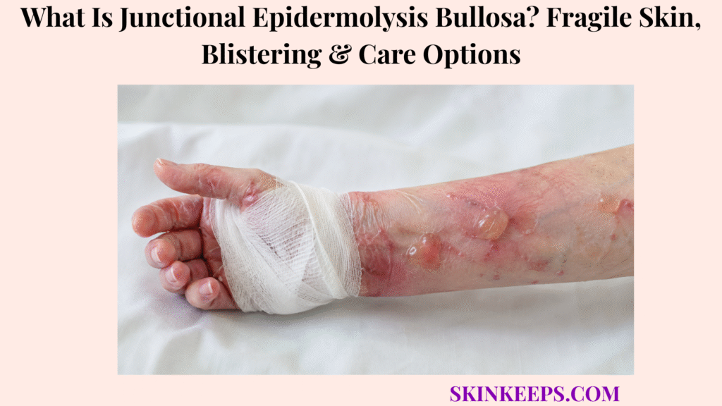
Junctional epidermolysis bullosa, abbreviated JEB, is a rare inherited condition in which skin and mucous membranes blister after little or no friction because attachment is weak near the boundary between the epidermis and dermis. Symptoms usually begin at birth or during the neonatal period, and JEB is not contagious.
Blistering can affect skin, mouth, eyes, upper airway, oesophagus and urinary tract and may occur with granulation tissue, enamel defects, nail changes, hair loss and poor growth. Care focuses on trauma prevention, non-adherent wound protection, pain control, infection prevention, nutrition and organ monitoring; Filsuvez can support eligible wounds but does not correct the inherited defect.
How Can You Recognize Junctional Epidermolysis Bullosa?
JEB is suspected when blisters and erosions appear from birth or early infancy after very minor trauma.
- Blistering at birth or soon afterward.
- Blisters after light rubbing, pressure or handling.
- Raw painful erosions after a blister roof separates.
- Oral or other mucosal lesions.
- Poorly healing wounds or excessive granulation tissue.
- Abnormal or absent nails.
- Sparse hair or scarring hair loss.
- Dental-enamel pitting.
- Hoarse cry or noisy breathing.
- Usually little primary scarring after uncomplicated blisters heal.
Junctional epidermolysis bullosa is one inherited type within the broader group of epidermolysis bullosa disorders.
Figure 1. JEB is suspected when blistering begins at birth or early infancy after minimal trauma, involves skin and mucosal surfaces, and is accompanied by granulation tissue, nail changes, hair loss or enamel defects. Severity spans generalized severe and generalized intermediate disease.
Where Does JEB Blistering Usually Develop?
JEB can affect friction-prone skin and internal mucosal surfaces, so assessment must extend beyond visible wounds.
| External Sites | Internal or Mucosal Sites |
|---|---|
| Back of head, face, nose, mouth and ears | Mouth, tongue and throat |
| Neck, trunk, buttocks and diaper area | Larynx and upper airway |
| Hands, fingers, feet and toes | Oesophagus |
| Knees, elbows and pressure sites | Eyes and conjunctiva |
| Areas beneath clothing or medical equipment | Urinary tract and genital mucosa |
How Does the Skin Separate in Junctional Epidermolysis Bullosa?
Essential adhesion proteins fail to secure the epidermis to the basement-membrane zone.
- Hemidesmosomes and connecting filaments normally resist mechanical force.
- JEB variants reduce or disrupt key adhesion proteins.
- Ordinary pressure creates separation in the junctional zone.
- Fluid collects and forms a blister.
- The same weakness affects mucosal surfaces.
Figure 2. JEB results when variants affecting laminin-332, collagen XVII or integrin-related attachment weaken the epidermal–dermal junction. EB simplex separates more superficially, while dystrophic EB separates below the basement membrane.
Which Genes Can Cause Junctional Epidermolysis Bullosa?
JEB is caused by variants in genes encoding proteins that anchor basal epidermal cells.
| Gene | Protein Relationship | Clinical Relevance |
|---|---|---|
| LAMA3, LAMB3, LAMC2 | Three chains of laminin-332 | Severe or intermediate generalized JEB |
| COL17A1 | Collagen XVII | Often intermediate phenotypes |
| ITGB4 | Integrin beta-4 | JEB spectrum and pyloric atresia |
| ITGA6 | Integrin alpha-6 | Pyloric-atresia-associated JEB |
| Multigene testing | Assesses several adhesion genes | Preferred when phenotype overlaps |
How Is JEB Inherited?
JEB is generally inherited in an autosomal-recessive pattern.
- An affected child usually has a pathogenic variant in both gene copies.
- Parents commonly carry one altered copy each.
- Carriers usually do not have skin fragility.
- When both parents carry variants in the same gene, each pregnancy typically has a 25% affected, 50% carrier and 25% unaffected-non-carrier chance.
- Each pregnancy has the same independent probabilities.
- Genetic counselling should use the confirmed familial variants.
How Are Generalized Severe and Generalized Intermediate JEB Different?
These categories form a severity spectrum influenced by gene, variant type and residual protein function.
What Characterizes JEB Generalized Severe?
- Blistering at birth or in the neonatal period.
- Extensive skin loss and mucosal involvement.
- Prominent granulation tissue.
- Airway and feeding complications.
- Growth failure, anaemia, infection and sepsis risk.
- Fluid and electrolyte imbalance.
- Life-limiting disease in many affected infants.
What Characterizes JEB Generalized Intermediate?
- Less extensive or more localized fragility.
- Survival beyond early childhood and often into adulthood.
- Hands, feet, knees and elbows commonly affected.
- Nail dystrophy, sparse hair and enamel pitting.
- Possible renal, ureteral or oesophageal involvement.
- Variable improvement with age in some people.
Older records may call these Herlitz and non-Herlitz JEB, but current wording is generalized severe and generalized intermediate.
What Is Excessive Granulation Tissue in JEB?
Excessive granulation tissue is raised, moist and friable repair tissue that persists above some JEB wounds.
- Bright-red or deep-red moist tissue.
- Easy bleeding and repeated crusting.
- Common around the nose, mouth, ears, fingers and toes.
- Can occur on scalp, buttocks and pressure sites.
- Internal airway granulation can obstruct breathing.
- Specialist options include wound optimization, selected potent topical steroid, silver nitrate, cautery or grafting.
How Is JEB Different From Epidermolysis Bullosa Simplex?
JEB separates at the epidermal–dermal junction, while EB simplex usually separates within epidermal keratinocytes.
| Feature | JEB | EB Simplex |
|---|---|---|
| Cleavage | Junctional zone | Within epidermis |
| Genes | Laminin-332, COL17A1 or integrin pathway | Often KRT5 or KRT14 |
| Mucosa | Common and sometimes severe | Less characteristic in common forms |
| Associated clues | Enamel defects and granulation tissue | Hands and feet often dominant |
| Inheritance | Usually recessive | Many common forms dominant |
How Is JEB Different From Dystrophic Epidermolysis Bullosa?
JEB separates at the junctional level, while dystrophic EB separates beneath the basement membrane.
| Feature | JEB | Dystrophic EB |
|---|---|---|
| Main protein pathway | Laminin-332, collagen XVII or integrin | Type VII collagen |
| Main gene | Several JEB genes | COL7A1 |
| Primary scarring | Usually limited after uncomplicated wounds | More prominent |
| Milia | Rare | Commoner |
| Contractures and pseudosyndactyly | Less characteristic | Important in severe disease |
How Is JEB Different From Ordinary Friction Blisters?
JEB blisters recur after exceptionally minor contact and may affect mucosa, unlike ordinary rubbing blisters.
Ordinary blisters usually follow repeated rubbing at one pressure site, while JEB may begin in a newborn after routine handling.
| Feature | JEB | Ordinary Friction Blister |
|---|---|---|
| Trigger | Minimal handling or pressure | Substantial repeated rubbing |
| Onset | Birth or early infancy | Any age after exposure |
| Distribution | Multiple skin and mucosal sites | Single mechanical site |
| Associated findings | Nail, hair, enamel or airway clues | None |
| Course | Recurrent inherited disorder | Stops when friction stops |
How Do Clinicians Diagnose Junctional Epidermolysis Bullosa?
Diagnosis begins with inherited skin-fragility clues and is confirmed mainly through molecular genetic testing.
- Onset, trauma threshold and blister distribution.
- Mouth, airway, eye, oesophageal and urinary symptoms.
- Granulation tissue.
- Nail, hair and dental findings.
- Feeding, growth and urine output.
- Family history, consanguinity and previous affected pregnancies.
- Biallelic pathogenic variants on targeted testing, multigene panel or sequencing.
When Are Skin Biopsy and Immunofluorescence Mapping Used?
Specialized biopsy testing is used when it can classify the blistering disorder or clarify genetic results.
- A sample is taken from carefully induced or very recent blister-edge skin.
- Immunofluorescence mapping assesses adhesion-protein location and amount.
- Electron microscopy identifies cleavage level and structural changes.
- Testing may provide rapid preliminary classification.
- Near-normal staining can occur in milder disease.
Why Is Rapid Genetic Testing Important in a Newborn?
Rapid testing can guide airway, feeding, pyloric, renal and prognosis decisions while a critically ill newborn is being stabilized.
- Separates JEB from other EB types.
- Identifies severe laminin-332 deficiency.
- Detects ITGA6 or ITGB4 pyloric-atresia pathways.
- Guides organ surveillance and family testing.
- Supports future prenatal or preimplantation testing.
- Reduces unnecessary or harmful procedures.
What Is JEB With Pyloric Atresia?
JEB with pyloric atresia combines skin and mucosal fragility with congenital blockage at the stomach outlet.
- Persistent non-bilious vomiting from birth.
- Inability to tolerate feeds.
- Dehydration and electrolyte disturbance.
- Abnormal abdominal imaging.
- Possible renal or ureteral abnormalities.
- Association with ITGB4 or ITGA6 variants.
Pyloric atresia is present at birth and differs from pyloric stenosis that develops later.
How Can JEB Affect the Mouth and Feeding?
Blisters and erosions in the mouth, throat or oesophagus can make sucking, chewing and swallowing painful.
- Oral, tongue or gum erosions.
- Painful feeding and reduced appetite.
- Avoidance of textured foods.
- Reflux or oesophageal blistering.
- Rare oesophageal narrowing.
- High calorie and protein needs from chronic wounds.
- Dietitian, feeding and swallowing support.
- Gastrostomy when oral intake cannot meet needs.
How Can JEB Affect the Upper Airway?
Blistering, scarring or granulation tissue can narrow the upper airway.
- Hoarse or weak cry.
- Persistent hoarseness.
- Noisy breathing or stridor.
- Recurrent croup-like episodes.
- Difficulty breathing.
- Laryngeal granulation tissue.
- Possible tracheostomy in severe obstruction.
Progressive hoarseness or stridor needs urgent assessment by an experienced airway team.
How Can JEB Affect the Eyes?
JEB can cause conjunctival irritation, corneal erosions and granulation tissue that threaten comfort or vision.
- Redness, tearing or light sensitivity.
- Eye pain or difficulty opening the eye.
- Corneal erosion.
- Eyelid or conjunctival granulation tissue.
- Reduced vision in severe disease.
- Preservative-free lubrication and ophthalmology care.
Sudden pain, marked light sensitivity or reduced vision requires urgent eye assessment.
How Can JEB Affect Teeth, Hair and Nails?
Enamel, hair and nail abnormalities are important diagnostic and monitoring clues.
| Structure | Possible JEB Findings | Care Need |
|---|---|---|
| Teeth | Thin enamel, pitting, sensitivity and early decay | Early preventive dentistry and fluoride |
| Hair | Sparse or fragile hair and scarring alopecia | Specialist scalp and wound care |
| Nails | Thickened, misshapen, partly lost or absent nails | Protection and pain control |
How Can JEB Affect the Urinary Tract and Kidneys?
Mucosal fragility and congenital anomalies can affect urethral, ureteral, bladder or kidney structures.
- Painful urination or weak stream.
- Urethral narrowing or urinary retention.
- Recurrent urinary infection.
- Hydronephrosis or hydroureter.
- Congenital renal or bladder abnormalities.
- Reduced urine during dehydration.
Why Can JEB Cause Anaemia and Poor Growth?
Chronic wounds, inflammation and painful feeding increase requirements while reducing intake.
- Blood loss and exudate from wounds.
- Iron deficiency and chronic inflammation.
- Oral pain and reduced intake.
- Higher protein and calorie needs.
- Infection and poor absorption.
- Protein-losing enteropathy in selected subtypes.
- Monitoring of growth, blood count, iron, albumin, zinc and vitamin D.
How Should New JEB Blisters Be Drained?
New blisters are generally drained carefully while preserving the roof as a protective layer.
- Wash hands and prepare clean equipment.
- Use the technique taught by the EB team.
- Pierce at one or more low points with a sterile needle when advised.
- Allow fluid to drain gently.
- Keep the intact roof in place.
- Use a non-adherent dressing.
- Monitor for fluid reaccumulation or infection.
Which Dressings Protect JEB Wounds?
JEB dressings should avoid sticking, absorb fluid and remain secured without adhesive trauma.
| Layer | Purpose | Examples |
|---|---|---|
| Primary contact layer | Prevents sticking and protects fragile tissue | Soft silicone or another non-adherent material |
| Secondary layer | Absorbs exudate and cushions | Foam, hydrofibre or alginate |
| Outer retention | Keeps layers in place without skin adhesive | Soft wrap or tubular retention |
A painful skin fissure or erosion in JEB needs atraumatic protection because ordinary rubbing can enlarge the wound.
How Can New Blisters and Skin Tears Be Prevented?
Prevention reduces shearing, pressure, heat and adhesive stripping.
- Lift rather than drag the body.
- Never lift a baby beneath the armpits.
- Use soft seamless clothing and bedding.
- Turn seams outward when useful.
- Pad pressure sites.
- Reduce heat and sweating.
- Protect knees, elbows, hands and feet.
- Avoid adhesive tape directly on skin.
- Use EB-safe device fixation and promptly adjust footwear or equipment.
How Should JEB Wounds Be Cleaned?
Wounds should be cleaned gently after pain preparation and dressing soaking.
- Premedicate when the care plan requires it.
- Soak stuck dressings before removal.
- Remove layers slowly.
- Use lukewarm water or prescribed cleansing solution.
- Do not rub with dry gauze.
- Allow fluid to lift debris.
- Remove only loose non-viable tissue when advised.
- Pat or air dry surrounding skin.
How Are Pain and Itching Managed in JEB?
Pain is assessed by source because acute blister, chronic wound, procedural, mucosal and neuropathic pain need different strategies.
- Regular pain scoring.
- Non-opioid or opioid analgesia according to severity.
- Neuropathic-pain medicines when appropriate.
- Selected topical anaesthetic.
- Cooling and itch-directed treatment.
- Positioning, distraction and relaxation.
- Psychological and specialist pain-team support.
How Are JEB Wound Infections Recognized and Treated?
Infection is suspected when a wound changes with increasing pain, warmth, swelling, drainage, odour or systemic illness.
- Spreading redness or warmth.
- Increasing pain or swelling.
- Pus or increased drainage.
- Strong or changing odour.
- Friable tissue or debris.
- Delayed healing.
- Fever or reduced feeding and activity.
- Culture and targeted topical, oral or intravenous treatment when indicated.
Spreading warmth, swelling and pain around a wound may suggest cellulitis rather than simple colonization.
How Does Filsuvez Support JEB Wound Healing?
Filsuvez is an approved birch-triterpene topical gel for eligible dystrophic and junctional EB wounds in adults and children aged 6 months or older.
- Applied to a cleansed partial-thickness wound or directly to the dressing.
- Used during dressing changes until the wound heals.
- Supports wound closure.
- Does not replace protective dressings or multidisciplinary care.
- Does not correct the underlying JEB gene or systemic disease.
- Use follows prescribing and EB-team guidance.
How Are JEB Granulation Tissue and Chronic Wounds Treated?
Persistent granulation tissue and chronic wounds need specialist assessment because friction, infection and cancer can resemble each other.
- Optimize non-adherent dressings and pressure control.
- Treat infection when clinically present.
- Use selected topical steroid, silver nitrate or cautery under specialist care.
- Consider surgical or grafting approaches for selected lesions.
- Use Filsuvez only for eligible partial-thickness wounds.
- Biopsy wounds that enlarge, harden, bleed repeatedly or fail to heal.
How Are Nutrition and Feeding Supported in JEB?
Nutrition support is part of wound treatment because healing substantially increases calorie and protein requirements.
- High-calorie and high-protein foods.
- Soft or blended textures.
- Oral supplements.
- Pain relief before meals.
- Reflux and constipation management.
- Iron and micronutrient replacement.
- Dietitian and swallowing support.
- Gastrostomy when needed for growth and healing.
How Is JEB Managed in Newborns?
Newborn care stabilizes skin, airway, fluids, temperature, feeding, pain and diagnosis at the same time.
- Neonatal admission with EB-experienced consultation.
- Atraumatic handling and no ordinary adhesives.
- Blister drainage with roof preservation.
- Non-adherent dressings.
- Fluid, electrolyte and temperature monitoring.
- Airway, mouth and feeding assessment.
- Pyloric atresia and renal evaluation.
- Rapid molecular testing.
- Prompt infection treatment.
- Family education and multidisciplinary decision-making.
How Are Dental Problems Prevented in JEB?
Dental prevention begins early because JEB enamel is structurally fragile.
- Early dental referral and frequent review.
- Fluoride treatment.
- Small soft toothbrush and non-traumatic technique.
- Dietary review.
- Early caries treatment.
- Pain control.
- Mouth-opening assessment.
- Procedure planning that avoids mucosal injury.
How Are Movement and Daily Function Protected?
Function is protected by reducing avoidable injury while maintaining safe movement.
- Gentle range-of-motion activity.
- Physiotherapy and occupational therapy.
- Soft protective footwear.
- Pressure redistribution and mobility aids.
- Hand and foot monitoring.
- Avoidance of prolonged immobility.
- Adapted school, writing and device equipment.
- Home and bathroom modifications.
- Individualized exercise.
Which Long-Term Complications Require Regular Surveillance?
Surveillance is severity-based and includes skin, blood, nutrition, bones, heart, airway, eyes, teeth and urinary systems.
| Area | Examples to Monitor |
|---|---|
| Wounds and infection | Chronic wounds, granulation tissue and recurrent infection |
| Blood and nutrition | Anaemia, iron, zinc, vitamin D and growth |
| Bone and heart | Osteopenia, osteoporosis and cardiomyopathy |
| Airway, eye and dental | Hoarseness, corneal disease and enamel decay |
| Renal and urinary | Obstruction, infection and structural abnormalities |
| Cancer and wellbeing | Changing wounds, pain, anxiety and depression |
Can Junctional Epidermolysis Bullosa Cause Skin Cancer?
Squamous cell carcinoma has been reported in JEB, especially in some long-term survivors with generalized intermediate disease.
- A tumour may resemble a chronic wound or granulation tissue.
- Warning signs include non-healing, rapid enlargement and increasing pain.
- Repeated bleeding, hard edges or unexplained drainage require review.
- Several biopsies may be needed when suspicion remains.
- Early diagnosis can permit treatment before wider spread.
How Does JEB Affect Mental Health and Family Life?
JEB affects the whole family through pain, dressing time, uncertainty, cost and social burden.
- Sleep disruption and fear of new injury.
- Visible wounds and stigma.
- School absence and limited play.
- Financial cost of dressings.
- Caregiver exhaustion and sibling impact.
- Anxiety, grief or depression.
- Psychology, social work, school planning and peer support.
- Palliative care alongside active treatment when symptom burden is high.
Which JEB Care Mistakes Should Be Avoided?
Routine handling or products can cause major injury when they create shear, adhesion or delayed escalation.
| Mistake | Mechanism of Harm | Safer Action |
|---|---|---|
| Drag a baby or lift beneath the arms | Shears weakly attached skin | Lift with head, neck and bottom supported |
| Apply ordinary adhesive tape | Strips epidermis | Use EB-safe non-adhesive fixation |
| Tear off a stuck dressing | Extends the wound | Soak and remove slowly |
| Deroof the blister | Removes natural protection | Drain and preserve roof |
| Rub with dry gauze | Creates friction and pain | Use fluid-assisted gentle cleansing |
| Ignore hoarseness or low urine | Delays airway or dehydration care | Escalate promptly |
| Treat every red wound repeatedly with antibiotics | Misses colonization or other causes | Assess clinical infection |
| Assume DEB gene therapy treats JEB | Targets the wrong gene/protein | Use subtype-specific specialist advice |
Adhesives and topical products can also cause allergic contact dermatitis, but direct tape trauma is a separate danger in JEB.
Inflamed itchy dermatitis / eczema may blister after scratching, but it does not explain neonatal minimal-trauma mucosal blistering.
Are Gene and Cell Therapies Available for JEB?
Gene and cell therapy for JEB remains highly specialized, while Filsuvez is approved wound therapy rather than gene correction.
- Gene-corrected LAMB3 epidermal grafts have restored durable local skin in exceptional patients.
- The process requires cell collection, laboratory correction, sheet expansion and surgery.
- Results demonstrate feasibility but are not routine worldwide care.
- Gene editing, nonsense suppression and protein replacement remain investigational.
- Vyjuvek targets COL7A1-related dystrophic EB and is not a JEB treatment.
Can Junctional Epidermolysis Bullosa Be Permanently Cured?
There is no broadly available permanent cure for inherited JEB.
- Supportive care reduces blistering, infection, pain and malnutrition.
- Some intermediate disease becomes less active with age.
- Filsuvez supports eligible wound closure without correcting systemic disease.
- Gene-corrected grafts can provide local correction in highly selected settings.
- Mucosal and organ complications may persist despite local skin improvement.
- Prognosis depends on gene, variants, protein expression and complication burden.
When Should JEB Symptoms Be Assessed Promptly?
Prompt review is needed when wound, feeding, urinary, airway, eye, dental, movement or emotional symptoms change.
- Increasing wound pain, odour, pus or spread.
- Fever or reduced activity.
- Poor feeding, weight loss or repeated vomiting.
- Reduced urine or painful urination.
- Eye pain or redness.
- New hoarseness.
- Persistent granulation or non-healing wound.
- Dental pain or loss of movement.
- Major emotional deterioration.
Which JEB Symptoms Require Emergency Medical Care?
Emergency care is required for airway obstruction, sepsis, severe dehydration, shock, vision threat or rapidly changing wounds.
- Stridor, breathing difficulty or chest retractions.
- Blue or grey lips.
- Sudden inability to swallow.
- Severe feeding failure in a newborn.
- Persistent vomiting with dehydration.
- Markedly reduced urine.
- High fever, confusion or reduced responsiveness.
- Rapidly spreading painful redness or foul wounds with systemic illness.
- Sudden eye pain with reduced vision.
- Heavy uncontrolled bleeding or shock.
- Rapidly enlarging suspicious wound.
Emergency route: stridor, breathing difficulty, blue lips, severe newborn feeding failure, high fever, low urine, reduced responsiveness, spreading painful infection, sudden vision loss, uncontrolled bleeding or shock requires hospital care.
Figure 3. JEB blister care generally drains expanding blisters while preserving the roof, uses layered non-adherent dressings and prepares pain control before cleansing. Filsuvez supports selected wounds but does not correct the gene; airway, sepsis, dehydration or vision signs require emergency care.
What Should You Remember About Junctional Epidermolysis Bullosa?
JEB is a rare inherited adhesion disorder causing skin and mucosal blistering after minimal trauma.
- Tissue separation occurs at the epidermal–dermal junction.
- Important genes include LAMA3, LAMB3, LAMC2, COL17A1 and ITGB4.
- Inheritance is usually autosomal recessive.
- Generalized severe disease can be life-limiting in infancy.
- Intermediate disease has variable skin and organ involvement.
- Granulation tissue, enamel pitting, nail dystrophy and alopecia are useful clues.
- Genetic testing is the preferred confirmation method.
- Drain blisters while preserving roofs.
- Use non-adherent dressings and prevent shear.
- Pain, nutrition, infection, airway, eye, urinary and dental care are integral.
- Filsuvez is an eligible wound treatment from age 6 months, not gene correction.
- Changing chronic wounds require cancer assessment.
- JEB requires lifelong subtype-specific multidisciplinary care.
Frequently Asked Questions About Junctional Epidermolysis Bullosa?
What causes junctional epidermolysis bullosa?
JEB is caused by inherited pathogenic variants that disrupt adhesion proteins at the epidermal–dermal junction. Important genes include LAMA3, LAMB3, LAMC2, COL17A1, ITGB4 and, in pyloric-atresia-associated disease, ITGA6 or ITGB4.
How is junctional epidermolysis bullosa diagnosed?
Diagnosis begins with neonatal or early minimal-trauma blistering, mucosal involvement, granulation tissue and nail, hair or dental clues. Confirmation usually relies on molecular genetic testing, with immunofluorescence mapping or electron microscopy used selectively.
How are junctional epidermolysis bullosa blisters and wounds cared for?
Care includes atraumatic handling, blister drainage while preserving the roof, non-adherent dressings, pain control, infection monitoring, nutrition support and avoidance of ordinary adhesive tape.
Which complications can junctional epidermolysis bullosa cause?
Complications can include wound infection, pain, anaemia, poor growth, feeding difficulty, enamel disease, eye problems, airway obstruction, urinary tract disease, chronic granulation tissue and squamous cell carcinoma in some long-term survivors.
Can junctional epidermolysis bullosa be cured?
There is no broadly available permanent cure. Supportive care reduces complications, Filsuvez supports eligible wound closure, and gene-corrected grafting remains specialized and limited rather than routine worldwide care.
Which Sources Support This Junctional Epidermolysis Bullosa Guidance?
GeneReviews — Junctional Epidermolysis Bullosa — Definition, generalized severe/intermediate categories, genes, molecular diagnosis, complications, wound care and surveillance.
DEBRA International — Neonatal Care in EB — Newborn diagnosis, hospital monitoring, wound care, pain, feeding, nutrition, family support and multidisciplinary planning.
DEBRA of America — Emergency Care for EB — Emergency skin and mucosal fragility, adhesive safety, dressing tolerance and EB-specific escalation.
DEBRA International — Skin and Wound Care in EB — Blister management, dressing principles, wound complications and global EB wound-care practice.
FDA — FILSUVEZ Drug Trials Snapshot — Approval for dystrophic and junctional EB wounds from age 6 months and the 223-patient trial.
DailyMed — FILSUVEZ Label — Current U.S. indication, cleansed-wound application, dressing-change use and topical-use limits.
FDA — VYJUVEK — Current indication for COL7A1-related dystrophic EB, clarifying that it is not a JEB therapy.
PubMed — Regeneration of the Entire Human Epidermis Using Transgenic Stem Cells — Specialized LAMB3 gene-corrected epidermal grafting in a patient with JEB.
This SkinKeeps article is educational and does not replace specialist or emergency care. Seek urgent help for widespread newborn blistering, raw skin, poor feeding, vomiting, low urine, fever, hoarse cry, noisy breathing, spreading infection, eye pain or a changing wound. Call emergency services for stridor, breathing difficulty, blue lips, inability to swallow, severe dehydration, reduced responsiveness, shock, uncontrolled bleeding or sudden vision loss. Do not drag or lift a baby beneath the arms, use ordinary tape, tear off dressings, remove blister roofs, rub wounds or use experimental therapy without specialist oversight.



