
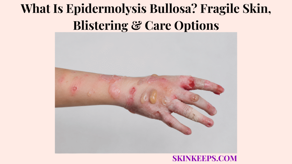
Epidermolysis bullosa (EB) is a group of rare conditions that make the skin fragile, so blisters, wounds, or tears can form after minor friction, rubbing, heat, pressure, or injury.
Most EB is inherited, while epidermolysis bullosa acquisita is a separate acquired autoimmune disorder. This page covers symptoms, locations, types, causes, diagnosis, wound care, infection, pain, nutrition, advanced treatment options, complications, mistakes, and urgent warning signs.
What Is Epidermolysis Bullosa and Why Does Skin Blister So Easily?
Epidermolysis bullosa is a group of rare conditions that weaken the attachments between skin layers, allowing blisters, erosions, or open wounds to form after trauma that would not normally damage healthy skin.
Different EB subtypes affect different structural proteins and skin levels. Mild disease may mainly involve the hands and feet, while severe disease can cause widespread wounds, scarring, feeding difficulty, eye problems, and long-term complications.
EB can affect both skin and mucous membranes, including the mouth, throat, esophagus, eyes, urinary tract, or genital area in some forms. Inherited EB is not contagious and should not be managed like an ordinary friction blister.
Why Does Minor Friction Cause EB Blisters?
Minor friction can cause EB blisters because weakened skin-adhesion proteins allow one skin layer to separate from another.
The separation creates a fluid-filled blister or a raw erosion. Repeated pressure from walking, gripping, clothing seams, medical tape, or routine handling can therefore cause new wounds.
How Can EB Range From Mild to Severe?
EB severity ranges from localized hand-and-foot blistering to generalized disease involving skin, wounds, nails, eyes, mouth, swallowing, nutrition, movement, and growth.
Subtype diagnosis matters because the depth of skin separation influences scarring, contractures, mucosal involvement, chronic wounds, and long-term monitoring needs.
Practical rule: Recurrent blisters after very minor friction, newborn skin loss, or wounds involving the mouth, eyes, or feeding should be medically assessed rather than treated as ordinary rubbing.
What Epidermolysis Bullosa Symptoms Appear on Skin?
Epidermolysis bullosa can cause fragile skin, blisters, raw wounds, erosions, crusting, scarring, milia, nail changes, thickened skin, pain, itching, and slow-healing sores.
Symptoms vary widely by subtype. Not every blister means EB, but repeated blistering after minimal trauma—especially from birth or early childhood—needs specialist evaluation.
What Do EB Blisters and Wounds Look Like?
EB blisters and wounds often appear after rubbing, pressure, heat, sweating, shoes, clothing seams, tape, or minor trauma that would not usually injure normal skin.
Blisters may rupture and leave painful raw erosions. Healing areas can develop crusting, color change, small white bumps called milia, scarring, nail thickening, or nail loss.
Some wounds heal slowly or reopen repeatedly because the surrounding skin remains fragile.
What Symptoms Suggest Severe EB or Complications?
Severe EB or its complications may cause widespread blistering, newborn skin loss, mouth or throat blisters, feeding difficulty, eye pain, recurrent infection, scarring, contractures, anemia, or chronic non-healing wounds.
A changing wound in severe dystrophic EB needs prompt review, especially if it becomes raised, hard, painful, thickened, bleeding, or persistently open.
| Symptom | EB Clue | Why It Matters |
|---|---|---|
| Blisters after minor friction | Classic skin-fragility pattern. | Needs friction reduction and diagnosis. |
| Raw wounds or erosions | Blister roof breaks easily. | Needs gentle wound protection. |
| Mouth or throat blisters | Mucosal involvement. | Can affect pain, feeding, and swallowing. |
| Nail changes | Nail-bed fragility or scarring. | May support subtype recognition. |
| Scarring or contractures | Deeper blistering pattern. | Can limit movement and function. |
| Chronic wound change | Severe DEB/RDEB concern. | Needs assessment for squamous cell carcinoma. |
| Fever, pus, odor, or red streaks | Possible wound infection. | Needs prompt medical review. |
Where Can Epidermolysis Bullosa Blisters Appear?
Epidermolysis bullosa blisters can appear on any area exposed to friction or pressure, but the hands, feet, elbows, knees, ankles, diaper area, back, mouth, throat, eyes, nails, and pressure points are especially important.
Areas under shoes, clothing seams, medical tape, dressings, or repeated gripping may blister. Some severe forms also affect the esophagus, digestive tract, urinary tract, or genital skin.
| Body Site | EB Risk | Care Issue |
|---|---|---|
| Hands | Grip and repeated friction. | Dressings, function, and hand therapy. |
| Feet and soles | Walking pressure and footwear. | Padding, shoe adjustment, and mobility. |
| Elbows and knees | Rubbing and pressure. | Soft clothing and protection. |
| Diaper area | Moisture, cleaning, and friction. | Gentle handling and barrier care. |
| Mouth and gums | Blistering during eating or oral care. | Dental care, pain control, and nutrition. |
| Throat or esophagus | Swallowing friction and scarring. | Specialist review for swallowing problems. |
| Eyes | Eyelid or corneal fragility. | Prompt eye care for pain or redness. |
| Nails | Repeated trauma or scarring. | Nail loss or thickening may occur. |
What Types of Epidermolysis Bullosa Are Important to Know?
Epidermolysis bullosa type matters because the depth of skin separation affects blister severity, scarring, mucous membrane problems, and long-term risks.
The four main inherited groups are EB simplex, junctional EB, dystrophic EB, and Kindler EB. Epidermolysis bullosa acquisita is acquired and autoimmune, so it follows a separate diagnostic and treatment pathway.
| EB Type | Blister Level or Main Clue | Why It Matters |
|---|---|---|
| EB simplex | Usually more superficial, often within the epidermis. | Can range from localized hand-and-foot blistering to generalized disease. |
| Junctional EB | Separation around the basement membrane zone. | Some forms are severe and can involve mucous membranes. |
| Dystrophic EB | Deeper separation involving collagen VII attachment. | Can cause scarring, contractures, and SCC risk in severe RDEB. |
| Kindler EB | Mixed blistering levels with photosensitivity. | Can create a broader skin-fragility pattern. |
| EB acquisita | Acquired autoimmune blistering. | Not the same as inherited EB; needs autoimmune evaluation. |
What Causes Epidermolysis Bullosa?
Most forms of epidermolysis bullosa are caused by inherited gene changes that affect proteins helping the skin layers attach securely to each other.
Different genes affect different structural proteins, so the level at which skin separates—and the severity of symptoms—varies by subtype.
How Do Genetic Changes Weaken Skin Adhesion?
Genetic changes weaken skin adhesion by altering structural proteins that normally anchor the epidermis, basement membrane zone, and dermis together.
When an attachment protein is missing or does not work normally, rubbing or pressure can split the skin at that level. Genetic testing can help confirm the subtype and support family counseling.
Is EB Contagious?
Inherited EB is not contagious and does not spread through touch, shared objects, dirt, or poor hygiene.
Open EB wounds can develop a secondary infection, but infection is a complication rather than the underlying cause. EB acquisita is also not contagious; it is an acquired autoimmune disorder.
| Cause Pathway | What Happens | Care Implication |
|---|---|---|
| Inherited gene change | A skin-adhesion protein is affected. | Subtype testing and genetic counseling. |
| Friction or pressure | Weak skin separates more easily. | Padding and skin protection. |
| Heat, sweat, or rubbing | Friction and blistering may worsen. | Cooling and clothing strategy. |
| Open wound burden | The skin barrier remains disrupted. | Pain and infection prevention. |
| Severe DEB/RDEB | Repeated deep wounds cause scarring. | Function and SCC surveillance. |
How Is Epidermolysis Bullosa Different From Other Blistering Conditions?
Epidermolysis bullosa can resemble ordinary friction blisters, burns, bacterial blistering, autoimmune blistering, or exposure-related dermatitis, so age of onset, trauma sensitivity, family history, examination, biopsy, and genetic testing matter.
Ordinary friction blisters usually follow a clear rubbing event and heal normally, while inherited EB can cause repeated wounds after very minor friction.
Bullous impetigo is a contagious bacterial infection that may cause fragile blisters, crusting, or pus rather than an inherited skin-adhesion disorder.
Bullous pemphigoid usually begins later in life and requires an autoimmune blistering workup.
Allergic contact dermatitis follows an exposure pattern and commonly causes itch and inflammation. Burns have a heat or chemical exposure history, while EB acquisita requires autoimmune evaluation.
| Condition | Main Clue | Why Confusion Happens | Safer Next Step |
|---|---|---|---|
| Inherited EB | Blisters after minimal trauma, often beginning early. | Blister overlap. | EB specialist assessment. |
| Friction blister | Clear rubbing or pressure event. | Similar fluid-filled lesion. | Protect and confirm normal healing. |
| Bullous impetigo | Contagious infection with crust or pus. | Fragile blister overlap. | Medical review and infection testing. |
| Bullous pemphigoid | Often older adult with tense blisters. | Generalized blistering. | Biopsy and immunofluorescence. |
| Burn | Heat, chemical, or radiation exposure. | Raw blistered skin. | Burn-specific care. |
| EB acquisita | Acquired autoimmune blistering. | Shared EB name. | Autoimmune testing. |
| Contact dermatitis | Exposure pattern with itch or inflammation. | Blisters may occur. | Trigger review and appropriate testing. |
How Is Epidermolysis Bullosa Diagnosed?
Epidermolysis bullosa is diagnosed through clinical examination, family history, blister pattern, specialized skin testing, and often genetic testing to identify the type and subtype.
A diagnosis should explain where the skin separates, which protein or gene is affected, how severe the condition may be, and which care plan fits the person.
What Tests Can Confirm EB Type?
Tests that can help confirm EB type include a skin biopsy with special mapping techniques and genetic testing.
Specialist centers may use immunofluorescence antigen mapping, electron microscopy in selected cases, and family testing. Blood tests alone do not classify inherited EB.
Why Does Newborn Blistering Need Careful Diagnosis?
Newborn blistering needs urgent, careful diagnosis because severe EB can appear at birth or shortly afterward, and routine handling, adhesives, or friction can worsen fragile skin.
Clinicians also need to rule out infection, trauma, other inherited blistering disorders, and serious neonatal conditions. Newborn skin loss should not be dismissed as diaper rash or ordinary rubbing.
Appointment preparation: Bring the age of onset, family history, trigger pattern, photographs, affected body sites, wound and infection history, feeding or eye symptoms, prior test results, current dressings, medicines, and details of any chronic wound that is changing.
- Age when blisters first appeared.
- Whether blistering started at birth or soon afterward.
- Photos of blisters, wounds, nails, mouth, or eye symptoms.
- Triggers such as friction, heat, pressure, shoes, clothing seams, tape, or dressings.
- Pain, itch, sleep disruption, or dressing-change problems.
- Fever, pus, odor, warmth, spreading redness, or worsening wounds.
- Feeding difficulty, poor growth, dehydration, constipation, or anemia symptoms.
- Family history and any prior biopsy, mapping, electron microscopy, or genetic testing.
What Care Options Help Epidermolysis Bullosa?
Epidermolysis bullosa care focuses on preventing new blisters, protecting wounds, reducing pain and itch, recognizing infection, supporting nutrition, and managing subtype-specific complications.
Care may involve dermatology, nursing, genetics, nutrition, dentistry, ophthalmology, gastroenterology, pain care, physical or occupational therapy, hand therapy, surgery, and psychosocial support.
| Care Need | Care Option | Main Goal |
|---|---|---|
| Blister prevention | Padding, soft clothing, cooling, and friction reduction. | Reduce new wounds. |
| Open wounds | Non-adherent dressings and gentle securement. | Protect healing skin. |
| Pain and itch | Individualized symptom plan. | Comfort, sleep, movement, and feeding. |
| Infection risk | Wound monitoring and targeted treatment. | Prevent local and systemic complications. |
| Nutrition | Dietitian and feeding support when needed. | Growth, energy, and wound healing. |
| Mouth and teeth | Gentle dental and oral care. | Reduce pain and preserve intake. |
| Mobility | PT, OT, hand care, and selected surgery. | Maintain function and limit contractures. |
| Severe DEB/RDEB wounds | Regular surveillance and prompt biopsy of suspicious changes. | Earlier cancer detection. |
How Should EB Blisters and Wounds Be Cared For?
EB wound care should protect fragile skin, use non-stick dressings, reduce pain, manage drainage, and prevent infection without creating additional trauma.
Why Must Dressings Be Gentle?
EB dressings must be gentle because sticky adhesives and rough removal can strip fragile skin and create new wounds.
Non-adherent dressings may reduce trauma, but the right material depends on wound depth, drainage, pain, infection risk, body site, and specialist guidance. This is not a one-product condition.
Why Should Blister Handling Be Taught by Specialists?
EB blister handling should be taught by specialists because casual popping can expand wounds, increase pain, and raise infection risk.
Some large blisters may need trained management to limit spread while preserving protective skin. The technique, equipment, and wound plan should come from an EB team rather than improvised home instructions.
- Protect fragile skin from pressure and rubbing.
- Use soft clothing, padding, and suitable footwear.
- Avoid sticky tape directly on fragile skin.
- Use non-adherent dressings selected for the wound.
- Remove dressings slowly and gently.
- Plan pain control around dressing changes.
- Watch for warmth, pus, odor, red streaks, fever, or worsening pain.
- Learn blister management from an EB specialist team.
- Keep regular wound and subtype follow-up.
How Do Infection, Pain, and Nutrition Affect EB Care?
Infection control, pain management, and nutrition are central to EB care because open wounds can be painful, slow to heal, and vulnerable to complications.
What Infection Signs Matter in EB Wounds?
Infection signs include increasing redness, warmth, swelling, worsening pain, pus, odor, fever, red streaks, and wounds that enlarge instead of healing.
Poor feeding or lethargy in an infant may also signal illness. An infected-looking wound should be assessed rather than treated repeatedly with antibiotics without guidance.
Impetigo is a separate bacterial infection; EB wounds can become infected, but infection is not the cause of inherited EB.
Why Does Nutrition Matter for Wound Healing?
Nutrition matters because chronic wounds increase healing demands while mouth, throat, or digestive involvement can make eating painful or difficult.
Children with severe EB may struggle with growth, and anemia or fatigue can complicate chronic wound burden. A dietitian can help match intake to swallowing ability, wound needs, growth, constipation, and overall health.
| Care Pillar | Why It Matters | What to Monitor |
|---|---|---|
| Infection | Open wounds can worsen or spread infection. | Fever, pus, odor, redness, streaks, pain. |
| Pain | Wounds and dressing changes may hurt. | Sleep, movement, mood, and feeding. |
| Nutrition | Healing needs energy and protein. | Weight, growth, intake, and anemia. |
| Itch | Scratching can create new wounds. | Sleep loss and skin damage. |
| Mobility | Blisters, scars, and contractures limit function. | Hands, feet, joints, and daily activities. |
Are There Advanced Treatment Options for Epidermolysis Bullosa?
Some advanced wound treatments are available for selected epidermolysis bullosa subtypes, but they do not replace daily wound care, pain control, infection prevention, nutrition support, or complication monitoring.
Eligibility depends on the EB subtype, gene findings, wound type, age, specialist assessment, treatment-center access, and local availability.
| Option | Best-Fit Context | Important Boundary |
|---|---|---|
| Specialist wound dressings | Many inherited EB types and wound patterns. | Daily care foundation, not a cure. |
| FILSUVEZ | Selected wounds associated with dystrophic or junctional EB. | Topical prescription wound treatment; specialist plan required. |
| VYJUVEK | DEB wounds with COL7A1 mutations. | Gene-based wound treatment with eligibility requirements. |
| ZEVASKYN | Wounds in recessive dystrophic EB. | Autologous cell-sheet gene therapy delivered through treatment centers. |
| Hand therapy or surgery | Contractures, fusion, or function loss. | Requires wound-risk and subtype planning. |
| Clinical trials | Selected patients and research protocols. | Eligibility and access vary. |
Reality check: An advanced therapy may help particular wounds or subtypes, but no single treatment applies to every person with EB or removes the need for long-term specialist care.
What Complications Can Epidermolysis Bullosa Cause?
Epidermolysis bullosa complications depend on type and severity, but severe disease can involve chronic wounds, infection, pain, scarring, contractures, feeding problems, anemia, eye disease, dental problems, and skin cancer risk.
Repeated deep wounds may cause fingers or toes to scar together, joints to tighten, nails to disappear, or swallowing passages to narrow. Mouth pain and wound burden can also affect nutrition and growth.
People with severe dystrophic EB, especially RDEB, need careful review of chronic wounds because a non-healing or changing wound may represent squamous cell carcinoma.
| Complication | Why It Happens | Monitoring Need |
|---|---|---|
| Infection | Open skin barrier and chronic wounds. | Fever, odor, drainage, redness, pain. |
| Scarring and milia | Repeated or deeper blistering. | Skin healing and subtype pattern. |
| Contractures or digit fusion | Repeated wounds and scar tightening. | PT, OT, hand care, and function. |
| Malnutrition and anemia | Painful intake plus high wound-healing demand. | Dietitian review, growth, and blood assessment. |
| Eye disease | Fragile eyelid or corneal tissue. | Prompt eye care for pain or redness. |
| Esophageal narrowing | Repeated mucosal injury and scarring. | Swallowing assessment. |
| Squamous cell carcinoma | Long-standing wounds in severe DEB/RDEB. | Urgent biopsy of suspicious wound change. |
What EB Care Mistakes Should You Avoid?
The biggest EB care mistake is treating fragile EB skin like ordinary skin, because adhesives, friction, squeezing, and rough dressing changes can create additional wounds.
Do not casually pop blisters, pull dressings quickly, scrub wounds, ignore pain, delay newborn assessment, or assume every wound needs repeated antibiotics.
Feeding difficulty, dehydration, anemia symptoms, eye pain, or a chronic wound that thickens, bleeds, becomes hard, or fails to heal should not be dismissed.
| Mistake | Why It Is Risky | Better Action |
|---|---|---|
| Sticky tape directly on skin | Can strip fragile skin. | Use specialist-approved non-adhesive securement. |
| Rough dressing removal | Creates new wounds and pain. | Remove slowly using the care plan. |
| Casual blister popping | Can enlarge injury or introduce infection. | Use specialist-taught management only. |
| Ignoring pain or itch | Reduces sleep, movement, and feeding; scratching damages skin. | Use an individualized symptom plan. |
| Repeating antibiotics blindly | May miss another cause and promotes unnecessary use. | Seek wound assessment and targeted treatment. |
| Ignoring nutrition | Weakens growth and wound healing. | Use dietitian and feeding support when needed. |
| Ignoring chronic wound change | Can delay SCC diagnosis in severe DEB/RDEB. | Arrange urgent specialist review and biopsy when indicated. |
When Should Epidermolysis Bullosa Symptoms Be Checked Urgently?
Epidermolysis bullosa symptoms need urgent assessment when blistering starts in a newborn, becomes widespread, involves the mouth, throat, eyes, or breathing, causes feeding or dehydration problems, shows infection signs, or creates severe pain.
A chronic wound also needs urgent review if it becomes raised, hard, thickened, painful, bleeding, rapidly changing, or persistently non-healing.
Seek urgent medical review if there is:
- Newborn blistering, skin loss, or rapidly spreading wounds.
- Fever, worsening redness, warmth, swelling, pus, odor, or red streaks.
- Severe or increasing wound pain.
- Poor feeding, dehydration, weight loss, or poor growth.
- Mouth, throat, swallowing, or esophageal symptoms.
- Eye pain, redness, light sensitivity, or vision change.
- Breathing difficulty, noisy breathing, or hoarseness.
- Severe fatigue, pallor, or suspected anemia.
- Hand or foot contractures that limit function.
- A chronic wound that becomes raised, hard, thickened, bleeding, painful, or non-healing.
- Known RDEB with any suspicious chronic wound change.
What Should You Remember About Epidermolysis Bullosa?
Epidermolysis bullosa is a rare fragile-skin blistering disorder group, so safe care depends on subtype diagnosis, gentle wound protection, infection prevention, nutrition support, and specialist follow-up.
- EB causes skin to blister or tear after minor trauma.
- Most EB is inherited; EB acquisita is a separate autoimmune condition.
- The main inherited groups are EB simplex, junctional EB, dystrophic EB, and Kindler EB.
- Severity ranges from localized hand-and-foot blistering to generalized multisystem disease.
- Diagnosis may include biopsy, immunofluorescence mapping, electron microscopy, and genetic testing.
- Care focuses on friction reduction, non-adherent wound protection, pain and itch control, infection recognition, nutrition, and function.
- Sticky adhesives and rough dressing changes can create new wounds.
- Selected advanced wound treatments exist, but eligibility is subtype-specific.
- Severe RDEB needs chronic-wound and SCC surveillance.
- Newborn blistering, infection signs, feeding difficulty, eye or throat involvement, or suspicious chronic wounds need urgent care.
Frequently Asked Questions About Epidermolysis Bullosa
Is epidermolysis bullosa contagious?
No. Inherited EB is a genetic skin-fragility disorder and does not spread by touch. Wounds can become infected, but infection is not the underlying cause.
What causes epidermolysis bullosa?
Most EB types are caused by inherited gene changes affecting proteins that help skin layers hold together. The affected gene, protein, and depth of skin separation differ by subtype.
What are the main types of epidermolysis bullosa?
The main inherited types are EB simplex, junctional EB, dystrophic EB, and Kindler EB. Epidermolysis bullosa acquisita is a separate acquired autoimmune disorder.
What does EB skin look like?
EB may cause fragile skin, blisters after minor friction, raw erosions, open wounds, crusting, scarring, milia, nail changes, mouth sores, eye symptoms, or chronic wounds, depending on subtype and severity.
How is epidermolysis bullosa diagnosed?
Diagnosis may include clinical examination, family history, blister pattern, skin biopsy with special mapping tests, electron microscopy in selected centers, and genetic testing to confirm the subtype.
What care options help EB?
Care may include friction reduction, non-adherent dressings, specialist-taught blister care, pain and itch control, infection treatment when needed, nutrition support, dental and eye care, physical or occupational therapy, and specialist EB follow-up.
Are there advanced treatments for EB?
Selected wound treatments exist for specific EB contexts, including FILSUVEZ for certain dystrophic and junctional EB wounds, VYJUVEK for dystrophic EB wounds with COL7A1 mutations, and ZEVASKYN for recessive dystrophic EB wounds. Eligibility requires specialist assessment.
When do EB symptoms need urgent care?
Urgent assessment is needed for newborn blistering, widespread wounds, fever, pus, odor, spreading redness, severe pain, dehydration, poor feeding, mouth or throat involvement, eye symptoms, breathing trouble, or a chronic wound that changes, bleeds, thickens, or does not heal.
Sources & Evidence About Epidermolysis Bullosa
NIAMS — Epidermolysis Bullosa: Overview, Symptoms & Causes was used for EB as a rare group of diseases causing fragile skin, the major inherited types, skin-layer adhesion, mucosal involvement, and the EB acquisita boundary.
NIAMS — Epidermolysis Bullosa: Diagnosis, Treatment & Steps to Take was used for clinical history, biopsy, genetic testing, non-adhesive bandages, infection warning signs, mobility care, and nutrition support.
DermNet — Epidermolysis Bullosa was used for inherited EB affecting skin and mucous membranes, friction-site blistering, subtype classification, and severe dystrophic EB complication context.
DEBRA UK — Skin and Wound Care for EB was used for specialist-led blister and wound management, pain and itch care, infection prevention, and EB clinical-practice guidance.
American Academy of Dermatology — Epidermolysis Bullosa Diagnosis and Treatment was used for specialist-team care, wound protection, nutrition, mobility, chronic wound complications, and available wound treatments.
FDA — FILSUVEZ Drug Trials Snapshot was used for FILSUVEZ as a topical treatment for wounds associated with dystrophic and junctional EB in eligible adult and pediatric patients.
FDA — VYJUVEK was used for VYJUVEK as a treatment for wounds in adult and pediatric patients with dystrophic EB and COL7A1 mutations.
FDA — ZEVASKYN was used for ZEVASKYN as an autologous cell sheet-based gene therapy for wounds in adult and pediatric patients with recessive dystrophic EB.
Educational Disclaimer: This SkinKeeps article is for educational purposes only and does not diagnose or replace medical care. Fragile skin, recurrent blisters, newborn blistering, skin loss, open wounds, mouth blisters, throat symptoms, feeding difficulty, poor weight gain, dehydration, eye pain, eye redness, vision symptoms, severe pain, severe itch, fever, pus, odor, warmth, swelling, spreading redness, red streaks, poor healing, anemia symptoms, contractures, nail loss, genital involvement, or chronic wounds that become raised, hard, painful, bleeding, thickened, or non-healing should be checked by a qualified healthcare professional. Seek urgent care for newborn blistering, rapidly spreading wounds, fever, dehydration, poor feeding, breathing trouble, swallowing trouble, eye symptoms, severe pain, or suspected infection. Do not self-diagnose EB, pop or lance EB blisters without specialist training, use sticky tape directly on fragile skin, pull dressings off quickly, scrub wounds, repeatedly use antibiotics without guidance, or start advanced treatment plans without specialist evaluation.



