
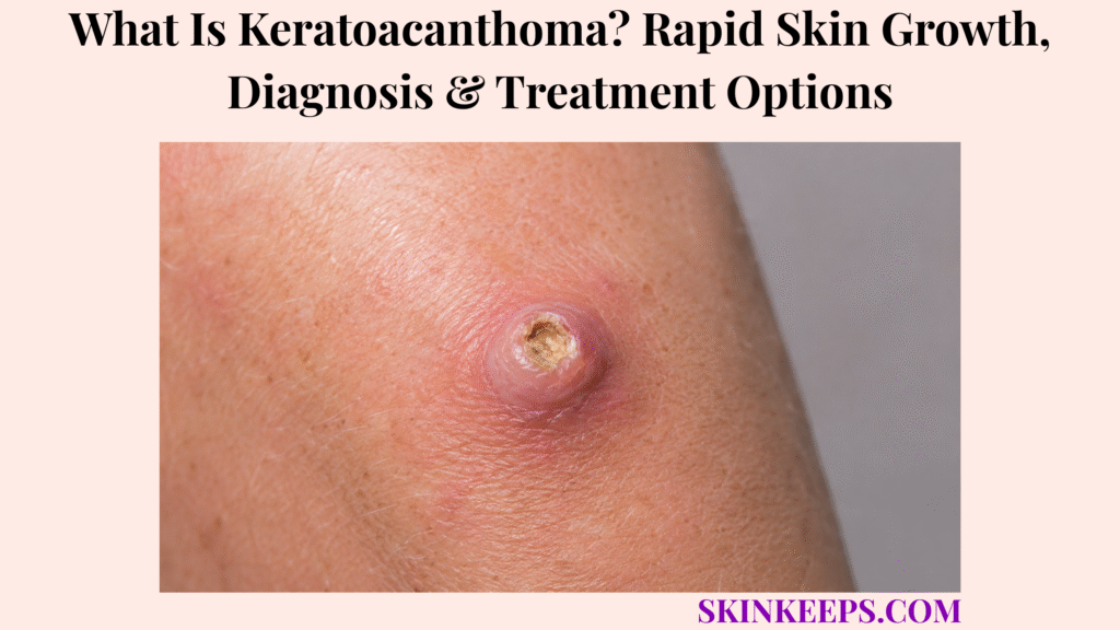
Keratoacanthoma, abbreviated KA, is a rapidly growing dome-shaped or crater-shaped keratinizing skin tumour that often forms around a central plug of keratin. It commonly develops on sun-exposed skin, including the face, ears, forearms and hands, and may reach a noticeable size within several weeks.
Some lesions later stop growing and regress, sometimes leaving a depressed scar, but the course cannot be predicted safely. KA closely overlaps with well-differentiated cutaneous squamous cell carcinoma, so adequate biopsy or removal is usually recommended instead of waiting at home.
How Can You Recognize a Keratoacanthoma?
Keratoacanthoma is usually recognized as a rapidly enlarging dome-shaped or crater-shaped nodule with a central keratin plug.
What Does a Typical Keratoacanthoma Look Like?
A typical keratoacanthoma appears as a firm round or oval nodule with a volcano-like profile and a central keratin-filled crater.
- Firm round or oval nodule.
- Dome-shaped or volcano-like profile.
- Smooth skin-coloured, pink, red, brown-red or purple-brown surface.
- Central crater with a hard yellow-white or brown keratin plug.
- Raised rolled or sloping edges.
- Often symmetrical in a classic lesion.
- Possible surrounding inflammation.
- Typical size around 1–2 cm, with uncommon larger lesions.
How Quickly Does a Keratoacanthoma Grow?
Keratoacanthoma classically grows quickly over several weeks, but its later behaviour cannot be predicted safely from appearance alone.
- A small papule appears.
- It enlarges rapidly over several weeks.
- A central keratin plug develops.
- The lesion may remain stable for several weeks.
- Some lesions regress gradually.
- Regression may leave an indented or discoloured scar.
A solitary lesion may move from first appearance to regression over roughly 4–6 months, but individual behaviour cannot be predicted at presentation.
Which Symptoms Can Occur?
Keratoacanthoma may be tender, painful, crusted, bleeding or completely asymptomatic despite rapid growth.
- Tenderness or pain.
- Itching or tingling.
- Bleeding after minor trauma.
- Crusting or ulceration.
- Pressure or tightness.
- No symptoms despite rapid enlargement.
Where Does Keratoacanthoma Usually Develop?
Keratoacanthoma most often develops on chronically sun-exposed skin.
- Central face, nose and cheeks.
- Lips and ears.
- Scalp and neck.
- Forearms and backs of hands.
- Lower legs.
- Less commonly beneath a nail, on mucosal or genital skin, or within a previous injury site.
Figure 1. Keratoacanthoma often grows from a small papule into a dome-shaped nodule with a central keratin-filled crater over several weeks. Pain, crusting, bleeding or ulceration can occur, and lesions on tissue-sensitive sites need prompt assessment.
Why Does Keratoacanthoma Develop?
Keratoacanthoma develops when keratinocytes proliferate rapidly and form a crateriform tumour with central keratin accumulation.
Which Skin Cells Produce a Keratoacanthoma?
Keratoacanthoma is a keratinizing tumour derived from epidermal keratinocytes and often appears related to the hair-follicle unit.
- Rapid keratinocyte proliferation creates the raised tumour.
- Keratin collects centrally and produces the crater-like plug.
- Molecular and microscopic findings overlap with cutaneous SCC.
- The precise biological boundary between KA and SCC remains controversial.
How Does Ultraviolet Exposure Affect Risk?
Ultraviolet exposure increases keratinocyte DNA damage and helps explain why many KAs arise on sun-exposed skin.
- Long-term UV exposure damages keratinocyte DNA.
- Most solitary lesions occur on chronically exposed sites.
- Cumulative sun damage becomes more relevant with age.
- Previous keratinocyte tumours or field damage raise diagnostic concern.
- KA can still occur on less-exposed skin.
Background sun damage and actinic keratosis can indicate a keratinocyte field in which KA-type and SCC-type tumours need careful evaluation.
Which Other Factors Can Trigger or Increase Risk?
Keratoacanthoma risk rises with older age, previous KA, skin injury, chronic sun damage, immune suppression and selected medication exposures.
- Older age.
- Previous keratoacanthoma.
- Surgery, burns, tattoos, insect bites or chronic scars.
- Immunosuppression or organ transplantation.
- Genetic predisposition.
- Selected cancer medicines.
Which Medicines Can Cause Keratoacanthoma-Like Tumours?
Some targeted cancer medicines can trigger keratoacanthoma-like cutaneous squamous tumours, so medication history matters.
- BRAF inhibitors such as vemurafenib or dabrafenib.
- Selected targeted cancer treatments.
- Other reported medicine associations.
Cancer or immune-suppressing medicine should not be stopped independently; dermatology and the prescribing specialist should coordinate care.
How Is Keratoacanthoma Different From Squamous Cell Carcinoma and Other Skin Growths?
Keratoacanthoma cannot be reliably separated from squamous cell carcinoma by appearance alone.
How Is Keratoacanthoma Different From Squamous Cell Carcinoma?
Keratoacanthoma may grow faster and look more symmetrical than many SCCs, but these clues are not reliable enough to replace tissue diagnosis.
| Feature | Keratoacanthoma Pattern | Cutaneous SCC Pattern |
|---|---|---|
| Growth | Often very rapid over weeks | Variable; weeks to months |
| Shape | Frequently symmetrical and crateriform | May be irregular, infiltrated or ulcerated |
| Centre | Prominent keratin plug | Keratin, scale, crust or ulcer can occur |
| Natural course | May regress | Does not predictably regress |
| Histology | Can closely resemble well-differentiated SCC | May show invasive malignant keratinocytes |
| Management | Usually removed because SCC cannot be excluded | Requires definitive cancer treatment |
How Is Keratoacanthoma Different From a Cutaneous Horn?
A cutaneous horn describes a cone of compact keratin, while keratoacanthoma is one possible tumour beneath or producing horn-like keratin.
A cutaneous horn is a shape rather than one diagnosis, so its base must be sampled because the underlying lesion may be benign, precancerous or malignant.
How Is Keratoacanthoma Different From an Inflamed Cyst or Boil?
An inflamed cyst or boil may look like a painful nodule, but it usually behaves more like an infection or blocked cyst than a firm keratin-filled crateriform tumour.
- Pus rather than a firm keratin crater.
- Fluctuation or soft movement under pressure.
- A pre-existing cyst opening.
- Drainage and inflammatory pain.
- Broader surrounding infection.
A lump with a central pore and recurrent drainage may suggest an epidermal cyst, but rapid crateriform growth still requires tumour assessment.
A painful pus-filled infection such as a boil can resemble an inflamed nodule, but a suspected KA should not be squeezed or punctured.
Which Other Lesions Can Resemble Keratoacanthoma?
Several benign, infectious and malignant lesions can resemble keratoacanthoma, so atypical or rapidly growing nodules need histologic assessment.
- Nodular basal cell carcinoma.
- Prurigo nodule.
- Wart or molluscum contagiosum.
- Amelanotic melanoma.
- Metastatic skin tumour.
- Foreign-body reaction.
- Deep fungal infection.
- Hypertrophic actinic keratosis.
- Other crateriform SCC variants.
A pearly or ulcerated nodule may suggest basal cell carcinoma, especially on chronically sun-exposed facial skin.
A firm small nodule may be a dermatofibroma, but rapid crater-like growth is not typical.
How Is Keratoacanthoma Diagnosed?
Keratoacanthoma is diagnosed by combining clinical growth history, examination, dermoscopy and an adequate tissue specimen for pathology.
Which Clinical Details Matter?
The most important clinical details are how quickly the growth appeared, where it is located and whether SCC risk factors are present.
- First appearance and speed of enlargement.
- Original and current size.
- Pain, itching, crusting or bleeding.
- Previous skin cancers or KAs.
- Sun-exposure history.
- Immune suppression or organ transplantation.
- New targeted medicines.
- Recent trauma or procedure.
- Family history of multiple similar tumours.
The clinician may also examine surrounding sun-damaged skin and regional lymph nodes.
Can Dermoscopy Confirm Keratoacanthoma?
Dermoscopy can support a keratinizing tumour pattern but cannot confirm benign keratoacanthoma or reliably exclude invasive SCC.
- Central keratin mass.
- White circles or keratin scales.
- Hairpin or irregular vessels.
- Blood spots.
- Pale or red structureless areas.
Which Biopsy Is Usually Preferred?
An architecture-preserving full-thickness specimen is preferred because KA diagnosis depends on the central crater, lateral lips and deep base.
- Complete excision often provides diagnosis and treatment.
- Deep saucerization or a designed incisional biopsy may be used when immediate excision is impractical.
- A small superficial shave or central punch can miss the edges or deepest invasion.
- Further excision may be required when sampling is incomplete or SCC cannot be excluded.
What Does the Pathologist Look For?
The pathologist looks for crateriform architecture while also checking for invasive SCC features.
- Symmetrical crateriform architecture.
- Central keratin-filled crater.
- Epidermal lips overhanging the crater.
- Well-differentiated squamous proliferation.
- Glassy keratinocytes.
- Depth and pattern of invasion.
- Cytologic atypia.
- Perineural or vascular invasion.
- Features favouring conventional SCC.
Figure 2. Keratoacanthoma and well-differentiated squamous cell carcinoma overlap clinically and dermoscopically. Diagnosis therefore relies on an architecture-preserving specimen that includes the central crater, both lateral lips and the deep base.
Why Is Keratoacanthoma Usually Treated Rather Than Observed?
Keratoacanthoma is usually treated because possible spontaneous regression does not safely exclude invasive squamous cell carcinoma.
Can Keratoacanthoma Disappear Without Treatment?
Some keratoacanthomas can regress, but spontaneous regression is not a dependable home-management strategy.
- Regression can take several months.
- A depressed or discoloured scar may remain.
- Growing lesions can damage tissue-sensitive sites.
- Future behaviour cannot be predicted from early appearance.
- A lesion presumed to be KA may instead be invasive SCC.
When Might Close Observation Be Considered?
Observation is reserved for exceptional clinician-supervised situations after diagnostic uncertainty and patient risk have been carefully weighed.
- Histology strongly supports a regressing KA.
- Surgery presents disproportionate medical risk.
- The patient is frail or has limited treatment tolerance.
- The site and behaviour can be monitored closely.
- The patient understands the uncertainty.
- Rapid access to treatment is available if growth continues.
Which Lesions Need Particularly Prompt Treatment?
Prompt treatment is especially important when the lesion is rapidly enlarging, symptomatic, high-risk by site or occurring in an immunosuppressed patient.
- Rapidly enlarging, painful, bleeding or ulcerated.
- Larger than expected or recurrent.
- On the lip, ear, eyelid, nose, finger or genital skin.
- Developing in a scar or nail unit.
- Associated with enlarged regional lymph nodes.
- Occurring in an immunosuppressed or transplant patient.
Which Surgical Treatments Are Used for Keratoacanthoma?
Surgical removal is commonly used because it can provide both definitive histology and treatment.
When Is Standard Surgical Excision Used?
Standard surgical excision is commonly preferred for a solitary accessible keratoacanthoma because it removes the tumour and provides a full pathology specimen.
- Removes the entire visible tumour.
- Provides a complete specimen for histology.
- Allows deep and lateral margin assessment.
- May be curative when completely removed.
- Can be followed by more treatment if pathology shows involved margins or higher-risk SCC features.
When Is Mohs Micrographic Surgery Considered?
Mohs surgery may be considered when tissue conservation and complete margin assessment are especially important.
- Facial lesions.
- Nose, lips, ears or eyelids.
- Large, recurrent or poorly defined tumours.
- Sites where tissue conservation matters.
- Lesions with aggressive SCC features.
- Immunosuppressed patients.
When Are Curettage and Electrosurgery Considered?
Curettage and electrosurgery may be considered only for selected small, typical, low-risk lesions when diagnostic uncertainty can still be managed safely.
- Small, typical lesions.
- Low-risk anatomical sites.
- Patients for whom formal excision is less suitable.
- Use by clinicians experienced in keratinocyte tumours.
Limitations include less complete margin assessment, possible residual deep tumour, difficulty excluding invasive SCC, scarring and the need for later excision.
Can Surgery Leave a Scar?
Surgery can leave a scar, but waiting for growth or regression can also cause scarring and tissue loss.
- Any full-thickness removal creates a scar.
- Scar size depends on tumour size, site, closure and healing.
- Regression can leave a depressed scar.
- Removing a smaller lesion may preserve more tissue than delayed surgery.
Which Non-Surgical Treatments Can Be Used in Selected Keratoacanthomas?
Non-surgical treatments are specialist options for selected keratoacanthomas when surgery is difficult, risky or impractical.
How Do Intralesional Methotrexate and 5-Fluorouracil Work?
Intralesional methotrexate and 5-fluorouracil are injected into selected lesions to reduce rapidly dividing keratinocytes.
- Injected directly into the tumour.
- Can shrink selected lesions.
- May preserve tissue in difficult locations.
- May be used before surgery in selected cases.
- Require repeated clinical review.
- Do not eliminate the need for adequate diagnostic tissue.
When Are Topical Treatments Considered?
Topical treatments are considered only in selected specialist-directed cases because inflammation, delay and missed SCC remain concerns.
- Topical 5-fluorouracil or imiquimod may be considered.
- Evidence is narrower than for surgery.
- Marked inflammation or ulceration can occur.
- Resolution can take longer.
- An SCC mistaken for KA could continue invading.
- Histologic confirmation and close follow-up remain necessary.
When Are Oral Retinoids Used?
Oral retinoids such as acitretin are reserved for selected multiple, eruptive, inherited or recurrent keratoacanthoma patterns.
- Numerous eruptive lesions.
- Inherited multiple-KA syndromes.
- Repeated tumours.
- Situations where repeated operations are impractical.
- Specialist monitoring for pregnancy prevention, liver, lipid and mucosal effects.
Can Radiation, Cryotherapy or Laser Be Used?
Radiation, cryotherapy and laser have selected roles but should not replace adequate diagnosis when SCC is possible.
- Radiation may be considered when surgery is unsuitable.
- Cryotherapy may treat selected small lesions but limits full histologic assessment.
- Laser and photodynamic approaches are not standard replacements for diagnostic excision.
- Treatment choice must preserve the ability to detect SCC.
What Do Multiple or Recurrent Keratoacanthomas Mean?
Multiple or recurrent keratoacanthomas may signal medication exposure, immune suppression, organ transplantation or an inherited tumour syndrome.
Which Multiple-Keratoacanthoma Patterns Can Occur?
Multiple KA patterns range from familial self-healing lesions to generalized eruptive disease with many small crateriform papules.
- Ferguson–Smith multiple self-healing squamous epitheliomas.
- Grzybowski generalized eruptive keratoacanthomas.
- Witten–Zak syndrome.
- Other familial multiple-KA patterns.
- Muir–Torre-associated keratoacanthomas.
- Medication-associated eruptive disease.
Grzybowski syndrome can involve hundreds to thousands of small KA-like papules on skin and sometimes mucosal surfaces.
When Should an Inherited Syndrome Be Considered?
An inherited syndrome should be considered when lesions are multiple, early-onset, familial, recurrent or associated with mucosal or internal-cancer clues.
- Several lesions at a young age.
- Multiple affected relatives.
- Repeated self-healing tumours.
- Numerous tumours erupting quickly.
- Oral or other mucosal lesions.
- Sebaceous tumours.
- Family history of Lynch-associated cancers.
What Should Be Reviewed When Several New Lesions Appear?
When several new lesions appear, clinicians should review immune status, transplant history, medicines, previous radiation and family tumour history.
- Immunosuppressive status.
- Organ-transplant history.
- BRAF or other targeted therapy.
- Previous radiotherapy.
- Chronic ultraviolet exposure.
- Personal skin-cancer history.
- Family tumour history.
How Are the Skin and Surgical Site Monitored After Treatment?
After keratoacanthoma treatment, follow-up focuses on pathology review, margin status, wound healing, recurrence and new keratinocyte tumours.
What Happens After Removal?
After removal, the pathology report determines the final classification and whether margins or SCC-risk features require further treatment.
- Tumour classification is reviewed.
- Margins are assessed.
- Additional excision may be recommended.
- Sutures and wound healing are monitored.
- Infection and bleeding precautions are followed.
- The scar remodels gradually.
- The treated site is checked for recurrence.
Can Keratoacanthoma Return?
Keratoacanthoma may not return after complete treatment, but recurrence or a new KA or SCC elsewhere remains possible.
- Recurrence can follow incomplete removal.
- Another KA or SCC can develop elsewhere.
- Multiple-KA syndromes produce repeated lesions.
- Immunosuppressed patients carry continuing risk.
- New thickening, pain, bleeding or rapid growth needs reassessment.
How Can Additional Tumours Be Reduced or Detected Earlier?
Additional tumours are reduced and detected earlier through sun protection, regular self-examination and scheduled professional skin checks.
- Broad-spectrum sun protection.
- Protective clothing and hats.
- Avoidance of tanning beds.
- Regular self-examination.
- Date-stamped photographs of new suspicious growths.
- Scheduled professional skin checks.
- Prompt assessment of any rapidly growing keratin-filled nodule.
- Treatment of surrounding actinic damage when appropriate.
Which Changes Require Prompt Reassessment?
Prompt reassessment is needed for new rapid growth, recurrent keratin plugging, ulceration, bleeding, pain, numbness, lymph-node enlargement or a wound that fails to heal.
- New rapid enlargement.
- Recurrent central keratin plug.
- Persistent ulceration or spontaneous bleeding.
- Increasing pain or firm fixation.
- Numbness or tingling.
- Enlarged regional lymph nodes.
- A wound that fails to heal.
Prompt route: rapid growth, bleeding, ulceration, pain, fixation, numbness, enlarged lymph nodes, non-healing skin or a lesion on a high-risk site requires medical assessment.
Figure 3. Surgical removal commonly provides diagnosis and treatment. Mohs is considered when tissue conservation or margin control is important, while nonsurgical therapies are selected by specialists. Follow-up reviews pathology, margins, healing, recurrence and the entire skin.
What Should You Remember About Keratoacanthoma?
Keratoacanthoma is a rapidly growing crater-shaped keratinizing tumour that is usually treated as possible SCC until pathology clarifies the diagnosis.
- A central keratin-filled crater is suggestive but not diagnostic.
- Most solitary lesions arise on sun-exposed skin.
- Some lesions regress, often with scarring.
- Clinical behaviour cannot be predicted safely.
- KA and well-differentiated SCC can be extremely difficult to separate.
- Dermoscopy cannot safely exclude invasive SCC.
- Architecture-preserving biopsy or complete excision is usually required.
- Standard excision commonly provides diagnosis and treatment.
- Mohs may suit facial, recurrent or tissue-sensitive lesions.
- Intralesional medicines are specialist-selected alternatives.
- Multiple lesions can indicate medicine, immune or inherited factors.
- Observation is reserved for exceptional supervised situations.
- Ongoing skin examination and sun protection support early detection.
Frequently Asked Questions About Keratoacanthoma?
Is keratoacanthoma a form of skin cancer?
Classification is controversial. Some sources describe keratoacanthoma as a benign follicular tumour, while many clinicians manage it as squamous cell carcinoma, keratoacanthoma type. Because well-differentiated SCC can look almost identical, a rapidly growing crater-shaped lesion needs medical assessment and histology.
How quickly does a keratoacanthoma grow?
Keratoacanthoma typically enlarges rapidly over several weeks before reaching a stable or possible regression phase. Its later course is unpredictable, so waiting at home is unsafe because SCC can have a similar appearance and may continue growing.
Why does keratoacanthoma need a biopsy?
Diagnosis depends on the tumour architecture and exclusion of invasive SCC. The pathologist needs to examine the central crater, lateral lips and deep base, which a superficial or very small partial sample may miss.
Which treatment is most commonly used for keratoacanthoma?
Complete surgical excision is commonly used for a solitary accessible lesion because it removes the tumour and supplies a full specimen for pathology. Mohs may be used for facial, recurrent, large, ill-defined or tissue-sensitive lesions.
Can keratoacanthoma return after treatment?
Recurrence can occur after incomplete removal, and another keratoacanthoma or SCC can develop elsewhere. Pathology review, treated-site monitoring, full-skin checks, sun protection and prompt reassessment of new rapid growth remain important.
Which Sources Support This Keratoacanthoma Guidance?
DermNet — Keratoacanthoma — Rapid crateriform morphology, SCC/KA-type controversy, typical growth cycle, biopsy limitations, treatment and outcome.
British Association of Dermatologists — Keratoacanthoma — Patient-facing recognition, sun-exposed distribution, skin-cancer resemblance, removal and sun-protection advice.
American Academy of Dermatology — Squamous Cell Carcinoma — SCC diagnosis and treatment context, including excision and Mohs surgery.
American Academy of Dermatology — Cutaneous SCC Clinical Guideline — Surgical-treatment priority and boundaries for selected nonsurgical approaches.
DermNet — Squamous Cell Carcinoma Dermoscopy — Central keratin, white structures and vascular clues that overlap across keratinocyte tumours.
PubMed — Intralesional Methotrexate Versus 5-Fluorouracil — Randomized specialist evidence for selected biopsy-proven keratoacanthomas.
DermNet — Generalised Eruptive Keratoacanthomas — Grzybowski syndrome, hundreds-to-thousands lesion burden, associated factors and systemic treatment context.
PMC — Keratoacanthoma Versus Squamous-Cell Carcinoma — Importance of complete architecture, full-thickness sampling and KA–SCC pathology overlap.
This SkinKeeps article is educational and does not replace dermatology, dermatopathology, Mohs, oncology, transplant, primary or emergency care. Seek prompt assessment for any rapidly enlarging, painful, bleeding, ulcerated, crusted or crater-shaped growth, especially on the lips, ears, eyelids, nose, fingers, genital skin, nail unit or scar, or in an immunosuppressed person. Seek urgent review for fixation, numbness, enlarged lymph nodes or a non-healing wound. Do not squeeze, puncture, scrape, burn, freeze or treat a suspected keratoacanthoma at home, and do not stop cancer or immune medicines independently.



