
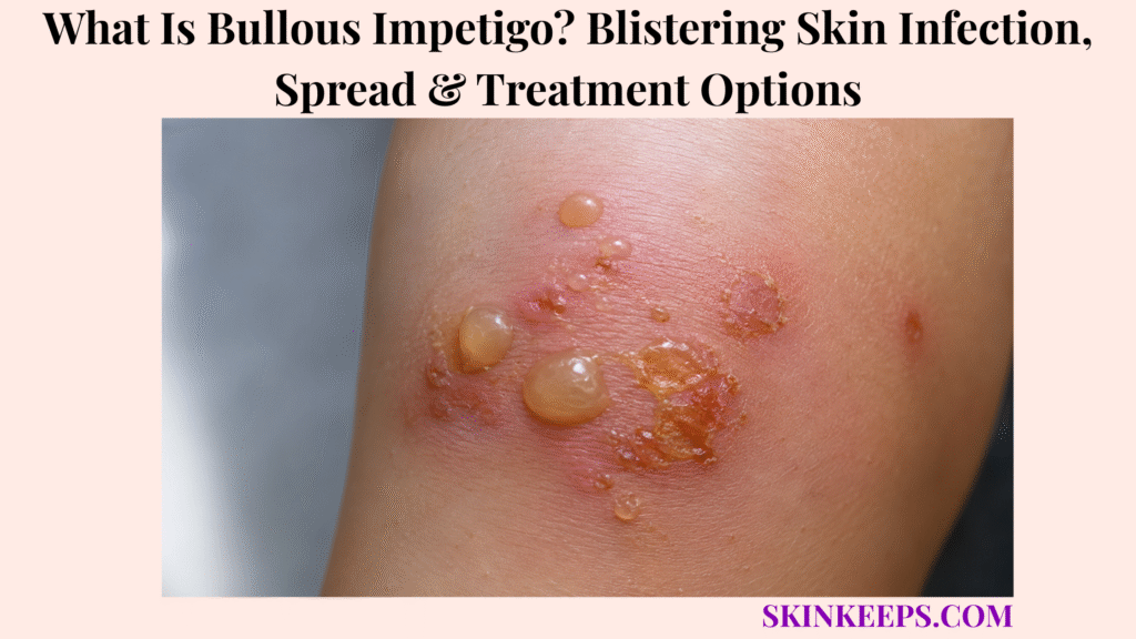
Bullous impetigo is a contagious bacterial skin infection that causes fragile fluid-filled blisters, usually because toxin-producing Staphylococcus aureus affects the top layers of the skin.
It should not be managed like a simple friction blister, and the blisters should not be popped or picked. This page covers blister appearance, causes, spread, diagnosis, antibiotic treatment, hygiene control, prevention, mistakes, and urgent signs.
What Is Bullous Impetigo and Why Does It Cause Blisters?
Bullous impetigo is a contagious bacterial skin infection that causes fragile fluid-filled blisters, usually because toxin-producing Staphylococcus aureus affects the top layers of the skin.
It is a blistering form of impetigo, a superficial bacterial skin infection that can spread through contact with infected skin, drainage, and shared items.
The word “bullous” refers to bullae, which are larger fluid-filled blisters. In bullous impetigo, the blister roof is often thin and fragile, so the blister can break easily and leave moist raw skin.
Why Is Bullous Impetigo Different From Non-Bullous Impetigo?
Bullous impetigo is different from non-bullous impetigo because it forms larger fragile blisters instead of mainly honey-colored crusted sores.
Non-bullous impetigo often forms honey-colored crusts after smaller sores break open. Bullous impetigo forms larger soft blisters that may rupture and leave raw shiny skin with a scaly rim.
Bullous impetigo is especially important in infants and young children because blisters can spread quickly and systemic signs need prompt attention.
Why Are the Blisters Fragile and Thin-Roofed?
The blisters are fragile and thin-roofed because Staphylococcus aureus toxin disrupts cell adhesion near the skin surface, allowing fluid to collect under a shallow roof.
This toxin effect creates a superficial split in the epidermis, the upper skin layer. Fluid then collects inside that split and forms a soft, flaccid bulla.
When the blister roof breaks, bacteria-containing fluid can spread to nearby skin, hands, towels, clothing, bedding, toys, or close contacts.
Practical rule: Bullous impetigo is a contagious bacterial blistering infection, so safe care focuses on antibiotic treatment when indicated, covering lesions, hygiene control, and prompt review when symptoms spread or a child looks unwell.
What Bullous Impetigo Symptoms Appear on Skin?
Bullous impetigo usually causes quickly appearing superficial blisters that may contain clear, yellow, cloudy, purulent, or darker fluid before they rupture and leave moist raw skin with a scaly rim.
The blisters may be small at first, then enlarge into soft thin-roofed bullae. Some cases have little surrounding redness at first, while others show spreading warmth, swelling, or darker color change as infection worsens.
What Do Early Bullous Impetigo Blisters Look Like?
Early bullous impetigo may begin as small vesicles that enlarge into thin-roofed, soft, flaccid blisters filled with clear or yellow fluid.
The fluid may later become cloudy, purulent, or darker. Blisters can appear over hours or days and may spread through scratching or contact.
Not every blister is bullous impetigo. A clinician checks the blister pattern, age, exposure, spread, fever, fluid, skin breaks, and similar lesions in close contacts.
What Does Ruptured Bullous Impetigo Look Like?
Ruptured bullous impetigo often leaves moist raw skin with yellow drainage and a scaly rim, sometimes called a collarette of scale.
The raw base may look red, pink, brown, purple-brown, or darker depending on skin tone and inflammation. Crusting can occur after rupture, but classic honey-colored crust is more typical of non-bullous impetigo.
Ruptured blisters should be covered where practical because drainage can spread bacteria to other skin areas or other people.
What Symptoms Suggest a More Serious Infection?
Bullous impetigo may need faster care when fever, malaise, swollen lymph nodes, spreading warmth, increasing pain, many blisters, or infant poor feeding and lethargy appear.
Rapid spread, many lesions, eye-area involvement, dehydration signs, no improvement after starting treatment, or recurrent infection should also prompt medical review.
Widespread peeling or scalded-looking skin is a separate urgent warning pattern because a more serious staph toxin illness may be involved.
| Blister Sign | What the Reader May Notice | Why It Matters |
|---|---|---|
| Small vesicles | Tiny fluid bumps at first | May enlarge into bullae |
| Flaccid bullae | Soft fragile blister roof | Bullous impetigo clue |
| Clear or yellow fluid | Fluid inside blister | Common early fluid pattern |
| Cloudy, purulent, or darker fluid | Fluid changes appearance | Infection burden may be higher |
| Ruptured raw base | Moist red, pink, brown, or darker skin after breakage | Needs covering and treatment |
| Scaly collarette | Rim of scale around raw area | Classic after rupture |
| Fever or malaise | Child or adult feels unwell | Medical review |
| Poor feeding or lethargy | Infant appears unwell | Prompt care |
| Widespread peeling | Scalded-looking skin | Urgent SSSS concern |
Where Does Bullous Impetigo Usually Appear?
Bullous impetigo can appear on the face, trunk, arms, legs, buttocks, and perineal or diaper area, and it may spread through scratching or contact.
It can occur around the mouth or nose, on the trunk, arms, legs, buttocks, diaper area, skin folds, or sites of minor trauma, insect bites, eczema, scratching, or broken skin.
Location alone cannot diagnose the infection. Blisters near the eyes, in infants, or spreading quickly need earlier care.
| Site | Common Clue | Spread / Safety Concern |
|---|---|---|
| Face / around nose or mouth | Contagious sores or ruptured blisters | Hand-to-face spread and close contact |
| Trunk | Flaccid blisters or raw patches | Clothing and bedding contamination |
| Arms / legs | Blisters after scratches or bites | Autoinoculation from scratching |
| Buttocks | Blisters or raw patches under clothing | Contact with clothing and bedding |
| Diaper / perineal area | Infant or young child involvement | Lower threshold for medical care |
| Skin folds | Warm, moist, rubbed areas | Spread and irritation risk |
| Broken skin / eczema sites | Infection over damaged barrier | Treat skin barrier and infection |
| Near eyes | Blisters close to eye area | Prompt medical review |
What Causes Bullous Impetigo?
Bullous impetigo is caused by toxin-producing Staphylococcus aureus, which disrupts superficial skin-cell adhesion and creates fragile blisters.
Staphylococcus aureus can live on skin or in the nose. Infection can develop when bacteria reach vulnerable skin and produce blister-forming toxin.
How Does Staphylococcus aureus Enter or Colonize Skin?
Staphylococcus aureus can live on skin or in the nose and can cause bullous impetigo when it reaches vulnerable skin and produces blister-forming toxin.
Minor cuts, scratches, insect bites, diaper irritation, shaving irritation, rubbed skin, or eczema-damaged skin can create easier entry points.
Atopic dermatitis can increase infection risk when the skin barrier is scratched, cracked, or inflamed.
Why Are Infants and Young Children Commonly Affected?
Infants and young children are commonly affected because their skin barrier may be more vulnerable and close-contact environments make spread easier.
Daycare, school, household exposure, diaper irritation, shared toys, close skin contact, and frequent touching can support spread.
Infants with fever, lethargy, poor feeding, widespread blisters, dehydration signs, or scalded-looking skin need prompt medical care.
Is Bullous Impetigo Caused by Poor Hygiene?
Bullous impetigo is not simply caused by poor hygiene; it is a bacterial infection, though hygiene and covering lesions help reduce spread.
The key issue is bacterial transmission, not moral blame. Handwashing, covering lesions, laundering items that touch drainage, and avoiding shared towels or toys help protect others.
Clean care matters, but it should not replace antibiotic treatment when a clinician recommends antibiotics.
How Does Bullous Impetigo Spread?
Bullous impetigo spreads when bacteria from blister fluid, drainage, skin contact, hands, towels, clothing, bedding, toys, or shared items reach another skin area or another person.
Scratching can move bacteria from one body site to another, which is called autoinoculation. Close household, daycare, school, sports, or crowded settings can also make spread easier.
Covering lesions, washing hands, changing dressings, and laundering contaminated items reduce spread while medical treatment clears the infection.
Who Is More Likely to Get Bullous Impetigo?
Bullous impetigo is more likely when skin is broken, irritated, scratched, infected, crowded, warm, humid, or exposed to close contact with someone who has impetigo.
Risk factors are practical clues, not blame. They help decide who needs faster evaluation and stronger spread-control steps.
How Is Bullous Impetigo Different From Other Blistering Skin Problems?
Bullous impetigo can resemble friction blisters, burns, eczema, herpes, chickenpox, hand-foot-and-mouth disease, bullous pemphigoid, or staphylococcal scalded skin syndrome, so pattern, age, spread, fever, and lesion distribution matter.
Correct diagnosis matters because bullous impetigo is contagious and antibiotic-led, while many other blistering conditions need different care.
How Is Bullous Impetigo Different From Non-Bullous Impetigo?
Bullous impetigo forms larger fragile blisters, while non-bullous impetigo more often forms honey-colored crusted sores.
Non-bullous impetigo may be caused by staph, strep, or both. Bullous impetigo is caused by toxin-producing Staphylococcus aureus.
How Is Bullous Impetigo Different From Friction or Burn Blisters?
Bullous impetigo is different from friction or burn blisters because it is contagious and infectious rather than caused only by rubbing, heat, sun, or chemical injury.
Blisters from friction often follow rubbing or pressure, while burn blisters follow heat, sun, chemical, or electrical injury.
Yellow drainage, multiple lesions, spread, fever, daycare or household exposure, or rapidly expanding raw areas should raise infection concern.
How Is Bullous Impetigo Different From Herpes or Chickenpox?
Bullous impetigo is different from herpes or chickenpox because it usually produces fragile superficial bullae and ruptured raw patches rather than grouped painful viral vesicles or widespread itchy crops at different stages.
Shingles usually follows a painful one-sided nerve-pattern blistering path, which is different from a contagious staph blistering infection.
Clinical review or viral testing may be needed when the pattern is unclear.
How Is Bullous Impetigo Different From Staphylococcal Scalded Skin Syndrome?
Bullous impetigo is usually a localized blistering infection, while staphylococcal scalded skin syndrome causes more widespread toxin-mediated skin tenderness, blistering, and peeling.
Widespread tenderness, fever, scalded-looking skin, peeling in sheets, or an unwell infant or young child needs urgent medical care.
This is a safety boundary, not a home diagnosis. A clinician should assess widespread peeling or scalded-looking skin quickly.
| Condition | Main Clue | Contagious? | Safer Next Step |
|---|---|---|---|
| Bullous impetigo | Fragile blisters, yellow fluid, raw base, collarette scale | Yes | Antibiotic treatment plus cover lesions |
| Non-bullous impetigo | Honey-colored crusts | Yes | Antibiotics plus hygiene control |
| Friction blister | Rubbing or pressure history | No | Protect and offload |
| Burn blister | Heat, sun, chemical, or electrical injury | No | Burn care |
| Herpes | Grouped painful vesicles | Yes | Clinical or viral evaluation if suspected |
| Chickenpox | Widespread itchy crops at different stages | Yes | Medical or public-health guidance |
| SSSS | Widespread tender peeling or scalded skin | Serious toxin illness | Urgent medical care |
| Bullous pemphigoid | Tense autoimmune blisters, often older adults | No | Dermatology diagnosis |
| Allergic contact dermatitis | Itchy blistering after plant, product, metal, or adhesive exposure | No | Trigger review and diagnosis |
How Is Bullous Impetigo Diagnosed or Checked?
Bullous impetigo is often diagnosed by physical examination, but bacterial culture may be used when infection is widespread, recurrent, resistant, severe, unclear, or when MRSA is a concern.
A clinician looks at the blister pattern, fluid, rupture stage, scaly collarette, spread, systemic symptoms, exposure history, and medical risk.
What Does a Clinician Check With Bullous Impetigo?
A clinician checks bullous impetigo by looking at blister size, number, fluid, ruptured areas, collarette scale, distribution, spread, systemic symptoms, exposure history, and medical risk.
The exam may include fever or malaise, swollen lymph nodes, eczema, cuts, insect bites, scratches, diaper irritation, household exposure, daycare exposure, school exposure, sports contact, previous impetigo, MRSA history, infant age, and immune risk.
Image comparison alone is not enough because burns, viral blisters, autoimmune blisters, and scalded-skin syndromes can overlap visually.
When Might Swab or Culture Be Needed?
Swab or culture may be needed when bullous impetigo is recurrent, severe, widespread, not responding to treatment, occurring in an outbreak, or associated with MRSA concern.
Culture can help identify the bacterial cause and guide antibiotic choice when standard treatment is not working or resistance is suspected.
Culture or further testing may also be considered when the diagnosis is unclear and herpes, burn injury, autoimmune blistering disease, or staphylococcal scalded skin syndrome is part of the differential.
What Treatment Options Are Used for Bullous Impetigo?
Bullous impetigo treatment usually requires antibiotics, with topical treatment for limited disease and oral antibiotics when lesions are widespread, numerous, severe, recurrent, or associated with systemic symptoms.
Antibiotic choice should be clinician-guided because severity, age, local resistance patterns, MRSA concern, recurrence, and medical risk affect treatment decisions.
When Are Topical Antibiotics Used?
Topical antibiotics may be used when bullous impetigo is limited, mild, localized, and a clinician confirms that topical treatment is appropriate.
This usually means few localized lesions, no systemic symptoms, no rapid spread, and reliable follow-up.
The full prescribed course should be completed. Cleaning and covering support treatment but do not replace medication when antibiotics are needed.
When Are Oral Antibiotics Used?
Oral antibiotics may be used when bullous impetigo has multiple lesions, widespread disease, rapid spread, systemic symptoms, treatment failure, outbreak risk, or high-risk patient factors.
Oral treatment is more likely to be considered with fever, malaise, swollen lymph nodes, many lesions, infant illness, immune suppression, recurrent disease, nonresponse, or MRSA concern.
Do not use leftover antibiotics or someone else’s prescription. Wrong medication can fail and may increase resistance risk.
Why Should Antibiotic Choice Be Clinician-Guided?
Antibiotic choice should be clinician-guided because bullous impetigo is Staphylococcus aureus-driven, resistance patterns vary, and infants or high-risk patients need careful assessment.
MRSA patterns vary by location and exposure history. Culture may be needed when infection is recurrent, severe, widespread, outbreak-related, nonresponsive, or clinically unclear.
Do not stop prescribed antibiotics early unless a clinician instructs you, and do not use steroid-only cream on suspected bacterial infection.
| Situation | Likely Treatment Direction | Key Caution |
|---|---|---|
| Few localized lesions | Topical antibiotic may be used | Confirm diagnosis and complete course |
| Multiple lesions | Oral antibiotic often considered | Needs clinician review |
| Widespread or rapidly spreading disease | Medical assessment and oral therapy | Watch for systemic illness |
| Fever, malaise, lymph nodes | Prompt medical review | Systemic symptoms matter |
| Recurrent or nonresponsive infection | Culture plus targeted treatment | MRSA or staph carriage possible |
| Infant or newborn | Prompt medical evaluation | Lower threshold for care |
| Eye-area lesions | Medical review | Avoid delay near eyes |
| Widespread peeling or scalded skin | Emergency evaluation | SSSS concern |
How Should Bullous Impetigo Lesions Be Cleaned and Covered?
Cleaning and covering bullous impetigo helps reduce spread, but it does not replace antibiotic treatment when antibiotics are needed.
Wash hands before and after care. Gently clean drainage as advised, avoid harsh scrubbing, and keep lesions covered with clean dressings where practical.
Change dressings when wet or dirty. Wash towels, clothing, bedding, and washcloths that touch lesions. Keep nails short to reduce scratching.
How Can Bullous Impetigo Be Prevented From Spreading or Coming Back?
Preventing bullous impetigo spread means treating active infection, covering lesions, reducing scratching, cleaning shared items, and addressing skin breaks that let bacteria enter.
Manage eczema, cuts, bites, scratches, and diaper irritation early. Keep nails trimmed and discourage scratching.
Recurrent episodes may need review for staph carriage, household transmission, MRSA risk, immune suppression, skin-barrier disease, or repeated close-contact exposure.
What Bullous Impetigo Mistakes Should You Avoid?
The biggest bullous impetigo mistake is popping or picking blisters and then touching other skin, because blister fluid can spread bacteria.
Do not use steroid cream alone on suspected bacterial infection. Do not use leftover antibiotics, and do not stop prescribed antibiotics early unless a clinician tells you to.
Do not send a child with uncovered spreading lesions into close-contact settings without clinician or local school guidance. Do not ignore fever, lethargy, poor feeding, eye-area lesions, or widespread peeling.
| Mistake | Why It Is Risky | Better Action |
|---|---|---|
| Popping blisters | Spreads bacteria and worsens raw skin | Cover and treat |
| Picking crusts or raw areas | Delays healing and spreads infection | Keep clean and covered |
| Steroid-only cream | Can worsen infection or delay care | Medical review |
| Sharing towels or toys | Spreads bacteria | Separate and wash items |
| Leftover antibiotics | Wrong drug or resistance risk | Use prescribed treatment only |
| Stopping antibiotics early | Treatment failure risk | Complete prescribed course |
| Ignoring systemic signs | Severe infection or SSSS concern | Prompt care |
| Treating it as friction blister | Delays antibiotic treatment | Confirm diagnosis |
When Should Bullous Impetigo Be Checked Urgently?
Bullous impetigo needs prompt medical care when blisters are widespread, rapidly spreading, associated with fever or malaise, affecting an infant, near the eyes, not improving with treatment, or accompanied by skin peeling, severe pain, lethargy, or dehydration signs.
Infants, newborns, immune-risk patients, and people with rapidly spreading blisters should not wait for the infection to “dry out” on its own.
Which Bullous Impetigo Signs Need Faster Care?
Faster care is needed when bullous impetigo causes fever, malaise, lethargy, poor feeding, rapid spread, many blisters, swollen lymph nodes, increasing pain, spreading warmth, eye-area lesions, dehydration signs, no improvement, or recurrence.
Spreading warmth, swelling, pain, redness, purple-brown change, or darker expanding color around lesions can suggest spreading bacterial infection such as cellulitis.
Which Signs May Suggest a More Serious Staph Toxin Problem?
Widespread skin tenderness, large areas of peeling skin, scalded-looking skin, fever with widespread blistering, or an unwell infant or young child may suggest a more serious staph toxin problem.
This may raise concern for staphylococcal scalded skin syndrome, which needs urgent medical assessment. Do not try to diagnose or treat widespread peeling at home.
Seek prompt medical care if bullous impetigo involves:
What Should You Remember About Bullous Impetigo?
The most important thing to remember about bullous impetigo is that it is a contagious bacterial blistering infection, so safe management depends on antibiotic treatment, covering lesions, hygiene control, and quick escalation when symptoms spread or a child looks unwell.
Fragile blisters, yellow drainage, raw patches, collarette scale, fever, rapid spread, or infant illness should be treated as infection clues rather than routine blister-care problems.
Frequently Asked Questions About Bullous Impetigo
Is bullous impetigo contagious?
Yes. Bullous impetigo can spread through contact with infected skin, blister fluid, hands, towels, clothing, bedding, toys, razors, and shared items. Antibiotic treatment and covering lesions reduce spread.
What causes bullous impetigo?
Bullous impetigo is caused by toxin-producing Staphylococcus aureus. The toxin disrupts superficial skin-cell adhesion, causing fragile fluid-filled blisters.
What does bullous impetigo look like?
It can cause thin-roofed fragile blisters, clear or yellow fluid, rupture, oozing, raw skin, and a scaly collarette rim. Honey-colored crust is more typical of non-bullous impetigo.
Is bullous impetigo the same as a normal blister?
No. A friction blister is caused by rubbing or pressure; bullous impetigo is a contagious bacterial infection that usually needs antibiotic treatment.
What is the best treatment for bullous impetigo?
There is no one best treatment for every case. Limited cases may use topical antibiotics, while widespread, multiple, severe, recurrent, systemic-symptom, infant, or MRSA-risk cases may need oral antibiotics and clinician review.
Should you pop bullous impetigo blisters?
No. Popping or picking can spread bacteria, worsen raw skin, and increase transmission; lesions should be covered and treated.
When can a child return to school or daycare after impetigo?
Local rules and clinician guidance matter. CDC guidance says people with impetigo can return to school or work at least 12 hours after starting appropriate antibiotic treatment when lesions are covered, with some scenarios requiring longer exclusion.
When should bullous impetigo need urgent care?
Prompt care is needed for infants, fever, lethargy, poor feeding, rapid spread, many blisters, eye-area lesions, severe pain, spreading warmth or color change, dehydration signs, no improvement, recurrence, or widespread peeling or scalded-looking skin.
Sources & Evidence About Bullous Impetigo
CDC — Clinical Guidance for Group A Streptococcal Impetigo was used for impetigo diagnosis by physical examination, bullous versus non-bullous causes, topical and oral antibiotic treatment direction, prevention through antibiotic treatment and covering lesions, laundry/no-sharing guidance, and return-to-school/work context.
CDC — About Impetigo was used for patient-facing impetigo overview, antibiotic treatment, topical versus oral antibiotic direction by number of sores, covering exposed sores, household spread, daycare/school risk, and hot/humid climate risk.
DermNet — Impetigo was used for bullous impetigo clinical features, thin-roofed bullae, yellow fluid, spontaneous rupture, scaly collarette, face/trunk/extremity/buttock/perineal sites, autoinoculation, and systemic symptoms being more likely in bullous impetigo.
MSD Manual Professional — Impetigo and Ecthyma was used for impetigo as a superficial bacterial infection, Staphylococcus aureus causing all bullous impetigo, exfoliative toxins causing bullae, clinical diagnosis, culture logic, topical and oral antibiotic treatment direction, MRSA culture context, and immunocompromise risk.
StatPearls / NCBI Bookshelf — Impetigo was used for bullous impetigo beginning as small vesicles that become flaccid bullae, exfoliative toxin A causing loss of superficial epidermal cell adhesion, clear/yellow fluid becoming purulent or dark, ruptured bullae leaving a rim of scale, honey-colored crust not being typical in bullous impetigo, culture use, and hygiene measures.
Merck Manual Professional — Staphylococcal Scalded Skin Syndrome was used for differentiating localized bullous impetigo from widespread toxin-mediated skin tenderness, bullae, epidermal sloughing, fever, systemic illness, and high concern in infants and children.
Educational Disclaimer: This SkinKeeps article is for educational purposes only and does not diagnose or replace medical care. Fragile spreading blisters, yellow drainage, fever, lethargy, poor feeding, swollen lymph nodes, eye-area lesions, rapid spread, many blisters, dehydration signs, recurrent infection, no improvement with treatment, immune-risk illness, infant illness, or widespread peeling/scalded-looking skin should be checked promptly by a qualified healthcare professional. Do not pop, pick, or intentionally burst suspected impetigo blisters.



