
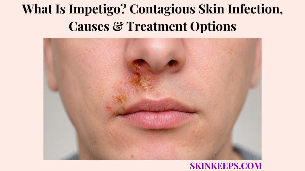
Impetigo is a highly contagious superficial bacterial skin infection that often begins as small sores, pustules or fragile blisters that break open and form golden or honey-coloured crusts. It is most common in young children but can affect adolescents and adults.
Bacteria often enter through scratching, eczema, bites, cuts or other barrier damage. A few localized non-bullous lesions may be treated topically, while numerous lesions, bullous impetigo, ecthyma or systemic illness often require oral treatment plus cleaning, coverage and transmission precautions.
How Can You Recognize Impetigo on the Skin?
Impetigo often appears as small sores, pustules or fragile blisters that rupture into moist erosions with honey-coloured crust.
- Red, pink, brown-red, purple-brown or skin-coloured bumps.
- Papules developing into pustules or superficial blisters.
- Clear fluid or pus leaking after rupture.
- Moist shallow erosions.
- Golden-yellow or honey-coloured crust.
- Itch with mild tenderness.
- Several nearby lesions or new lesions after scratching.
Pustules from folliculitis can resemble early impetigo, but impetigo more often progresses into moist erosions and honey-coloured crusts.
Figure 1. Non-bullous impetigo classically progresses from a papule to a pustule or fragile blister, rupture, moist erosion and honey-coloured crust; bullous disease forms larger blisters, while ecthyma extends deeper and can scar.
Where Does Impetigo Usually Appear?
Impetigo commonly appears around the nose, mouth and exposed limbs but can develop anywhere bacteria enter damaged skin.
- Around the nose, mouth, chin and cheeks.
- Arms, hands and legs.
- Sites of insect bites, scratches and cuts.
- Areas affected by eczema or another itchy rash.
- Trunk, folds and diaper region in bullous disease.
- Any body area after transfer from an infected lesion.
How Does Non-Bullous Impetigo Develop?
Non-bullous impetigo usually begins as a small papule that becomes a pustule or fragile blister before rupturing into honey-coloured crust.
- Small papule or sore.
- Vesicle or pustule formation.
- Fragile surface rupture.
- Moist shallow erosion.
- Thick adherent golden crust.
- Merging nearby lesions.
- Mild nearby lymph-node enlargement in some cases.
- Fever usually absent in uncomplicated disease.
It may be caused by S. aureus, group A streptococcus or both; appearance does not reliably identify the organism.
How Does Bullous Impetigo Look Different?
Bullous impetigo creates larger flaccid fluid-filled blisters and broad erosions rather than mainly small honey-crusted sores.
Larger fragile blisters may suggest bullous impetigo, especially in infants or young children with trunk, fold or diaper-area lesions.
- Large clear or cloudy blisters.
- Thin fragile blister roof.
- Rapid rupture.
- Broad moist erosion.
- A thin collar of scale around the erosion.
- Less thick honey crust than non-bullous disease.
- Possible fever, diarrhoea or illness when extensive.
Bullous impetigo is caused by toxin-producing S. aureus.
What Is Ecthyma—and How Is It Related to Impetigo?
Ecthyma is a deeper ulcerative infection related to impetigo and can leave scars.
- Thick firmly attached crust.
- Punched-out ulcer beneath the crust.
- Surrounding redness and swelling.
- Pain or tenderness.
- Common lower-leg involvement.
- Slow healing and scar risk.
Ecthyma generally needs oral antimicrobial treatment rather than topical care alone.
Which Bacteria Cause Impetigo?
Impetigo is usually caused by Staphylococcus aureus, group A Streptococcus pyogenes or both.
| Clinical Type | Likely Organism Pattern | Important Boundary |
|---|---|---|
| Non-bullous | S. aureus, group A strep or mixed | Appearance cannot identify which organism |
| Bullous | Toxin-producing S. aureus | Large flaccid blisters |
| Ecthyma | S. aureus and/or streptococci | Deeper infection needing systemic care |
| Selected resistant cases | Possible MRSA | Use local epidemiology and culture |
How Do the Bacteria Enter the Skin?
Bacteria enter through scratches, bites, cuts, inflamed rashes or microscopic barrier damage.
A skin fissure can create an entry point when dry or inflamed skin is repeatedly scratched.
- Scratches, cuts and abrasions.
- Insect bites.
- Eczema and other itchy rashes.
- Scabies or chickenpox lesions.
- Burns or shaving irritation.
- Cracked skin.
- Microscopic injury that is not easily visible.
Who Is More Likely to Develop Impetigo?
Anyone can develop impetigo, but young children and people with damaged or itchy skin are affected more often.
- Children approximately 2–5 years old.
- Close contact with an infected person.
- Crowded households or childcare.
- Contact sports.
- Warm humid climates.
- Eczema, scabies or insect bites.
- Cuts, abrasions or chronic scratching.
- Immune suppression, diabetes or chronic skin disease.
Is Impetigo Contagious?
Impetigo is highly contagious and spreads through lesion fluid, direct contact, hands and shared items.
- Skin-to-skin contact.
- Fluid from a blister or sore.
- Scratching and touching another person.
- Shared towels, bedding or clothing.
- Razors, toys and sports equipment.
- Transfer to another body area.
Risk is highest while lesions are untreated, moist, draining or uncovered.
How Long After Exposure Can Impetigo Appear?
Impetigo can appear days after exposure; group A streptococcal impetigo commonly has an incubation period of about 10 days.
- Timing varies with the bacterial cause and barrier damage.
- Staphylococci may already be carried on skin or in the nose.
- The most recent contact is not always the source.
- Household cases may begin on different days.
How Does Scratching Spread Impetigo?
Scratching moves bacteria beneath the nails and into new breaks in nearby skin, a process called autoinoculation.
- Bacteria collect beneath fingernails.
- Scratching creates new barrier breaks.
- New lesions form in clusters or lines.
- Hands transfer bacteria to face, toys, surfaces and people.
- Repeated scratching delays healing.
How Is Impetigo Different From a Cold Sore?
Impetigo is bacterial, while a cold sore is a recurrent herpes simplex virus blister pattern.
A recurrent grouped blister with tingling near the lip may be a cold sore, while impetigo more often forms moist bacterial erosions and honey-coloured crust.
| Feature | Impetigo | Cold Sore |
|---|---|---|
| Prodrome | Usually none | Tingling or burning common |
| Lesion | Papule, pustule, erosion and crust | Grouped small vesicles |
| Recurrence | Not fixed to one site | Often same lip area |
| Cause | Bacterial | HSV |
| Treatment | Antibacterial | Antiviral when indicated |
How Is Impetigo Different From Eczema?
Eczema is an inflammatory itchy rash, while impetigo is a contagious bacterial infection that can develop on top of eczema.
Active dermatitis / eczema can become impetiginized after scratching damages the barrier.
| Feature | Eczema | Impetigo |
|---|---|---|
| Main change | Dry itchy inflammation | Pustules, erosions and honey crust |
| Course | Flares and remissions | Contagious infection |
| Surface | Scale, cracks or weeping | Moist crusted lesions |
| Treatment | Barrier and anti-inflammatory care | Antimicrobial care |
| Overlap | Can become secondarily infected | May arise within eczema |
How Is Impetigo Different From Ringworm?
Impetigo forms moist crusted erosions, while ringworm usually forms a slowly expanding scaly ring.
| Feature | Impetigo | Ringworm |
|---|---|---|
| Surface | Moist erosion and crust | Dry scaly border |
| Shape | Irregular cluster | Circular or oval ring |
| Centre | No central clearing required | Central clearing often present |
| Cause | Bacterial | Fungal |
| Spread | Contact and lesion fluid | Fungal contact spread |
How Is Impetigo Different From Chickenpox or Other Viral Blisters?
Impetigo is usually localized or clustered, while chickenpox often causes widespread crops of blisters at several stages.
| Feature | Impetigo | Chickenpox |
|---|---|---|
| Distribution | Localized face or limbs common | Widespread, often trunk-predominant |
| Lesion stage | Similar local sequence | Multiple stages at once |
| Systemic symptoms | Usually absent if uncomplicated | Fever or illness common |
| Crust | Honey-coloured bacterial crust | Viral vesicles crust as they age |
Scratched viral blisters can become secondarily impetiginized.
How Is Impetigo Different From Scabies or Insect Bites?
Scabies and insect bites cause itching and barrier breaks that may later become infected with impetigo.
| Feature | Impetigo | Scabies | Insect Bites |
|---|---|---|---|
| Main clue | Pustule, erosion, honey crust | Night itch and burrows | Discrete itchy papules |
| Distribution | Face and damaged skin | Webs, wrists, waist or genital sites | Exposure-related sites |
| Cause | Bacteria | Mite | Arthropod bite |
| Overlap | Can follow scratching | Can become impetiginized | Can become impetiginized |
How Is Impetigo Different From Cellulitis?
Impetigo is superficial, while cellulitis is a deeper spreading infection with diffuse warmth, redness and swelling.
Rapidly spreading warmth, redness, swelling and pain may suggest cellulitis, which is deeper than ordinary impetigo.
| Feature | Impetigo | Cellulitis |
|---|---|---|
| Depth | Superficial surface infection | Deeper skin and soft tissue |
| Visible lesion | Erosion, blister or crust | Diffuse expanding redness |
| Warmth/swelling | Limited | Prominent |
| Pain | Mild tenderness | Increasing tenderness |
| Systemic illness | Usually absent | May include fever or weakness |
How Do Clinicians Diagnose Impetigo?
Clinicians usually diagnose impetigo by examining lesion type, distribution, spread and risk factors.
- Honey-coloured crust or flaccid blisters.
- Face, limb, fold or barrier-injury distribution.
- Speed of spread.
- Contact with an infected person.
- Eczema, scabies, bites or injury.
- Fever or systemic symptoms.
- Previous antibiotics and recurrence.
Appearance cannot reliably distinguish staphylococcal from streptococcal non-bullous disease.
When Is a Bacterial Culture Needed?
Culture is useful when results could change diagnosis or antibiotic choice.
- Uncertain diagnosis.
- Treatment failure or worsening during therapy.
- Recurrent infection.
- Several linked cases or an outbreak.
- Suspected MRSA.
- Immune suppression.
- Extensive disease or ecthyma.
- Important local resistance concerns.
Pus or exudate culture can identify the organism and susceptibility; typical limited cases may be treated without testing.
Figure 2. Impetigo begins when bacteria enter a barrier break and can spread through scratching, direct contact and shared items. Typical limited disease is diagnosed clinically; culture helps when disease is uncertain, recurrent, resistant, extensive or linked to MRSA or an outbreak.
Does Impetigo Require Treatment?
Impetigo should usually be treated to shorten infection, reduce spread and lower the risk of deeper disease.
- Shortens the infectious period.
- Reduces self-spread and household transmission.
- Limits new lesion formation.
- Lowers risk of deeper infection.
- Supports faster healing.
- Allows a safer return to school or work.
When Is Topical Treatment Used for Impetigo?
Topical treatment is generally used for a few localized non-bullous lesions in a person who is otherwise well.
- Only a few lesions.
- Localized disease.
- No systemic illness.
- No ecthyma.
- No extensive bullous disease.
- Reliable application and lesion coverage.
How Are Mupirocin, Retapamulin and Ozenoxacin Used?
These topical antibiotics are applied directly to localized impetigo lesions when locally approved and clinically appropriate.
- Mupirocin covers common staphylococcal and streptococcal organisms.
- Retapamulin is an option for eligible limited disease in some countries.
- Ozenoxacin is approved for eligible patients in selected regions.
- Approved ages and licensing differ by country.
- Use only for the prescribed short course.
- Wash hands before and after application.
- Do not reuse indefinitely for every crusted rash.
When Is Hydrogen Peroxide Cream Considered?
NICE permits hydrogen peroxide 1% medicinal cream for selected localized non-bullous impetigo when the person is not systemically unwell or at high complication risk.
- This is a formulated 1% medicinal cream.
- It is a regulated medicinal 1% formulation, not a home-use peroxide product.
- Do not dilute or improvise stronger solutions.
- It may be unsuitable near the eyes.
- A short topical antibiotic course may be used if it is unsuitable or ineffective.
- Availability and recommendations differ by country.
When Are Oral Antibiotics Used for Impetigo?
Oral antibiotics are used for numerous lesions, several body areas, bullous disease, ecthyma, systemic illness or failed local treatment.
- Numerous or widespread lesions.
- Bullous impetigo.
- Ecthyma.
- Fever or systemic illness.
- Rapid spread or outbreak.
- Topical treatment impractical.
- Higher complication risk.
- Failure of appropriate topical care.
Which Oral Antibiotics May Treat Impetigo?
Oral antibiotic selection depends on likely organisms, local resistance, allergy history, age, pregnancy, kidney function and MRSA risk.
| Clinical Context | Possible Clinician-Selected Class | Key Boundary |
|---|---|---|
| Typical MSSA/strep coverage | Antistaphylococcal penicillin or first-generation cephalosporin | Local susceptibility matters |
| True penicillin allergy | Macrolide or another suitable alternative | Resistance and pregnancy matter |
| Selected resistant infection | Clindamycin or another culture-guided option | Use susceptibility data |
| Suspected MRSA | Doxycycline, clindamycin or trimethoprim-sulfamethoxazole in eligible patients | Ensure streptococcal coverage when needed |
Leftover or shared antibiotics should not be used, and this page does not provide dosing.
Why Should Antibiotic Resistance Be Considered?
Unnecessary or repeated antibiotic exposure can make later infections harder to treat.
- Confirm the eruption fits impetigo.
- Use topical treatment only for suitable localized disease.
- Use the narrowest appropriate medicine.
- Complete the prescribed short course.
- Culture recurrent or treatment-resistant disease.
- Avoid saving antibiotics for future self-treatment.
- Reassess failure for resistance, poor adherence or wrong diagnosis.
How Should Impetigo Sores Be Cleaned and Covered?
Sores should be cleaned gently, treated as prescribed and covered to reduce bacterial transfer.
- Wash hands before care.
- Soften crust with warm water.
- Use mild soap or cleanser as directed.
- Remove only crust that separates easily.
- Pat dry with a clean disposable or dedicated towel.
- Apply prescribed medicine.
- Cover with clean gauze or dressing.
- Wash hands again.
Should Impetigo Blisters Be Popped or Crusts Be Picked Off?
Blisters should not be popped and firmly attached crusts should not be forcefully removed.
- Do not pop bullae or squeeze pustules.
- Do not scrape with sharp tools.
- Do not peel adherent crust.
- Do not shave across lesions.
- Gentle soaking is enough to loosen detachable crust.
Picking spreads bacteria, creates wounds, delays healing and increases scar or pigment-change risk.
How Can Itching Be Controlled Without Spreading Impetigo?
Itch control reduces scratching, self-spread and barrier injury.
- Keep fingernails short.
- Use cool compresses.
- Cover lesions securely.
- Wear loose clothing over affected sites.
- Ask about age-appropriate itch medicine.
- Treat underlying eczema or scabies.
- Clean hands after accidental contact.
When Can Someone With Impetigo Return to School or Work?
Return is generally appropriate when the person is well, has started effective treatment, can cover lesions and can maintain hygiene.
- CDC threshold: at least 12 hours after appropriate antibiotics.
- The person is well enough to participate.
- Lesions remain covered.
- Hand hygiene can be maintained.
- Some schools, childcare settings or outbreaks require at least 24 hours.
- Draining lesions that cannot be covered may require longer absence.
- Contact sports follow sport-specific medical clearance.
How Can Impetigo Be Prevented From Spreading at Home?
Household spread is reduced by covering lesions, washing hands and separating personal items.
- Avoid direct contact with sores.
- Keep lesions covered.
- Do not share towels, washcloths, clothes or razors.
- Separate toys and sports equipment.
- Use a clean towel each time.
- Wash used clothing, linens and towels daily.
- Clean frequently touched surfaces.
- Trim nails and treat itchy rashes.
- Wash hands before food preparation after lesion care.
Why Does Impetigo Sometimes Keep Returning?
Recurrent impetigo may reflect bacterial carriage, untreated barrier disease, household spread or resistance.
- Nasal S. aureus carriage.
- Bacteria carried on the skin.
- Untreated eczema or scabies.
- Persistent scratching or repeated bites.
- Shared contaminated items.
- Incomplete treatment.
- Antibiotic resistance.
- Contact-sport exposure.
- Diabetes or immune impairment.
When Is Decolonization Considered for Recurrent Impetigo?
Decolonization is considered only for confirmed recurrent disease after clinical assessment.
- Nasal or skin culture where useful.
- Nasal antibiotic treatment.
- Antiseptic body washing.
- Carefully measured clinician-directed dilute bleach baths.
- Household-member assessment.
- Eczema or scabies control.
- Cleaning shared items.
It is not required for every first episode, and nasal antibiotics or bleach baths should not be repeatedly self-directed.
How Is Impetigo Managed When Eczema Is Also Present?
Treatment must clear bacteria and repair the itchy inflamed barrier that allowed infection to begin.
When atopic dermatitis is present, antibiotics treat infection while anti-inflammatory and barrier care reduce scratching and reinfection.
- Identify which areas are infected.
- Use prescribed topical or oral antimicrobial treatment.
- Clean and cover infected lesions.
- Moisturize non-infected dry skin.
- Use clinician-directed eczema anti-inflammatory care.
- Avoid several irritating medicines on raw skin.
- Reassess widespread weeping or rapid worsening.
How Is Bullous Impetigo Managed in Babies?
Bullous impetigo in babies needs prompt assessment because infants can become unwell quickly.
- Examine blister distribution and extent.
- Check feeding, hydration and temperature.
- Assess for widespread disease.
- Use oral or systemic treatment when indicated.
- Clean gently and dress erosions.
- Monitor rapid spread.
- Consider staphylococcal scalded skin syndrome.
- Review nursery or household transmission.
How Is Impetigo Different From Staphylococcal Scalded Skin Syndrome?
Bullous impetigo is usually localized, while staphylococcal scalded skin syndrome causes widespread toxin effects with tender redness and peeling.
| Feature | Bullous Impetigo | SSSS |
|---|---|---|
| Extent | Localized blisters | Widespread tender redness |
| Bacteria | Present in lesion material | Toxin spreads systemically |
| Peeling | Defined erosions | Large superficial peeling areas |
| Systemic illness | May be mild | Fever, irritability and illness common |
| Care setting | Prompt clinical treatment | Often hospital treatment |
How Long Does Impetigo Take to Heal?
New lesion formation and spread should begin improving within several days of appropriate treatment, while crusts and colour changes may take longer.
- Existing crust separates gradually.
- Superficial disease usually heals without permanent scar.
- Temporary red, brown or pale marks may remain.
- Ecthyma heals more slowly.
- Persistent drainage, new lesions or worsening redness needs reassessment.
- Complete the prescribed course unless the prescriber changes it.
Does Impetigo Leave Scars or Dark Marks?
Superficial impetigo usually heals without scars, but ecthyma, picking or deeper infection can leave permanent or temporary changes.
- Non-bullous and uncomplicated bullous disease usually do not scar.
- Ecthyma can scar.
- Scratching and picking increase tissue injury.
- Post-inflammatory hyperpigmentation or hypopigmentation may follow.
- Colour changes can persist longer in darker skin.
- Use sun protection only after exposed lesions have closed.
Which Complications Can Impetigo Cause?
Most impetigo is mild, but infection can deepen, spread or cause toxin- and immune-mediated complications.
- Cellulitis.
- Ecthyma.
- Abscess.
- Lymph-node or lymphatic inflammation.
- Staphylococcal scalded skin syndrome.
- Post-streptococcal glomerulonephritis.
- Rare systemic infection.
- Scarring after deep disease.
Post-streptococcal glomerulonephritis can appear one to two weeks after the skin infection resolves.
Which Kidney Symptoms Can Occur After Streptococcal Impetigo?
Kidney symptoms can begin after the original sores have improved or healed.
- Dark tea- or cola-coloured urine.
- Blood in urine.
- Reduced urination.
- Swelling around the eyes.
- Swollen hands, feet or ankles.
- Headache or high blood pressure.
- Fatigue or nausea.
These symptoms need prompt medical assessment.
Which Impetigo Treatment Mistakes Should Be Avoided?
Incorrect lesion handling and antibiotic use can spread infection, injure skin or promote resistance.
| Mistake | Why It Fails | Potential Harm | Safer Action |
|---|---|---|---|
| Pop blisters or pick crust | Moves bacteria and opens skin | Spread, bleeding and scarring | Soften gently |
| Share towels or clothing | Transfers bacteria | Household outbreak | Separate and wash |
| Take antibiotics saved from an earlier illness | May be wrong or incomplete | Failure and resistance | Use prescribed treatment |
| Stop treatment early | Bacteria may remain | Relapse | Complete the course |
| Use steroid alone | Does not treat bacteria | Worsening infection | Assess and treat infection |
| Use household bleach/peroxide | Wrong strength and formulation | Chemical injury | Use clinician-approved products only |
| Treat ecthyma topically only | Does not reach deeper infection | Scarring and spread | Use systemic assessment |
When Should Possible Impetigo Be Checked by a Clinician?
Assessment is recommended for honey-crusted sores, rapid spread, large blisters, eye-area lesions, recurrence or treatment failure.
- Several rapidly spreading lesions.
- Large or flaccid blisters.
- Lesions near the eyes.
- Uncertain diagnosis.
- Crusted or pustular eczema.
- Newborn or young-infant blisters.
- Immune suppression, diabetes or poor circulation.
- Recurrent infection.
- Several household or team cases.
- No improvement with treatment.
When Does Impetigo Require Urgent Medical Care?
Urgent care is needed when signs suggest deeper infection, toxin-mediated disease, eye involvement, kidney complications or systemic illness.
- Fever, severe pain or rapidly spreading redness.
- Marked swelling, red streaking, pus or foul drainage.
- Facial infection spreading toward the eye.
- Difficulty opening the eye.
- Extensive blistering or widespread tender peeling skin.
- Extreme sleepiness, poor responsiveness or poor feeding in a baby.
- Reduced urination, dark urine or swelling around the eyes or feet.
- Deep ulcer or infection in an immunocompromised person.
- Worsening despite appropriate antibiotics.
Urgent route: eye spread, extensive blistering, tender peeling skin, severe pain, fever, poor feeding, dark urine or rapidly spreading redness requires prompt medical care.
Figure 3. A few localized non-bullous lesions may follow a topical route, while numerous lesions, bullous disease, ecthyma or systemic illness usually need broader prescribing. Cleaning, covering and separating personal items reduce spread; deeper, toxin-mediated, eye or kidney signs require prompt care.
What Should You Remember About Impetigo?
Impetigo is a highly contagious superficial bacterial infection that commonly forms pustules, erosions and honey-coloured crusts.
- Non-bullous disease can involve S. aureus, group A strep or both.
- Bullous impetigo is toxin-producing S. aureus disease.
- Ecthyma is deeper and may scar.
- Scratching spreads bacteria to new barrier breaks.
- Typical diagnosis is clinical; culture helps recurrent or resistant cases.
- Few localized lesions may use topical treatment.
- Numerous lesions, bullous disease and ecthyma often need oral therapy.
- Antibiotic choice depends on local resistance and MRSA risk.
- Gentle cleaning, treatment and coverage support healing.
- Hand hygiene and separate personal items reduce spread.
- Spreading redness, peeling, fever, eye involvement or kidney symptoms need prompt care.
Frequently Asked Questions About Impetigo?
Is impetigo contagious—and how does it spread?
Yes. Impetigo spreads through direct skin contact, fluid from sores or blisters, scratching, contaminated hands and shared towels, bedding, clothing, toys, razors or sports equipment.
What do impetigo sores look like?
They may begin as small sores, pustules or fragile blisters that break open into moist shallow erosions with golden-yellow or honey-coloured crust. Bullous impetigo causes larger flaccid blisters.
Which bacteria cause impetigo?
Non-bullous impetigo can be caused by Staphylococcus aureus, group A Streptococcus pyogenes or both. Bullous impetigo is caused by toxin-producing S. aureus.
How is impetigo treated?
Treatment depends on type and extent. A few localized lesions may be treated topically, while numerous lesions, bullous disease, ecthyma, systemic illness, outbreaks or failed topical treatment often require oral antibiotics.
How long does impetigo remain contagious?
Risk is highest while lesions are untreated, moist, draining or uncovered. CDC permits return to school or work after at least 12 hours of appropriate antibiotics when the person is well, lesions are covered and hygiene is maintained, but local policies may require longer.
Which Sources Support This Impetigo Guidance?
CDC — Clinical Guidance for Group A Streptococcal Impetigo — Bacterial causes, 10-day incubation, clinical diagnosis, culture, topical versus oral treatment, transmission, 12-hour return threshold and kidney complication.
American Academy of Dermatology — Impetigo Overview — Contagiousness, children and athletes, bacterial entry through scratches, bites and rashes, and shared-item transmission.
American Academy of Dermatology — Impetigo Signs and Symptoms — Non-bullous lesion progression, honey-coloured crust, bullous pattern and usual healing without scars.
American Academy of Dermatology — Impetigo Diagnosis and Treatment — Clinical diagnosis, lesion culture when needed, topical and oral antibiotics, crust soaking, coverage and recurrent-disease care.
American Academy of Dermatology — Impetigo Self-Care — Covering lesions, hand hygiene, separate personal items, laundry, surface cleaning, nail trimming and itch control.
IDSA — Skin and Soft Tissue Infection Guidelines — Ecthyma depth, culture principles, mupirocin or retapamulin for limited disease, oral treatment and MRSA-active options when suspected.
NICE NG153 — Impetigo Antimicrobial Prescribing Recommendations — Hydrogen peroxide 1% medicinal cream for selected localized non-bullous disease, oral treatment for bullous disease and resistance-aware prescribing.
NICE NG153 — Impetigo Prescribing Rationales — Treatment importance, localized versus widespread decisions and antimicrobial-stewardship rationale.
This SkinKeeps article is educational and does not diagnose or replace dermatology, pediatric, infectious-disease, pharmacy, pregnancy, kidney, eye or emergency care. Seek urgent care for fever, rapidly spreading redness, severe pain, swelling, red streaking, eye-area spread, extensive blistering, tender peeling skin, poor feeding in a baby, dark or reduced urine, swelling around the eyes or feet, deep ulcer, immune suppression, or worsening despite treatment. Do not pop blisters, pick crusts, share personal items, take saved antibiotics, stop treatment early, apply steroid alone, use household bleach or peroxide, or treat ecthyma with topical cream alone.



