
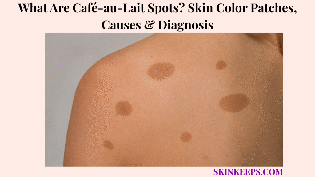
Café-au-lait spots are flat light-brown to dark-brown skin patches that are often present at birth or become visible in early childhood.
One or a few spots are often benign, but multiple large spots or associated signs need evaluation. This page covers appearance, causes, number and size meaning, diagnosis, NF1-related warning patterns, treatment limits, monitoring, and when to seek care.
What Are Café-au-Lait Spots and Why Do They Look Brown?
Café-au-lait spots are flat light-brown to dark-brown skin patches, often present at birth or appearing in early childhood, and their name means “coffee with milk” because of their typical color.
The medical term is café-au-lait macules, often shortened to CALMs. A macule is a flat area of color change, not a raised bump.
These patches are a form of localized hyperpigmentation. That means the affected skin has more visible pigment than nearby skin.
Why Are Café-au-Lait Spots Called Macules?
Café-au-lait spots are called macules because they are flat areas of color change rather than raised bumps.
They are pigment patches, not lumps, warts, scabs, or thick plaques. Typical café-au-lait spots are usually painless, not itchy, not scaly, and not crusted.
Some become more noticeable as a child grows or after sun exposure changes the contrast between the patch and surrounding skin.
Why Does the Color Range From Light Brown to Dark Brown?
The color ranges from light brown to dark brown because café-au-lait spots contain increased melanin pigment compared with nearby skin.
Melanin is the pigment that gives skin its brown tones. A café-au-lait spot may look tan, coffee-colored, light brown, medium brown, or dark brown.
Visibility depends on natural skin tone, lighting, and contrast with surrounding skin. The patch is usually darker than nearby skin, regardless of ancestry or skin color.
Practical rule: A single stable café-au-lait spot is usually not the same as a syndrome diagnosis. Count, measure, and check associated signs before drawing conclusions.
What Do Café-au-Lait Spots Look Like on Skin?
Café-au-lait spots usually look like flat, evenly colored tan or brown patches with smooth or sometimes irregular borders.
They may be round, oval, or less regular in outline. Their color is usually even across the patch rather than sharply multi-colored.
What Color and Shape Can Café-au-Lait Spots Have?
Café-au-lait spots can be light brown, medium brown, or dark brown, and they are often round, oval, or smoothly bordered flat patches.
Some have smooth edges, while others have more irregular borders. Size can range from a few millimeters to many centimeters.
Flat surface and even pigment are more important clues than exact shade alone.
What Are “Coast of California” and “Coast of Maine” Patterns?
“Coast of California” usually describes smoother café-au-lait spot borders, while “coast of Maine” describes more irregular or jagged borders.
These terms help clinicians describe border shape. They should not be used alone to diagnose NF1, McCune-Albright syndrome, or any other condition.
Number, size, age, distribution, associated signs, family history, and examination matter more than border nickname alone.
What Symptoms Are Not Typical for Café-au-Lait Spots?
Pain, itching, bleeding, crusting, scaling, ulceration, raised lumps, or rapid color change are not typical features of an ordinary café-au-lait spot.
These signs should prompt review because another diagnosis may be present. A café-au-lait spot should not behave like an inflamed rash, scab, wound, or raised mole.
A new changing adult pigmented lesion should not automatically be called a café-au-lait spot.
| Feature | Typical Café-au-Lait Clue | When to Check |
|---|---|---|
| Surface | Flat patch | Raised, bumpy, thick, or scaly |
| Color | Even tan, light brown, medium brown, or dark brown | Multiple colors or rapid change |
| Symptoms | Usually painless and not itchy | Pain, itch, bleeding, crusting, or ulceration |
| Border | Smooth or irregular but stable | Changing border |
| Number | One or a few can be benign | Multiple large patches |
| Timing | Birth or early childhood common | New changing adult lesion |
Where Do Café-au-Lait Spots Usually Appear?
Café-au-lait spots can appear anywhere on the body, commonly on the trunk, back, chest, abdomen, arms, legs, and buttocks.
Less visible areas may be found only during a full skin exam. Parents may first notice spots during bathing, clothing changes, or routine pediatric visits.
Skin-fold freckling in the armpits or groin should be considered separately from larger café-au-lait macules because tiny freckle-like spots can change the evaluation pathway.
| Body Area | Common Clue | What Clinician May Check Next |
|---|---|---|
| Trunk / back | Flat brown patch noticed in childhood | Count total spots |
| Chest / abdomen | Smooth tan or brown macule | Measure larger patches |
| Arms | Visible flat pigment patch | Look for symmetry or segmental pattern |
| Legs | One or multiple flat patches | Track number over time |
| Buttocks | Often less visible to parents or patients | Full skin exam may help |
| Armpit / groin freckling | Tiny freckle-like spots, not large CALMs | NF1-related evaluation clue |
What Causes Café-au-Lait Spots?
Café-au-lait spots are caused by localized increased skin pigment, but the exact reason they form is often unknown unless they are part of an underlying genetic condition.
They are not caused by dirt, infection, allergy, poor hygiene, or something a parent did wrong.
How Does Melanin Create Café-au-Lait Color?
Melanin creates café-au-lait color because it is the pigment that gives skin its brown tones, and café-au-lait spots have more visible pigment than nearby skin.
Melanocytes are cells that make melanin. In café-au-lait spots, the brown color reflects localized pigmentation rather than inflammation or infection.
This is why stable café-au-lait spots usually do not itch, hurt, crust, or drain.
Are Café-au-Lait Spots Inherited?
Café-au-lait spots can be isolated or familial, and multiple spots may sometimes be linked to inherited or new genetic changes.
Some people have one or a few café-au-lait spots without a syndrome. Some families may have isolated pigment patterns.
When multiple café-au-lait spots meet evaluation criteria or occur with other findings, clinicians may consider NF1, Legius syndrome, McCune-Albright syndrome, Noonan syndrome with multiple lentigines, or other rare conditions. Absence of family history does not fully rule out a genetic condition because some variants can arise newly.
Can Café-au-Lait Spots Appear After Birth?
Café-au-lait spots may be present at birth, appear in early childhood, or become more noticeable as a child grows.
Some spots are subtle at first and easier to see later. Size may increase as the child’s body grows, and new spots may become visible over time.
That is why counting, measuring, and photos can be more useful than relying on memory alone.
| Pigment Factor | Visible Clue | Diagnosis Meaning |
|---|---|---|
| Increased melanin | Brown patch darker than surrounding skin | Pigment change, not infection |
| Early visibility | Present at birth or childhood | Common timing for CALMs |
| Family pattern | Similar spots in relatives | May be isolated familial or syndromic |
| Multiple spots | More than one body area | Count and measure |
| Associated signs | Freckling, bumps, eye, bone, or development clues | Needs broader evaluation |
When Do Multiple Café-au-Lait Spots Suggest a Genetic Condition?
One or a few café-au-lait spots can be harmless, but multiple spots—especially six or more with age-based size criteria—should prompt evaluation for NF1 or related genetic syndromes.
This is the most important safety balance: one small spot should not create panic, but many large spots should not be ignored.
Why Does the Number of Café-au-Lait Spots Matter?
The number matters because one or two café-au-lait spots are common, while six or more large spots are part of the evaluation criteria for NF1.
Six or more café-au-lait macules larger than 5 mm before puberty or larger than 15 mm after puberty are part of NF1 diagnostic criteria.
These thresholds are evaluation criteria, not an automatic diagnosis. Other signs, family history, eye findings, genetics, or follow-up may be needed.
What Other Signs Should Be Checked With Café-au-Lait Spots?
Other signs that matter include armpit or groin freckling, soft skin bumps, plexiform neurofibroma-like growths, eye findings, bone differences, developmental concerns, and family history.
Clinicians may look for axillary or inguinal freckling, neurofibromas, eye findings such as Lisch nodules, optic pathway concerns, bone changes, scoliosis, learning concerns, developmental delay, and family history of NF1.
Vision symptoms, headaches with visual symptoms, rapidly enlarging lumps, bone bowing, scoliosis, or developmental concerns should move the pathway beyond cosmetic spot observation.
Which Other Syndromes Can Have Café-au-Lait Spots?
NF1 is not the only condition linked with multiple café-au-lait spots; Legius syndrome, McCune-Albright syndrome, Noonan syndrome with multiple lentigines, and other rare conditions may also be considered.
Legius syndrome can overlap with NF1 pigment signs but lacks some tumor, optic, ocular, and bone features typical of NF1.
McCune-Albright syndrome, Noonan syndrome with multiple lentigines, Watson syndrome, segmental or mosaic pigmentary disorders, and rare specialist contexts may be considered when the overall pattern fits.
| Finding | Why It Matters | Suggested Next Step |
|---|---|---|
| 1–2 spots | Often benign | Routine observation |
| Six or more spots | NF1/Legius evaluation trigger | Clinical/genetic review |
| Large spots by age threshold | Part of NF1 criteria | Measure accurately |
| Armpit/groin freckling | NF1 criterion clue | Full skin exam |
| Soft skin bumps | Neurofibroma concern | Dermatology/genetics review |
| Eye/vision signs | Optic pathway concern | Pediatric/eye evaluation |
| Bone/development signs | Syndromic clue | Multidisciplinary review |
| Strong family history | Inherited condition possible | Genetics counseling |
How Are Café-au-Lait Spots Different From Other Brown Skin Patches?
Café-au-lait spots can resemble moles, Becker’s nevus, post-inflammatory hyperpigmentation, lentigines, congenital melanocytic nevus, nevus spilus, melasma, or fungal discoloration patches, so texture, timing, borders, symptoms, and pattern matter.
Not every brown patch is a café-au-lait spot. The safest comparison uses whether the lesion is flat or raised, present early or acquired later, evenly pigmented or changing, symptomatic or silent, and isolated or part of a wider pattern.
How Are Café-au-Lait Spots Different From Moles?
Café-au-lait spots are flat pigment patches, while moles may be raised, textured, or show a different melanocytic structure.
Congenital melanocytic nevi are different from café-au-lait macules. A raised, hairy, bumpy, or changing lesion should not automatically be labeled as a café-au-lait spot.
How Are Café-au-Lait Spots Different From Becker’s Nevus?
Café-au-lait spots are usually flat and often appear early, while Becker’s nevus more often becomes noticeable around adolescence and may become hairy or thicker.
Hair growth, acne-like changes, thickening, and puberty-onset visibility point away from a typical café-au-lait macule.
How Are Café-au-Lait Spots Different From Post-Inflammatory Hyperpigmentation?
Café-au-lait spots are often birthmarks or early childhood patches, while post-inflammatory hyperpigmentation usually follows acne, rash, injury, burn, bite, or irritation.
History is key. A mark that appears after inflammation may fade gradually, while many café-au-lait spots persist.
How Are Café-au-Lait Spots Different From Melanoma-Like Patches?
Café-au-lait spots are usually flat and evenly colored, while melanoma concern rises with asymmetry, changing borders, multiple colors, bleeding, pain, ulceration, or evolution.
A new, rapidly changing, raised, painful, bleeding, ulcerated, multi-colored, or irregular adult pigmented lesion should be checked rather than assumed to be a café-au-lait spot.
| Condition | Main Clue | Timing Clue | Safer Next Step |
|---|---|---|---|
| Café-au-lait spot | Flat even tan/brown patch | Birth/childhood common | Count and monitor |
| Mole / nevus | Raised or mole-like structure | Birth or later | Check if changing |
| Becker’s nevus | Brown patch plus hair/thickening | Puberty/adolescence common | Dermatology if uncertain |
| PIH | Dark mark after inflammation | After rash/injury/acne/burn | Treat cause and monitor fading |
| Lentigo | Small brown sun/age spot | Later life/sun areas | Assess pattern |
| Melasma | Facial pigment patches | Hormone/UV pattern | Pigment-disorder evaluation |
| Melanoma | Changing, irregular, multi-color, symptomatic | New/evolving | Prompt evaluation |
How Are Café-au-Lait Spots Diagnosed?
Café-au-lait spots are usually diagnosed by clinical examination, but multiple or large spots require counting, measuring, full skin exam, family history, and assessment for associated signs.
Diagnosis is not based on color alone. The clinician checks the patch surface, border, number, size, distribution, age of appearance, and whether other signs are present.
What Does a Clinician Check During Examination?
A clinician checks café-au-lait spots by counting them, measuring them, reviewing their borders, checking color uniformity, and looking for associated signs.
The exam may include number of spots, size of each spot, border pattern, color uniformity, age when spots appeared, growth in number or size, distribution on both sides or one segment, and less visible body areas.
The clinician may also look for armpit or groin freckling, soft skin bumps, neurofibroma-like lesions, eye findings, bone findings, neurologic or developmental concerns, and family history.
When Is Genetic Testing Considered?
Genetic testing may be considered when multiple café-au-lait spots, size thresholds, family history, or associated clinical signs make NF1 or a related syndrome uncertain.
Testing may be considered in a young child with evolving features, unclear NF1 versus Legius syndrome, a strong family history, or associated findings that change the pathway.
Specialist-directed testing is better than random testing because different conditions can share pigment findings.
When Might Wood Lamp Examination Help?
Wood lamp examination may help when very light café-au-lait spots are difficult to see clearly during counting and mapping.
A Wood lamp can improve visibility in some exams, especially when subtle light patches are hard to count.
It helps with identification and mapping; it does not diagnose NF1 by itself.
Do Café-au-Lait Spots Need Treatment or Removal?
Café-au-lait spots usually do not need medical treatment because they are benign and symptom-free, but laser treatment may be discussed for cosmetic reasons with realistic expectations.
Treatment is not medically necessary for a stable isolated spot that has been properly identified and has no concerning associated signs.
When Is No Treatment the Best Option?
No treatment is often the best option when café-au-lait spots are single or few, stable, symptom-free, clearly diagnosed, and not causing cosmetic distress.
Observation avoids unnecessary irritation, pigment change, and scarring risk. The main medical task is not removal; it is correct identification and appropriate evaluation when multiple spots or associated signs are present.
Can Laser Fade Café-au-Lait Spots?
Laser treatment may lighten some café-au-lait spots, but results are inconsistent and pigment can return or change.
Some spots may partially lighten. Others may not clear completely or may recur. Risks can include darker pigmentation, lighter pigmentation, irritation, and scarring.
Bleaching creams, harsh acids, and home lightening methods are not recommended because they can irritate skin and worsen pigment problems.
| Option | Goal | Limitation |
|---|---|---|
| Observation | Avoid unnecessary treatment | Requires review if multiple, large, or increasing |
| Photography | Track number and size changes | Does not diagnose syndrome |
| Laser | Cosmetic lightening | Inconsistent; recurrence and pigment/scar risk |
| Genetic/specialist care | Evaluate associated syndrome | Only needed when indicated |
| Harsh lightening products | Not recommended | Irritation and pigment worsening risk |
How Should Café-au-Lait Spots Be Monitored Over Time?
Monitoring café-au-lait spots means counting them, measuring larger patches, photographing them consistently, and watching for associated signs rather than checking color alone.
Count spots on the full body, measure larger patches, and note when new spots appear. In children, spots larger than 5 mm are especially relevant when multiple spots are present before puberty.
Watch for armpit or groin freckling, soft bumps, fast-growing lumps, vision concerns, bone findings, learning or developmental concerns, and family history. Avoid daily anxious checking unless a clinician recommends closer tracking.
What Café-au-Lait Spot Mistakes Should You Avoid?
The biggest café-au-lait spot mistake is treating every spot as NF1—or ignoring multiple large spots that meet evaluation criteria.
Do not panic over one small flat brown spot. Do not diagnose NF1 from café-au-lait spots alone.
Do not ignore six or more spots, skip measurement, confuse CALMs with other brown patches, use harsh lightening products, or expect laser to permanently remove every spot.
| Mistake | Why It Fails | Better Action |
|---|---|---|
| Assuming one spot means NF1 | Causes unnecessary fear | Count and monitor |
| Ignoring many spots | Delays syndrome evaluation | Measure and refer |
| Skipping measurement | Misses age-based criteria | Measure largest spots |
| Bleaching at home | Irritation and pigment risk | Avoid harsh products |
| Calling every brown patch CALM | Mimics exist | Clinical exam |
| Expecting laser cure | Results are inconsistent | Discuss realistic outcomes |
| Ignoring associated signs | Misses broader diagnosis | Check eyes, skin, bones, development, family history |
When Should Café-au-Lait Spots Be Checked by a Doctor?
Café-au-lait spots should be checked when a child has multiple spots, six or more spots, large spots meeting age-based thresholds, spots increasing in number, or associated signs such as armpit/groin freckling, skin bumps, vision problems, bone changes, or developmental concerns.
Review is also appropriate when the diagnosis is uncertain, the pattern is segmental or unusual, there is a strong family history, or a brown patch becomes raised, painful, bleeding, scaly, ulcerated, multi-colored, or rapidly changing.
Which Café-au-Lait Patterns Need Medical Evaluation?
Medical evaluation is important when there are six or more café-au-lait spots, more spots appearing over time, large spots by age threshold, bilateral multiple spots, segmental clusters, family history, or uncertain diagnosis.
Large CALMs mean greater than 5 mm before puberty or greater than 15 mm after puberty in NF1 evaluation criteria.
These findings should prompt clinical review, not a home diagnosis.
Which Associated Signs Need Faster Review?
Faster review is needed when café-au-lait spots occur with armpit or groin freckling, soft skin bumps, rapidly enlarging lumps, vision symptoms, bone changes, developmental concerns, early puberty signs, or a painful or changing lesion.
Eye or vision concerns, headaches with visual symptoms, bone bowing, scoliosis, learning concerns, developmental delay, early puberty signs, and a family history of NF1 or related syndromes should be mentioned at the visit.
Seek medical review if there are:
What Should You Remember About Café-au-Lait Spots?
The most important thing to remember about café-au-lait spots is that they are usually harmless flat brown pigment patches, but their number, size, and associated findings determine whether medical or genetic evaluation is needed.
One or a few stable patches are often benign. Multiple large patches, armpit or groin freckling, skin bumps, vision concerns, bone findings, developmental concerns, or family history should shift the plan toward clinical evaluation.
Frequently Asked Questions About Café-au-Lait Spots
Are café-au-lait spots dangerous?
Usually no. One or a few café-au-lait spots are generally benign, but multiple spots may need evaluation for NF1 or other genetic syndromes.
What do café-au-lait spots look like?
Café-au-lait spots look like flat light-brown to dark-brown patches, often round or oval, with smooth or sometimes irregular borders and usually no pain, itching, scale, crust, or bleeding.
Do café-au-lait spots mean neurofibromatosis?
Not always. Many people have one or a few café-au-lait spots without NF1, but six or more spots meeting age-based size thresholds should be evaluated because they are part of NF1 diagnostic criteria.
How many café-au-lait spots are concerning?
Six or more are concerning, especially if larger than 5 mm before puberty or larger than 15 mm after puberty. These thresholds should be framed as evaluation criteria, not automatic diagnosis.
Can café-au-lait spots appear later?
Yes. They are often present at birth or early childhood, but they may become more visible as a child grows or after sun exposure.
Can café-au-lait spots be removed?
They usually do not need treatment. Laser may lighten some spots for cosmetic reasons, but results are inconsistent and pigment change, recurrence, or scarring can occur.
Are café-au-lait spots the same as moles?
No. Café-au-lait spots are flat pigment patches, while moles may be raised or have a different melanocytic structure. Changing, raised, bleeding, painful, or multi-colored spots need evaluation.
When should café-au-lait spots be checked?
They should be checked for six or more spots, increasing number, large spots, uncertain diagnosis, armpit/groin freckling, skin bumps, vision problems, bone changes, developmental concerns, family history, or any spot becoming raised, lumpy, painful, bleeding, scaly, ulcerated, or changing.
Sources & Evidence About Café-au-Lait Spots
DermNet — Café-au-Lait Macule was used for CALM definition, hyperpigmented patch framing, birth or early infancy timing, melanin basis, systemic associations, clinical diagnosis, and laser-treatment limitations.
StatPearls / NCBI Bookshelf — Café Au Lait Macules was used for flat hyperpigmented lesion description, isolated benign framing, NF1 number and size thresholds, differential diagnosis, and Wood lamp examination context.
GeneReviews / NCBI Bookshelf — Neurofibromatosis 1 was used for NF1 diagnostic criteria, six-or-more café-au-lait macules, age-based size thresholds, associated clinical signs, and Legius syndrome differential context.
International Consensus Recommendation — Revised Diagnostic Criteria for NF1 and Legius Syndrome was used for updated NF1 and Legius syndrome diagnostic boundaries, including CALM and freckling criteria.
Cleveland Clinic — Café-au-Lait Spots was used for patient-friendly appearance descriptions, common benign framing, more-than-six warning, common body sites, treatment usually being unnecessary, and laser limitations.
American Academy of Dermatology — Birthmarks: Signs and Symptoms was used for parent-facing guidance about flat café-au-lait birthmarks and dermatologist review when a child has six or more spots or freckle-like spots around them.
Educational Disclaimer: This SkinKeeps article is for educational purposes only and does not diagnose or replace medical care. Multiple café-au-lait spots, six or more spots, large spots meeting age-based thresholds, increasing spot number, armpit or groin freckling, soft skin bumps, rapidly enlarging lumps, vision symptoms, bone changes, developmental or learning concerns, family history of NF1 or related syndromes, or any brown patch that becomes raised, painful, bleeding, crusted, scaly, ulcerated, multi-colored, or rapidly changing should be checked by a qualified healthcare professional.



