
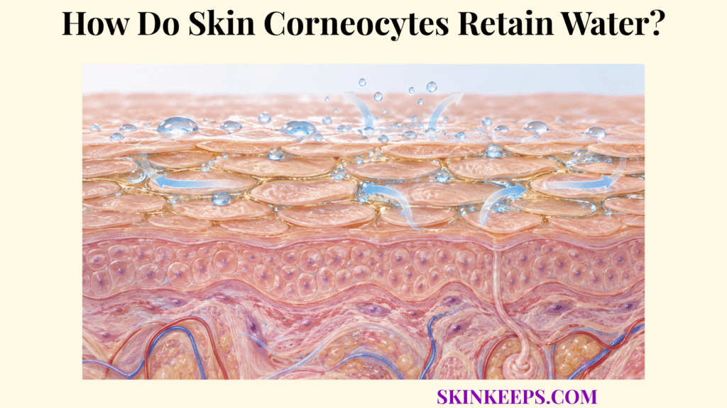
Skin corneocytes retain water through a protein-rich intracellular matrix containing natural moisturizing factor, whose hygroscopic molecules increase water-holding capacity while the surrounding stratum-corneum lipid barrier slows that water from escaping.
Corneocyte hydration therefore depends on several linked processes: NMF composition, regulated filaggrin breakdown, water–protein interactions, surrounding lipid resistance, and the environmental drying gradient. These mechanisms maintain physiological flexibility and desquamation without making maximal hydration the biological goal.
Where Do Skin Corneocytes Hold Water?
Skin corneocytes hold water throughout their keratin- and natural-moisturizing-factor-rich intracellular matrix within the stratum corneum rather than in a dedicated fluid compartment.
Where Are Skin Corneocytes Located?
Corneocytes form multiple overlapping layers throughout the stratum corneum, the outermost epidermal layer. Their intracellular hydration supports the broader physiology of skin water-loss prevention, while whole-layer permeability belongs to the skin water barrier.
Is Water Stored in a Special Compartment Inside Skin Corneocytes?
No. Water is distributed through the corneocyte’s intracellular protein and solute matrix, where it associates with keratin, free amino acids, NMF components, ions, and other polar molecules rather than filling a vacuole or membrane-bound reservoir.
How Does Keratin Contribute to Skin Corneocyte Hydration?
Keratin and associated proteins interact with water, and increasing hydration changes their molecular mobility and mechanical behavior. Water therefore plasticizes the protein-rich corneocyte matrix; it does not dissolve keratin or turn the cell into a simple sponge.
Why Does Corneocyte Hydration Matter to the Whole Stratum Corneum?
Because corneocytes form most of the structural volume of the stratum corneum, their hydration strongly influences tissue flexibility, swelling, mechanical resilience, and surface texture. The hydration gradient is not uniform: deeper stratum-corneum layers are generally more hydrated than the surface.
How Do Skin Corneocytes Use Natural Moisturizing Factors?
Skin corneocytes use natural moisturizing factor as a concentrated mixture of small hygroscopic solutes that associate with water and maintain intracellular hydration across changing environmental conditions.
What Makes NMF Hygroscopic?
NMF molecules contain polar and ionic chemical groups that interact strongly with water and contribute substantial osmotic strength inside mature corneocytes. The result is greater intracellular water-holding capacity rather than formation of an external oily seal.
Which NMF Components Are Most Relevant to Skin Corneocytes?
Major components include free amino acids, pyrrolidone carboxylic acid (PCA), urocanic acid (UCA), lactate, urea, and inorganic ions, with additional small water-soluble molecules contributing to the total pool. The broader role is covered by skin natural moisturizing factors, while detailed composition belongs to skin natural moisturizing factor components.
How Does PCA Support Skin Corneocyte Hydration?
PCA is a strongly hygroscopic amino-acid-derived NMF component that contributes to the osmotic and water-binding environment inside corneocytes. It is produced mainly from glutamine-related products generated during filaggrin breakdown and later metabolism.
How Do Lactate and Urea Support Skin Corneocyte Hydration?
Lactate and urea are small water-soluble NMF constituents that add hygroscopic capacity and help maintain the physicochemical environment of the cornified layer. Their presence in NMF does not mean that they arise primarily from filaggrin.
Does Every NMF Molecule Come From Filaggrin?
No. Filaggrin is a major source of free amino acids, PCA, and UCA-related NMF, while lactate, urea, inorganic salts, sugars, and other small compounds have additional physiological origins. The detailed hygroscopic and osmotic mechanisms belong to skin natural moisturizing factor water binding.
| NMF component | Main origin / context | Water-retention role | Functional effect |
|---|---|---|---|
| Free amino acids | Largely filaggrin degradation | Polar / osmotic water association | Supports corneocyte hydration |
| PCA | Mainly filaggrin / glutamine-derived | Strong hygroscopic contribution | Increases water-holding capacity |
| Urocanic acid | Filaggrin / histidine-derived | NMF and acidification roles | Supports SC physicochemical environment |
| Lactate | Epidermal / metabolic contribution | Hygroscopic small molecule | Supports SC hydration |
| Urea | Systemic / epidermal NMF pool | Hygroscopic water association | Supports hydration and flexibility |
| Inorganic ions | Multiple physiological sources | Osmotic contribution | Supports intracellular water balance |
| Other small solutes | Mixed origins | Add total NMF osmotic capacity | Broadens hydration buffering |
NMF composition varies with epidermal differentiation, environment, body site, age, genetics, and barrier state; these are major functional classes rather than a fixed universal formula.
How Do Skin Corneocytes Gain Natural Moisturizing Factors?
Skin corneocytes gain much of their natural moisturizing factor through a regulated maturation pathway in which profilaggrin is converted into filaggrin and later degraded into hygroscopic amino acids and derivatives.
Where Does Filaggrin Begin?
Profilaggrin is stored in keratohyalin granules of granular-layer keratinocytes before terminal differentiation produces the mature cornified cell. It is a large, highly phosphorylated precursor rather than NMF itself.
What Happens When Profilaggrin Becomes Filaggrin?
During the granular-to-cornified transition, profilaggrin is dephosphorylated and proteolytically processed into filaggrin monomers. Filaggrin then associates with keratin intermediate filaments and promotes cytoskeletal aggregation and compaction during corneocyte maturation.
When Does Filaggrin Become a Source of NMF?
After its keratin-organizing phase, filaggrin is chemically modified—including deimination—and progressively degraded by regulated proteolysis into free amino acids. Further metabolism produces major NMF derivatives such as PCA from glutamine-related precursors and UCA from histidine.
Why Is Filaggrin Processing Important for Water Retention?
This maturation sequence links epidermal differentiation to later hydration capacity: early filaggrin contributes to keratin organization, while later filaggrin degradation generates a concentrated amino-acid-derived pool that increases the mature corneocyte’s ability to retain water.
Can Environmental Humidity Influence Filaggrin Breakdown?
Yes. Reconstructed-human-epidermis experiments show that lower relative humidity can increase filaggrin deimination, degradation, and measured NMF generation within that model. This adaptive response does not mean dry environments cannot dehydrate human skin or that increased NMF always fully compensates for environmental drying.
How Do Skin Corneocytes Work With Barrier Lipids?
Skin corneocytes work with barrier lipids through complementary functions: corneocytes use NMF and hydrated proteins to retain water internally, while extracellular ceramide-, cholesterol-, and free-fatty-acid-rich lamellae restrict outward water diffusion between the cells.
What Do the “Bricks” Represent in the Stratum Corneum?
In the brick-and-mortar analogy, corneocytes are the structural bricks that contain keratin and NMF and therefore carry much of the stratum corneum’s intracellular water. The analogy is incomplete because real corneocytes are dynamic, hydrated biological structures rather than dry impermeable blocks.
What Does the “Mortar” Represent?
The mortar represents organized extracellular lipid lamellae composed mainly of ceramides, cholesterol, and free fatty acids that surround corneocytes and restrict diffusion. Their detailed packing and phase behavior belong to skin intercellular lipids.
Why Is NMF Not Enough to Prevent Water Loss?
Even a highly hygroscopic corneocyte can lose water if surrounding permeability resistance is weak, so effective hydration requires both intracellular water-holding capacity and intact extracellular lipid organization. Because many NMF molecules are small and water-soluble, an intact barrier also helps reduce their loss from the cornified layer.
Why Are Lipids Not Enough to Maintain Corneocyte Hydration?
An intact lipid barrier slows water escape but does not itself supply the intracellular hygroscopic solutes or hydrated protein environment needed to maintain optimal corneocyte water content and flexibility. Water-loss resistance and water-holding capacity therefore solve different problems.
How Do Both Systems Affect TEWL?
NMF supports the water content of individual corneocytes, while lipid lamellae provide the stronger control over whole-stratum-corneum diffusion and therefore the outward flux measured as skin transepidermal water loss.
How Do Skin Corneocytes Maintain Flexibility When Hydrated?
Skin corneocytes maintain flexibility when hydrated because water plasticizes the keratin-rich intracellular matrix, increases molecular mobility, and supports hydration-dependent enzymatic processes required for orderly stratum-corneum turnover.
How Does Water Make Skin Corneocytes More Flexible?
Water interacting with keratin and associated proteins increases molecular mobility and reduces the stiffness of the cornified matrix, making hydrated corneocytes more pliable under ordinary mechanical deformation. Biophysical studies link increasing hydration with changes in keratin dynamics, swelling, and macroscopic mechanical behavior.
Why Do Dehydrated Skin Corneocytes Become Stiffer?
When corneocyte water content falls, keratin and associated protein structures become less plasticized, increasing stratum-corneum stiffness and brittleness. This raises susceptibility to visible roughness, microfissuring, and cracking under mechanical stress.
How Does Hydration Support Normal Desquamation?
Appropriate water content supports several proteolytic enzymes involved in progressive corneodesmosome degradation, allowing mature corneocytes to detach individually from the surface. Enzyme activity also depends on local pH, inhibitors, substrate availability, and location.
What Happens to Desquamation When Corneocytes Become Too Dry?
Excessive dryness can make corneodesmosome processing less effective, allowing corneocytes to remain attached in larger groups that become visible as roughness, flaking, or scaling.
Does Maximum Hydration Produce the Strongest Stratum Corneum?
No. Physiological hydration improves flexibility, but excessive hydration can increase swelling, permeability, and friction susceptibility. The goal is balanced water content, not the greatest possible amount of water.
What Reduces Water Retention in Skin Corneocytes?
Water retention in skin corneocytes falls when NMF concentration decreases, filaggrin processing is impaired, the surrounding permeability barrier becomes disorganized, or environmental conditions create a stronger drying challenge than intracellular and extracellular defenses can offset.
How Does Loss of NMF Reduce Skin Corneocyte Hydration?
Lower NMF concentration reduces the intracellular pool of hygroscopic solutes available to associate with water, decreasing corneocyte water-holding capacity and contributing to lower stratum-corneum hydration and flexibility.
How Can Impaired Filaggrin Processing Reduce Water Retention?
Reduced filaggrin production or abnormal processing can reduce the amino-acid-derived fraction of NMF and therefore lower hydration capacity. This mechanism does not allow dry or flaky skin to diagnose an FLG variant, atopic dermatitis, ichthyosis, or any other specific condition.
How Can Excessive Cleansing Reduce Corneocyte Water Retention?
Repeated harsh cleansing can remove water-soluble NMF components while also interacting with keratin, proteins, and surrounding barrier lipids, reducing both intracellular hydration capacity and extracellular water-loss resistance. Effects depend on surfactant chemistry, concentration, contact time, frequency, rinsing, and baseline barrier state.
How Does Low Humidity Challenge Skin Corneocyte Hydration?
Low humidity increases the environmental drying gradient and can reduce water content in the outer stratum corneum even though adaptive filaggrin deimination, breakdown, and NMF generation may rise in some experimental systems. Adaptation can therefore occur without completely preventing dehydration.
How Does Barrier Disruption Reduce Skin Corneocyte Hydration?
When extracellular lipid lamellae become disorganized, water escapes from the tissue more rapidly, increasing the burden on intracellular water-retention mechanisms and making it harder for corneocytes to maintain normal water content. The larger failure pathway belongs to skin barrier disruption.
What Happens When Skin Corneocytes Lose Too Much Water?
Poorly hydrated corneocytes become less flexible and more prone to abnormal cohesion, visible scaling, roughness, and fissuring as the stratum corneum loses normal mechanical and desquamatory function. These signs are nonspecific and do not identify one disease by themselves.
What Are the Key Takeaways About Skin Corneocytes?
The key fact about skin corneocytes is that they retain water mainly through intracellular natural moisturizing factor and hydrated protein structures, while surrounding intercellular lipids perform the complementary task of slowing that water from escaping.
Filaggrin has two distinct phases: it first helps organize keratin and is later degraded into amino acids and derivatives that form much of the NMF pool. Physiological hydration plasticizes the corneocyte matrix and supports orderly desquamation, while low NMF, strong environmental drying, or barrier disruption can reduce water retention.
- Skin corneocytes are specialized terminally differentiated cells within the stratum corneum.
- Water is distributed through the corneocyte’s protein- and solute-rich interior rather than stored in a fluid-filled compartment.
- Natural moisturizing factor is concentrated mainly inside corneocytes.
- NMF contains free amino acids, PCA, UCA, lactate, urea, salts, and other small soluble compounds.
- NMF increases intracellular water-holding capacity through hygroscopic and osmotic effects.
- Much of the NMF amino-acid fraction comes from filaggrin degradation.
- Not every NMF component is derived from filaggrin.
- Profilaggrin is processed into filaggrin during terminal differentiation.
- Filaggrin first helps organize keratin before it is later degraded.
- PCA and UCA are important filaggrin-derived NMF components.
- Environmental humidity can influence filaggrin processing and NMF generation.
- Keratin hydration contributes to corneocyte mechanical flexibility.
- Water acts as a plasticizer within the stratum corneum.
- Intercellular lipids perform a different function from NMF.
- NMF retains water inside cells; lipid lamellae restrict water escape between cells.
- Ceramides, cholesterol, and free fatty acids form the surrounding extracellular permeability barrier.
- Adequate hydration supports normal enzyme activity and desquamation.
- Excessively dry corneocytes become stiffer and can shed abnormally.
- Reduced NMF can decrease corneocyte water-holding capacity.
- Impaired filaggrin production or processing can reduce NMF.
- Harsh repeated cleansing can disturb NMF, proteins, and extracellular barrier lipids.
- Low humidity challenges hydration but can also trigger adaptive changes in filaggrin metabolism.
- Barrier disruption increases the demand placed on intracellular water-retention mechanisms.
- Maximum hydration is not the goal; excessive water can also alter stratum-corneum structure and permeability.
- Healthy stratum-corneum hydration requires cooperation between corneocytes and extracellular lipids.
What Common Questions Do People Ask About Skin Corneocytes?
Common questions about skin corneocytes focus on where they hold water, how natural moisturizing factor works, whether filaggrin creates NMF, why lipids remain necessary, and what happens when corneocytes become dehydrated.
Where Do Skin Corneocytes Store Water?
Skin corneocytes do not use a dedicated water reservoir; water is distributed throughout their keratin- and NMF-rich intracellular matrix, where proteins and hygroscopic solutes maintain corneocyte hydration.
What Makes Natural Moisturizing Factor Hold Water in Skin Corneocytes?
NMF contains small polar and ionic molecules—including amino acids, PCA, lactate, urea, and salts—that interact with water and increase the osmotic and hygroscopic water-holding capacity of the corneocyte.
Does All Natural Moisturizing Factor Come From Filaggrin?
No. Filaggrin breakdown supplies much of the free-amino-acid fraction and major derivatives such as PCA and UCA, while lactate, urea, inorganic ions, and other NMF constituents have additional sources.
Why Do Skin Corneocytes Need Intercellular Lipids if They Already Have NMF?
NMF helps individual corneocytes retain water internally, whereas extracellular ceramide-, cholesterol-, and free-fatty-acid-rich lipids slow water diffusion out of the stratum corneum, so the two systems provide complementary hydration control.
What Happens When Skin Corneocytes Become Too Dry?
Excessive dehydration makes the stratum corneum less flexible, can impair normal hydration-dependent desquamation, and promotes roughness, scaling, stiffness, and fissuring when the drying challenge exceeds water-retention and barrier capacity.
Sources & Evidence
Medical/Educational Disclaimer
This page explains normal stratum-corneum hydration physiology and is not medical advice. Seek professional evaluation for persistent severe dryness, painful or bleeding fissures, weeping skin, marked inflammation, or suspected infection.



