
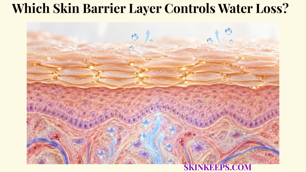
The stratum corneum is the primary skin barrier layer controlling water loss because its stacked corneocytes and highly organized extracellular lipid lamellae create strong resistance to outward water diffusion. It slows normal water escape rather than making skin waterproof.
Corneocytes provide structure, extracellular lipids provide the principal continuous permeability resistance, and natural moisturizing factor retains water inside corneocytes. Tight junctions in the granular layer also contribute to epidermal water homeostasis, but they do not replace the stratum corneum as the principal permeability barrier.
Which Skin Barrier Layer Provides the Main Water-Loss Control?
The skin barrier layer that provides the main control of water loss is the stratum corneum, the outermost epidermal layer where corneocytes and organized extracellular lipids produce most of the skin’s resistance to transepidermal water movement.
Where Is the Main Skin Barrier Layer Located?
The stratum corneum sits at the outer surface of the epidermis, directly between the living epidermis and the external environment. Its inside-out role is central to skin water-loss prevention, while its wider anatomy is covered by the skin stratum corneum barrier page.
Why Is the Stratum Corneum the Main Water Barrier?
The stratum corneum combines stacked protein-rich corneocytes with a continuous extracellular lipid matrix that forces water to cross a highly resistant and tortuous pathway before reaching the surface. The “brick-and-mortar” analogy is useful, but incomplete because envelopes, cohesion, NMF, lipid phase organization, and hydration gradients also contribute.
Are Other Epidermal Layers Involved in Water Control?
Yes. Tight junctions in the stratum granulosum and water-transport processes in viable epidermal cells contribute to water homeostasis, but the stratum corneum remains the dominant barrier to transepidermal water loss.
How Does the Skin Barrier Layer Use Corneocytes to Retain Water?
The skin barrier layer uses corneocytes as strong, flattened structural units that create a long diffusion path, contain water-binding molecules, and provide the framework within which the extracellular lipid barrier is organized.
What Makes Corneocytes Strong Structural Units?
Corneocytes are terminally differentiated, flattened, anucleate epidermal cells containing densely organized keratin within a cross-linked cornified envelope. They are specialized structural elements rather than inert waste.
How Do Stacked Corneocytes Slow Water Movement?
Multiple overlapping corneocyte layers increase the distance and complexity of the route that water must traverse before reaching the skin surface. This tortuous pathway adds resistance without making each corneocyte an impermeable block.
Does Keratin Alone Prevent Water Loss?
No. Keratin-rich corneocytes contribute mechanical structure and intracellular hydration properties, but the extracellular lipid lamellae provide the major continuous resistance to water diffusion between cells.
How Does Corneocyte Structure Support the Lipid Barrier?
The cornified protein envelope gives each corneocyte mechanical integrity, while the distinct corneocyte lipid envelope forms an interface that helps organize surrounding extracellular lamellae. Detailed intracellular hydration is covered by skin corneocyte water retention.
How Does the Skin Barrier Layer Use Lipids to Reduce Water Escape?
The skin barrier layer reduces water escape mainly through highly ordered extracellular lamellae composed principally of ceramides, cholesterol, and free fatty acids that restrict diffusion through the spaces between corneocytes.
Where Are Water-Barrier Lipids Located?
The key permeability lipids occupy extracellular spaces between corneocytes, where they form continuous lamellar structures rather than ordinary phospholipid cell-membrane bilayers. This extracellular lipid path is the principal continuous route that diffusing water must cross.
How Do Ceramides Reduce Water Permeability?
Ceramides contribute long hydrophobic chains and specialized molecular structures that support dense lamellar organization and low permeability. Their subclasses, chain lengths, abundance, and packing influence barrier performance, but ceramides do not function effectively in isolation.
Why Are Cholesterol and Free Fatty Acids Also Necessary?
Cholesterol and free fatty acids modify phase behavior, packing, flexibility, and hydrophobic organization, allowing the multicomponent lipid matrix to maintain effective permeability resistance. Free fatty-acid chain composition also influences lipid packing and surface acidity.
Why Does Lipid Organization Matter as Much as Lipid Quantity?
Water permeability depends on how lipids are arranged and packed, so abnormal ceramide subclasses, chain lengths, ratios, lateral packing, or lamellar organization can weaken the barrier even when lipid material remains present. The detailed lipid architecture belongs to skin intercellular lipids.
How Does the Skin Barrier Layer Keep Water Inside Corneocytes?
The skin barrier layer keeps water within corneocytes partly through natural moisturizing factor, a mixture of small hygroscopic molecules that bind and retain water inside the cornified cells.
What Is Natural Moisturizing Factor?
Natural moisturizing factor is a mixture of water-soluble hygroscopic molecules concentrated within corneocytes, much of it derived from filaggrin breakdown during terminal differentiation. Important components include free amino acids, pyrrolidone carboxylic acid (PCA), urocanic-acid-related compounds, lactate, urea, and electrolytes.
How Does NMF Retain Water?
NMF components attract and associate with water molecules, increasing the ability of the corneocyte interior to remain hydrated when environmental conditions favor drying. This is intracellular water holding rather than formation of the continuous extracellular permeability barrier.
Why Does Corneocyte Hydration Matter?
Adequate hydration supports corneocyte flexibility, enzyme activity, orderly desquamation, and resistance to surface cracking. Excessive hydration can also alter stratum-corneum structure, so the physiological objective is balanced hydration rather than maximal water content.
Does NMF Form the Main Water-Loss Seal?
No. NMF primarily regulates water content within corneocytes, while the extracellular lipid matrix provides the main resistance to water movement across the stratum corneum. The detailed composition and filaggrin-derived chemistry belong to skin natural moisturizing factors.
How Does the Skin Barrier Layer Control Transepidermal Water Loss?
The skin barrier layer controls transepidermal water loss by providing enough diffusion resistance that only a limited amount of water moves passively from deeper tissue through the stratum corneum and evaporates from the surface.
Where Does Water Move From Before It Becomes TEWL?
Water is more abundant in deeper viable tissue and moves outward toward the relatively dry external environment along concentration, activity, and vapor-pressure gradients. The skin does not actively pump this water toward the surface.
Where Is the Greatest Resistance to Outward Water Movement?
The largest permeability resistance occurs in the stratum corneum, especially within its organized extracellular lipid domains. Corneocyte architecture and intracellular hydration add complementary resistance and water-holding effects.
What Happens When Water Reaches the Skin Surface?
Water that reaches the outer surface evaporates into surrounding air, producing the passive flux measured as transepidermal water loss. TEWL is therefore a rate of water movement, not a direct measurement of how much water is stored inside the stratum corneum.
Is TEWL the Same as Sweating?
No. TEWL is passive, largely insensible water diffusion through epidermal tissue, whereas sweating is active fluid secretion through eccrine sweat glands and ducts. Active sweating can also complicate instrumental interpretation of TEWL.
Is Higher TEWL Always Proof of Disease?
No. Elevated TEWL can reflect reduced permeability-barrier integrity, but values also vary with body site, age, temperature, humidity, sweating, acclimatization, and measurement technique. Detailed interpretation belongs to skin transepidermal water loss.
| Barrier component | Water-control mechanism | Functional result | Important boundary |
|---|---|---|---|
| Corneocytes | Structural / tortuous pathway | Slows diffusion and provides architecture | Not the main continuous seal alone |
| Keratin matrix | Mechanical support / intracellular structure | Supports corneocyte resilience | Not waterproof |
| Cornified envelope | Cross-linked structural boundary | Stabilizes corneocyte | Distinct from lipid envelope |
| Corneocyte lipid envelope | Interface for extracellular lipids | Supports barrier organization | Not identical to free lipid lamellae |
| Ceramides | Dense lamellar packing | Restricts extracellular water diffusion | Subclass and chain length matter |
| Cholesterol | Lipid phase organization | Supports matrix packing and flexibility | Works with CER / FFA |
| Free fatty acids | Hydrophobic packing | Supports low permeability | Chain composition matters |
| NMF | Intracellular hygroscopic binding | Maintains corneocyte hydration | Not the main extracellular seal |
| Tight junctions | Granular-layer paracellular barrier | Supports epidermal water homeostasis | Secondary to SC for TEWL control |
| Intact whole SC | Combined architecture | Limits TEWL | Does not stop all evaporation |
What Weakens the Skin Barrier Layer and Increases Water Loss?
The skin barrier layer becomes more permeable when its lipid organization, corneocyte proteins, NMF content, cohesion, or inflammatory homeostasis is disrupted, allowing water to move outward more easily.
How Does Lipid Depletion or Disorganization Increase Water Loss?
Loss or abnormal organization of ceramides, cholesterol, or free fatty acids creates a less effective extracellular diffusion barrier and can increase permeability to water. The wider failure process belongs to skin barrier disruption.
How Can Excessive Cleansing Weaken the Skin Barrier Layer?
Repeated exposure to harsh surfactants can disturb lipid packing, interact with keratin and other proteins, remove NMF components, and increase irritation. The magnitude depends strongly on surfactant chemistry, concentration, pH, contact time, and frequency; not all cleansing systems disrupt the barrier equally.
How Do Irritants and Solvents Increase Water Loss?
Irritants and organic solvents can extract or disorganize extracellular lipids, alter corneocyte proteins, and weaken cohesion, lowering stratum-corneum resistance and increasing permeability. These are mechanistic examples rather than exposure recommendations.
How Do Dry Environmental Conditions Affect the Skin Barrier Layer?
Low ambient humidity can reduce stratum-corneum hydration and increase roughness or dryness, but its direct effect on measured TEWL varies with vapor-pressure gradients, intrinsic barrier condition, temperature, and measurement conditions. Low humidity therefore does not always produce a higher measured TEWL value.
How Does Inflammation Weaken the Skin Barrier Layer?
Inflammatory signaling can alter keratinocyte differentiation, lipid synthesis and processing, filaggrin-related hydration pathways, and barrier recovery. Barrier disruption can in turn increase exposure to irritants, allergens, and microbial products, so inflammation and barrier dysfunction can reinforce one another without identifying a specific disease.
What Happens After Water-Loss Resistance Falls?
Reduced barrier resistance can increase TEWL, lower stratum-corneum hydration, and contribute to dryness, roughness, fissuring, and greater irritant susceptibility. These downstream consequences are treated in more depth under skin water loss and irritation.
What Are the Key Takeaways About the Skin Barrier Layer?
The key fact about the skin barrier layer is that the stratum corneum provides the main control of water loss through the combined action of structural corneocytes, highly organized extracellular lipids, and intracellular water-binding molecules.
Corneocytes create a mechanically strong, tortuous framework; ceramide-, cholesterol-, and free-fatty-acid lamellae provide the principal continuous resistance to water diffusion; and NMF maintains intracellular hydration. TEWL is the limited passive water flux that remains, and it rises when barrier resistance falls, although measurement conditions also matter.
- The stratum corneum is the main skin barrier layer controlling transepidermal water loss.
- The stratum corneum is the outermost epidermal layer.
- Corneocytes provide structural strength and create a tortuous diffusion pathway.
- Keratin-filled corneocytes are important but do not form the main continuous permeability seal by themselves.
- Extracellular lipid lamellae provide the principal continuous resistance to outward water diffusion.
- Ceramides, cholesterol, and free fatty acids are the major lipid classes forming that matrix.
- Lipid organization and packing matter in addition to lipid quantity.
- NMF is concentrated mainly within corneocytes.
- Filaggrin breakdown contributes many NMF components.
- NMF retains water inside corneocytes rather than forming the main extracellular water barrier.
- Adequate hydration supports corneocyte flexibility and normal desquamation.
- TEWL is passive water diffusion and evaporation through the epidermis.
- TEWL is not the same process as eccrine sweating.
- TEWL and stratum-corneum hydration are related but different measurements.
- Tight junctions contribute to epidermal water homeostasis but do not replace the stratum corneum as the primary barrier.
- Harsh surfactants can affect proteins, lipids, NMF, and barrier organization.
- Low humidity can reduce stratum-corneum hydration, but measured TEWL changes remain environment-dependent.
- Barrier inflammation and barrier disruption can reinforce one another.
- Increased TEWL can indicate barrier impairment but is not a diagnosis by itself.
- An intact stratum corneum slows water loss; it does not completely stop normal evaporation.
What Common Questions Do People Ask About the Skin Barrier Layer?
Common questions about the skin barrier layer focus on which epidermal layer prevents water loss, whether corneocytes or lipids provide the main seal, how natural moisturizing factor holds water, what TEWL means, and why barrier damage causes dryness.
Which Skin Barrier Layer Prevents Most Water Loss?
The stratum corneum provides the primary resistance to transepidermal water loss because stacked corneocytes are embedded in a highly organized extracellular lipid matrix that greatly slows outward water diffusion.
Do Corneocytes or Lipids Provide the Main Water Barrier?
Both are necessary, but the extracellular ceramide-, cholesterol-, and free-fatty-acid-rich lipid lamellae provide the main continuous permeability resistance, while corneocytes provide structural support and increase the diffusion path.
How Does Natural Moisturizing Factor Help the Skin Barrier Layer?
Natural moisturizing factor contains hygroscopic molecules inside corneocytes that bind water and maintain cell hydration, flexibility, and normal cornified-layer function rather than forming the main extracellular seal.
Is Transepidermal Water Loss the Same as Sweating?
No. TEWL is passive water movement through the epidermis followed by surface evaporation, whereas sweat is actively secreted through eccrine sweat glands and ducts.
Why Does Skin Barrier Damage Increase Water Loss?
Barrier damage can disorganize extracellular lipids, alter corneocyte proteins and cohesion, and reduce water-holding capacity, lowering the stratum corneum's resistance to outward diffusion and allowing TEWL to rise.
Sources & Evidence
1. The Skin Barrier: An Extraordinary Interface With an Exceptional Lipid Organization
2. Using Molecular Simulation to Understand the Skin Barrier
3. Skin Lipid Barrier: Structure, Function and Metabolism
4. Physiological, Pathological, and Circadian Factors Impacting Skin Hydration
5. The Discovery and Function of Filaggrin
6. Transepidermal Water Loss (TEWL): Environment and Pollution—A Systematic Review
7. Epidermal Tight Junctions in Health and Disease
8. Cleansing Formulations That Respect Skin Barrier Integrity
Medical/Educational Disclaimer
This page explains normal skin-barrier physiology and is not medical advice. Seek professional evaluation for persistent severe dryness, deep fissures, weeping, marked inflammation, or suspected infection.



