
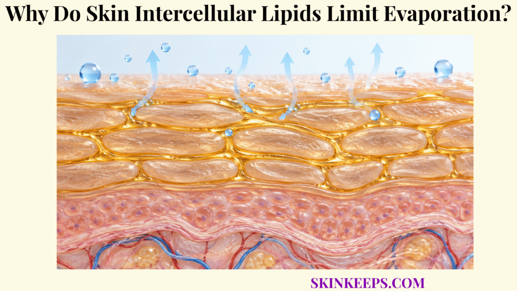
Skin intercellular lipids limit evaporation by forming highly organized extracellular lamellae between stratum-corneum corneocytes that strongly resist outward water diffusion, reducing the amount of water that reaches the surface to evaporate.
Ceramides, cholesterol, and free fatty acids provide this resistance through lamellar organization, long hydrophobic chains, and dense molecular packing. The lipids primarily control diffusion through the stratum corneum rather than the atmospheric evaporation step itself.
Where Do Skin Intercellular Lipids Form the Water Barrier?
Skin intercellular lipids form the water barrier in the extracellular spaces between stratum-corneum corneocytes, where ceramides, cholesterol, and free fatty acids assemble into organized lamellar structures.
Where Are Skin Intercellular Lipids Located Relative to Corneocytes?
Intercellular barrier lipids surround corneocytes throughout the stratum corneum, forming the continuous extracellular matrix between its cellular structural units. Their physiological purpose sits within skin water-loss prevention and the larger skin water barrier.
Which Lipids Dominate the Intercellular Matrix?
Ceramides, cholesterol, and free fatty acids dominate the mature stratum-corneum extracellular lipid matrix. Classical descriptions often place them near roughly 50%, 25%, and 10–15% by weight, respectively, but these are approximate reference proportions rather than fixed values for every site, age, condition, or analytical method.
Are Skin Intercellular Lipids the Same as Sebum?
No. Stratum-corneum barrier lipids arise from epidermal differentiation and lamellar-body processing, whereas sebum is produced mainly by sebaceous glands and delivered through follicles to the surface. Sebum contains a different lipid profile and is not the principal continuous TEWL barrier.
How Are Intercellular Barrier Lipids Delivered to the Stratum Corneum?
Upper epidermal keratinocytes package lipid precursors and processing enzymes into lamellar bodies, which secrete their contents at the granular-to-cornified transition. Those intracellular organelles are not the mature barrier lamellae: after secretion, precursor lipids are enzymatically processed and reorganized into extracellular ceramide-, cholesterol-, and free-fatty-acid-rich structures. The broader architecture belongs to the skin stratum corneum barrier.
How Do Skin Intercellular Lipids Seal Water Between Corneocytes?
Skin intercellular lipids “seal” water between corneocytes by forming densely packed multilamellar domains that create a low-permeability extracellular route rather than an open aqueous space. The effect is strong resistance, not complete waterproofing.
How Does the Brick-and-Mortar Model Explain the Lipid Barrier?
The brick-and-mortar model treats corneocytes as structural bricks and the organized extracellular lipid matrix as mortar, highlighting that water cannot move freely through open spaces between cells. The analogy is incomplete because both components are dynamic, molecularly specialized, hydrated biological structures rather than inert masonry.
Do Skin Intercellular Lipids Simply Fill Empty Spaces?
No. Ceramides, cholesterol, and free fatty acids self-organize into ordered molecular structures whose composition, chain architecture, lamellar phase behavior, and lateral packing determine permeability. They occupy extracellular space but function as an organized molecular barrier rather than passive filler.
How Does Dense Lipid Packing Reduce Water Escape?
Long hydrophobic lipid chains interact closely through van der Waals and hydrophobic forces, producing domains in which polar water has low solubility and restricted molecular mobility. Denser organization generally corresponds with lower permeability, although packing is not identical throughout every region of the stratum corneum.
Why Does Lipid Composition Matter to the Seal?
Changing ceramide subclass, ceramide or free-fatty-acid chain length, unsaturation, relative ratios, or cholesterol balance can alter lamellar phases and lateral packing even when total lipid remains present. Barrier quality therefore depends on molecular architecture and organization as well as quantity.
How Do Skin Intercellular Lipids Slow Water Diffusion?
Skin intercellular lipids slow water diffusion because water crossing the extracellular stratum-corneum route must repeatedly partition into and move through densely packed hydrophobic lipid domains.
Why Does Water Move Slowly Through Hydrophobic Lipid Domains?
Water is polar and partitions poorly into long hydrocarbon-rich regions, making highly ordered barrier lipids energetically and physically unfavorable pathways compared with free aqueous diffusion. This lowers the effective diffusion and partitioning of water through the extracellular route.
How Do Multiple Lipid Lamellae Increase Diffusion Resistance?
Repeated lamellar domains force water to cross multiple interfaces and ordered hydrophobic regions before it can progress toward the surface. The repeated layered structure, combined with the tortuous route around corneocytes, lengthens and complicates the transport pathway.
How Does Lipid Chain Length Affect Water Permeability?
Longer and appropriately matched ceramide and free-fatty-acid chains generally support stronger hydrophobic interactions and denser organization, while an increased proportion of shorter chains can produce more permeable experimental lipid systems. Model-membrane findings explain mechanism but do not define universal human TEWL values.
How Does Lateral Lipid Packing Affect Permeability?
More ordered lateral packing restricts molecular motion within the lipid matrix, while less ordered states can increase molecular mobility and permeability. No single packing state exists uniformly across every depth or microdomain of the stratum corneum.
How Does Slower Diffusion Affect TEWL?
Greater lipid resistance lowers the rate at which internal water reaches the surface, thereby reducing the passive flux measured as skin transepidermal water loss. Once water reaches the surface, ambient humidity, temperature, and vapor-pressure gradients also influence evaporation.
How Do Skin Intercellular Lipids Work With Corneocytes?
Skin intercellular lipids work with corneocytes by controlling extracellular water escape while corneocytes provide the structural framework and retain water internally through keratin and natural moisturizing factor.
What Water-Control Role Do Corneocytes Perform?
Corneocytes provide the keratin-rich structural units of the stratum corneum and contain hygroscopic natural moisturizing factor that supports intracellular water retention. Their detailed water-holding physiology belongs to skin corneocyte water retention.
What Water-Control Role Do Intercellular Lipids Perform?
Intercellular lipids restrict the movement of water through the continuous extracellular pathway surrounding corneocytes. Their lamellar continuity provides much of the stratum corneum’s resistance to water and many external molecules.
How Does NMF Differ From Skin Intercellular Lipids?
NMF increases water-holding capacity within corneocytes, whereas intercellular lipids determine much of the resistance to water escaping between those cells. The composition and broader physiology of these intracellular solutes belong to skin natural moisturizing factors.
Why Does Normal Hydration Require Both Systems?
Corneocytes need sufficient intracellular water-holding capacity, while the extracellular lipid matrix must remain resistant enough to prevent that water from diffusing outward too rapidly. Water holding and water-loss resistance therefore cooperate without performing the same molecular task.
| Barrier component | Main water-control role | Functional result | Key boundary |
|---|---|---|---|
| Corneocyte | Structural unit + intracellular water holding | Supports SC hydration / flexibility | Not main continuous diffusion barrier |
| Keratin matrix | Protein hydration / mechanics | Supports pliability | Not waterproof |
| NMF | Hygroscopic intracellular solutes | Retains water in corneocyte | Not extracellular seal |
| Ceramides | Major lamellar lipid family | Supports dense low-permeability organization | Many subclasses / chain lengths |
| Cholesterol | Modulates phase behavior | Supports normal lipid organization | Not inert filler |
| Free fatty acids | Hydrophobic packing | Supports low water permeability | Chain length / composition matter |
| Corneocyte lipid envelope | Lipid scaffold / interface | Supports extracellular lamella organization | Distinct from free lamellae |
| Whole lipid matrix | Continuous extracellular resistance | Reduces outward diffusion / TEWL | Not completely impermeable |
What Disrupts Skin Intercellular Lipids and Increases Evaporation?
Skin intercellular lipids become less effective when surfactants, solvents, repeated washing, inflammatory signaling, or other barrier stresses alter lipid amount, chain architecture, molecular packing, or lamellar organization.
How Can Harsh Cleansers Disrupt Skin Intercellular Lipids?
Some surfactants can penetrate the stratum corneum and interact with both lipids and proteins, extracting selected components or disturbing molecular organization enough to increase permeability and irritation. The larger multi-component failure process belongs to skin barrier disruption.
How Does Repeated Washing Affect the Lipid Barrier?
Frequent washing can repeatedly expose the stratum corneum to surfactants, water, friction, and changing hydration states, producing cumulative changes in barrier function depending on formulation, water temperature, frequency, contact time, and individual skin. Challenge studies with sodium lauryl sulfate should not be generalized to every routine wash.
How Can Solvents Disrupt Skin Intercellular Lipids?
Solvents that partition into or extract stratum-corneum lipids can alter lipid composition and molecular organization, reducing resistance to water and chemical permeation. Different solvents have different partitioning and extraction behavior, so the effect cannot be generalized to all solvents.
Do Dry Environmental Conditions Directly Remove Barrier Lipids?
Not necessarily. Low humidity primarily increases the hydration challenge and changes water activity, enzyme function, desquamation, and adaptive epidermal responses; it should not be described simply as extracting intercellular lipids. Measured TEWL responses to climate and humidity are not uniformly directional.
How Can Inflammation Disrupt Skin Intercellular Lipids?
Inflammatory cytokine signaling can alter keratinocyte lipid synthesis, fatty-acid elongation, ceramide profiles, and lamellar-body biology, producing compositions that may form a less effective permeability barrier. The exact pattern depends on cytokine environment, site, lesion status, and biological context rather than a universal fall in every lipid class.
What Happens When Skin Intercellular Lipids Become Deficient?
When skin intercellular lipids become deficient or abnormally organized, the extracellular pathway becomes more permeable, allowing faster outward water movement and easier penetration of selected environmental substances.
Does Skin Intercellular Lipid Deficiency Always Mean Fewer Total Lipids?
No. Barrier failure can result from reduced abundance of selected lipids, but it can also arise from abnormal ceramide subclasses, shorter lipid chains, altered ratios, or disordered lamellar and lateral packing. Normal barrier function requires adequate composition and correct organization.
Why Does Lipid Deficiency Increase TEWL?
A less organized or incomplete lipid matrix reduces resistance along the extracellular route, allowing more water to diffuse toward the surface per unit time. The downstream rise in surface water availability can increase passive evaporative loss.
How Does Increased Water Escape Affect the Stratum Corneum?
Faster outward water loss can reduce stratum-corneum hydration and contribute to stiffness, roughness, scaling, and fissuring when intracellular water retention cannot compensate for the increased diffusion gradient.
Why Does Lipid Deficiency Increase Irritant Susceptibility?
The same permeability barrier that limits inside-out water loss also restricts outside-in chemical penetration, so lipid disorganization can increase access of selected irritants and allergens to deeper epidermal structures. The water-loss consequences are explored further under skin water loss and irritation.
Can Barrier Damage and Inflammation Reinforce Each Other?
Yes. Greater permeability can increase environmental exposure and inflammatory signaling, while inflammation can alter lipid synthesis and organization further, creating a bidirectional barrier–inflammation cycle in susceptible contexts. This mechanism does not diagnose atopic dermatitis, psoriasis, contact dermatitis, or any other condition from dryness or TEWL alone.
What Are the Key Takeaways About Skin Intercellular Lipids?
The key fact about skin intercellular lipids is that they form the highly organized extracellular matrix that provides much of the stratum corneum’s resistance to outward water diffusion, thereby limiting the amount of water available for surface evaporation.
Ceramides, cholesterol, and free fatty acids must be present in appropriate molecular forms and organized into effective lamellar and lateral packing states. Corneocytes provide structure and intracellular water holding, while the lipid matrix supplies the principal continuous extracellular resistance; disruption of either amount or organization can increase permeability.
- Skin intercellular lipids are located between corneocytes in the stratum corneum.
- They are different from surface sebum.
- Ceramides, cholesterol, and free fatty acids are the dominant extracellular barrier-lipid classes.
- Barrier lipids form organized lamellae rather than a random oily mixture.
- The extracellular lipid matrix forms the main continuous permeation pathway through the stratum corneum.
- Intercellular lipids limit evaporation primarily by restricting water diffusion before water reaches the surface.
- The lipid matrix is highly resistant but not completely impermeable.
- Ceramides are a structurally diverse lipid family.
- Ceramide subclass and chain length influence barrier organization.
- Free-fatty-acid chain length and composition also influence permeability.
- Cholesterol contributes to normal lipid phase behavior and barrier homeostasis.
- Lipid molecular packing matters in addition to lipid quantity.
- Lamellar organization matters in addition to total lipid quantity.
- Long hydrophobic chains contribute to dense low-permeability lipid domains.
- Corneocytes provide structural and intracellular water-retention functions.
- NMF retains water inside corneocytes rather than replacing the extracellular lipid barrier.
- Lamellar bodies deliver precursors and enzymes required to form mature extracellular barrier lipids.
- The corneocyte lipid envelope and free extracellular lipid lamellae are distinct structures.
- Surfactants can interact with both lipids and proteins rather than simply “stripping oil.”
- Repeated washing effects depend on formulation, temperature, frequency, contact time, and barrier state.
- Low humidity challenges hydration but does not simply extract intercellular lipids.
- Inflammatory signaling can alter lipid synthesis and molecular composition.
- Barrier impairment may result from lipid deficiency or abnormal lipid organization.
- Lipid disruption can increase TEWL and environmental permeability.
- Increased TEWL is a physiological sign of altered barrier function, not a diagnosis by itself.
What Common Questions Do People Ask About Skin Intercellular Lipids?
Common questions about skin intercellular lipids focus on where they are located, how they slow water loss, whether ceramides alone form the barrier, how they differ from corneocytes and NMF, and what happens when lipid organization is disrupted.
Where Are Skin Intercellular Lipids Located?
Skin intercellular lipids occupy the extracellular spaces between corneocytes in the stratum corneum, where ceramides, cholesterol, and free fatty acids form highly organized lamellar structures.
How Do Skin Intercellular Lipids Reduce Water Evaporation?
They primarily reduce the diffusion of water through the stratum corneum, so less internal water reaches the skin surface per unit time to evaporate into the surrounding air.
Do Ceramides Alone Form the Skin Intercellular Lipid Barrier?
No. Ceramides are major barrier lipids, but effective permeability control depends on their interaction with cholesterol and free fatty acids as well as appropriate chain lengths, ratios, lamellar organization, and molecular packing.
How Are Skin Intercellular Lipids Different From Natural Moisturizing Factor?
Intercellular lipids form the extracellular diffusion barrier between corneocytes, while natural moisturizing factor consists mainly of small hygroscopic molecules inside corneocytes that increase intracellular water retention.
What Happens When Skin Intercellular Lipids Are Disrupted?
Disrupted lipid amount or organization lowers extracellular diffusion resistance, which can increase transepidermal water loss, reduce stratum-corneum hydration, and increase susceptibility to dryness, scaling, fissuring, and penetration of irritants.
Sources & Evidence
1. The Skin Barrier: An Extraordinary Interface With an Exceptional Lipid Organization
2. Using Molecular Simulation to Understand the Skin Barrier
3. Role of Lipids in the Formation and Maintenance of the Cutaneous Permeability Barrier
4. Lipids and the Permeability and Antimicrobial Barriers of the Skin
5. Skin Lipid Barrier: Structure, Function and Metabolism
6. Cleansing Formulations That Respect Skin Barrier Integrity
7. Transepidermal Water Loss (TEWL): Environment and Pollution—A Systematic Review
8. Cytokines and Epidermal Lipid Abnormalities in Atopic Dermatitis: A Systematic Review
Medical/Educational Disclaimer
This page explains normal epidermal lipid-barrier physiology and is not medical advice. Seek professional evaluation for persistent severe dryness, painful fissures, weeping or bleeding skin, widespread inflammation, or suspected infection.



