
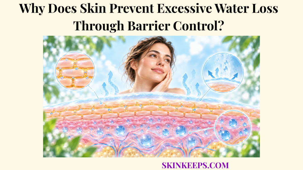
Skin prevents excessive water loss because life in a dry external environment requires the epidermis to restrict passive evaporation while retaining enough water in the stratum corneum for normal flexibility, enzyme activity, cell maturation, and controlled shedding. Deeper skin contains substantially more water than the surrounding air, so a natural outward water gradient exists even when the barrier is healthy.
This article explains how water normally moves through skin, how the stratum corneum slows that movement, how extracellular lipids and natural moisturizing factor perform different hydration functions, what tight junctions add deeper in the epidermis, what changes when barrier resistance falls, and how normal barrier recovery can be supported.
Why Is Water-Loss Control Essential to Healthy Skin Function?
Water-loss control is essential because the stratum corneum requires adequate hydration to remain flexible, maintain normal enzymatic activity, support orderly corneocyte maturation and shedding, and resist cracking under mechanical stress. Control of water loss is therefore one of the body’s important skin functions, not simply a cosmetic feature of surface softness.
Water acts as a plasticizer within the outer epidermis, allowing keratin-rich corneocytes to remain sufficiently pliable during ordinary movement and friction. When the stratum corneum becomes markedly dry, these cells can become stiffer and more prone to roughness or microscopic fissuring.
Hydration also supports hydrolytic enzymes involved in corneocyte maturation and controlled breakdown of corneodesmosomes, the adhesive structures that help hold corneocytes together. These water-dependent reactions contribute to orderly desquamation, or shedding of superficial corneocytes.
When stratum-corneum hydration falls too far, normal desquamatory activity can become less efficient and superficial cells may shed in visible aggregates. Roughness, scaling, and cracking can follow, especially when dryness is combined with mechanical or chemical stress.
Why Does the Epidermis Need Adequate Water to Remain Flexible and Functional?
Adequate epidermal and stratum-corneum water keeps corneocytes sufficiently plastic and flexible to tolerate normal movement and friction without becoming excessively rigid or prone to microscopic cracking. This mechanical effect depends on water within the outer tissue, but more water is not always better because prolonged soaking or overhydration can also perturb barrier structure.
How Does Water Retention Support Normal Enzyme Activity and Skin-Cell Turnover?
Normal stratum-corneum hydration supports water-dependent enzymatic reactions that regulate corneodesmosome breakdown, corneocyte maturation, and orderly desquamation. Proteases and other hydrolytic enzymes involved in shedding work within a local chemical environment whose water content and pH influence activity.
Why Must Skin Limit Water Loss While Still Allowing Physiological Evaporation?
Skin must limit rather than eliminate water loss because a small continuous outward water flux occurs normally across healthy epidermis, while the barrier prevents that physiological flux from becoming excessive. Healthy skin is therefore water-resistant, not perfectly waterproof.
| Water-dependent function | Why hydration matters | Effect of excessive water loss |
|---|---|---|
| Corneocyte flexibility | Water plasticizes keratin-rich corneocytes and supports pliability. | Excessive dryness can increase rigidity and cracking. |
| Enzyme activity | Several stratum-corneum enzymes require an appropriate hydration environment. | Reduced hydration can disturb normal maturation and desquamation. |
| Desquamation | Hydration supports controlled breakdown of corneodesmosomes. | Dry stratum corneum can shed in visible aggregates, producing roughness and scaling. |
| Barrier continuity | Flexible hydrated tissue tolerates routine movement better. | Severe dryness can promote fissuring and irritation. |
How Does Transepidermal Water Loss Occur Through the Skin?
Transepidermal water loss occurs when water from deeper, more hydrated skin compartments passively diffuses outward through the epidermis and stratum corneum and then evaporates from the surface into the surrounding air. This normal, largely insensible process is called transepidermal water loss, or TEWL.
The water originates from deeper tissue and living epidermal compartments that contain more water than the superficial stratum corneum and the external environment. That difference creates a concentration gradient that drives passive diffusion toward the surface; water is not actively pumped through the epidermis.
How Does Water Move From Deeper Epidermal Layers Toward the Surface?
Water moves outward because living epidermal tissue contains substantially more water than the superficial stratum corneum and external environment, creating a concentration gradient that drives passive diffusion toward the surface. As water approaches the outer epidermis, the stratum corneum becomes the major resistance step controlling how quickly that movement continues.
What Is Transepidermal Water Loss, or TEWL?
Transepidermal water loss, or TEWL, is the continuous insensible flux of water that diffuses from deeper skin through the epidermis and stratum corneum into the surrounding environment. It differs from sweating, which is active glandular secretion used partly for thermoregulation.
TEWL is widely used as an experimental and clinical indicator of epidermal permeability-barrier function, but a measured value depends on body site, age, skin condition, temperature, humidity, acclimatization, device type, and protocol. There is no single consumer cutoff that defines a healthy or damaged barrier in every person and setting.
Why Does TEWL Increase When Barrier Resistance Decreases?
TEWL can rise when barrier resistance decreases because disrupted corneocyte organization, extracellular lipid structure, or junctional control creates less resistance to outward water diffusion. The same outward concentration gradient is present, but the pathway opposing diffusion becomes less effective.
Barrier disruption lowers the resistance step, which can permit greater outward flux.
How Does the Stratum Corneum Restrict Excessive Water Escape?
The stratum corneum restricts excessive water escape through a two-compartment structure in which hydrated, keratin-rich corneocytes provide the cellular framework while highly ordered extracellular lipids create the main low-permeability pathway between those cells. This outer skin water barrier provides the principal resistance to passive water diffusion.
How Do Corneocytes Form the Structural Framework of the Water Barrier?
Corneocytes form the structural framework of the water barrier as flattened, keratin-rich cells surrounded by reinforced cornified envelopes that provide mechanical resilience while retaining water-binding molecules inside the cell. Their hydration and flexibility are central to corneocyte water retention, but corneocytes are not the main molecular diffusion seal between cells.
Corneodesmosomes connect neighboring corneocytes and help maintain tissue cohesion until controlled proteolysis allows superficial cells to detach. This structural system must preserve both strength and orderly turnover.
How Does the “Brick-and-Mortar” Arrangement Slow Water Movement?
The “brick-and-mortar” model describes corneocytes as structural bricks surrounded by an extracellular lipid “mortar,” with the ordered lipid pathway providing much of the resistance that slows water diffusion through the stratum corneum. It is a teaching analogy, not literal microscopic architecture.
How Does Controlled Shedding Preserve Barrier Continuity?
Controlled desquamation preserves barrier continuity by removing superficial corneocytes gradually while newly differentiated cells replace them from below, maintaining a relatively stable stratum-corneum thickness and organization. Normal shedding is part of barrier homeostasis rather than evidence of barrier failure.
How Do Skin Lipids and Natural Moisturizing Factors Maintain Hydration?
Skin lipids and natural moisturizing factors maintain hydration through complementary mechanisms: extracellular lipids reduce water permeability between corneocytes, while NMF molecules bind and retain water inside the corneocytes themselves. These mechanisms should be separated because water sealing and intracellular water binding are not the same function.
How Do Ceramides, Cholesterol, and Fatty Acids Reduce Water Permeability?
Ceramides, cholesterol, and free fatty acids organize into extracellular lamellar structures between corneocytes that create the stratum corneum’s principal low-permeability pathway and slow outward water diffusion. These skin intercellular lipids work as a cooperating mixture rather than as isolated ceramides acting alone.
Granular-layer keratinocytes package lipid precursors and processing enzymes in lamellar bodies. When these organelles release their contents near the stratum granulosum–stratum corneum interface, extracellular processing helps create the hydrophobic lamellar matrix that limits water diffusion.
How Do Natural Moisturizing Factors Attract and Retain Water Inside Corneocytes?
Natural moisturizing factors retain water inside corneocytes because their hygroscopic amino acids, amino-acid derivatives, urea, lactate, and electrolytes bind water within the outer epidermal cells. These skin natural moisturizing factors function intracellularly rather than forming the extracellular lipid seal.
Profilaggrin is processed to filaggrin during epidermal differentiation, and later filaggrin breakdown produces free amino acids and derivatives such as pyrrolidone carboxylic acid and urocanic acid that contribute to NMF. This natural moisturizing factor water binding increases the water-holding capacity of corneocytes.
How Do Tight Junctions Provide Additional Control in Deeper Epidermal Layers?
Tight junctions provide additional water-control capacity by sealing spaces between keratinocytes in the stratum granulosum and restricting paracellular movement beneath the stratum corneum. Claudin proteins, including claudin-1, are important junctional components, but tight junctions are not the principal outer permeability barrier.
| Component | Location | Main mechanism | Effect on water retention |
|---|---|---|---|
| Corneocytes | Stratum corneum | Provide hydrated keratin-rich structural units. | Support flexible, mechanically stable outer tissue. |
| Ceramide/cholesterol/FFA lamellae | Between corneocytes | Create an ordered hydrophobic diffusion barrier. | Slow outward water movement. |
| Natural moisturizing factor | Inside corneocytes | Hygroscopic molecules bind water. | Maintain intracellular corneocyte hydration. |
| Tight junctions | Mainly stratum granulosum | Restrict paracellular water and solute movement. | Provide additional deeper epidermal control. |
| Corneodesmosomes/desquamation system | Between corneocytes | Regulate cohesion and orderly shedding. | Maintain barrier continuity and thickness. |
What Causes Skin Barrier Failure and Excessive Water Loss?
Skin barrier failure can increase water loss when chemical, mechanical, environmental, genetic, or inflammatory stress disrupts stratum-corneum lipids, corneocyte organization, differentiation, or deeper junctional control enough to reduce permeability resistance. Skin barrier disruption describes reduced barrier performance; dryness, stinging, or scaling alone do not prove a specific barrier defect.
How Do Harsh Cleansers, Solvents, and Repeated Washing Disrupt Water Control?
Harsh cleansers, solvents, frequent washing, and prolonged hot-water exposure can worsen water control by extracting or disrupting surface and stratum-corneum lipids, altering proteins, and increasing dryness or irritation. Normal cleansing is not inherently harmful; the effect depends on cleanser formulation, frequency, temperature, contact time, friction, and baseline skin condition.
How Do Dry Air and Low Humidity Increase Barrier Stress?
Dry air and low humidity increase evaporative drying pressure on the skin surface, which can reduce stratum-corneum hydration and contribute to roughness, tightness, flaking, and cracking when water retention cannot compensate adequately. Measured TEWL does not rise in a simple universal way under every low-humidity condition because measurement and environmental effects are context-dependent.
How Can Inflammation or Skin Disease Weaken Barrier Organization?
Inflammatory and inherited skin disorders can weaken water-barrier organization by altering keratinocyte differentiation, structural proteins, lipid composition, junctional proteins, or inflammatory signaling. Atopic dermatitis, psoriasis, and ichthyosis are examples of conditions in which barrier abnormalities may occur, but diagnosis and detailed disease mechanisms belong on disease-specific pages.
What Happens When Increased Water Loss Leads to Dryness, Scaling, Irritation, and Sensitivity?
When water loss exceeds the barrier’s ability to preserve stratum-corneum hydration, corneocytes become less flexible, normal desquamation can become disorganized, and the surface may become rough, scaly, tight, cracked, or more easily irritated. These visible findings do not by themselves prove elevated TEWL or uncomplicated barrier damage.
| Barrier stressor | Structural / functional change | Water-loss effect | Possible visible outcome |
|---|---|---|---|
| Harsh or repeated cleansing | Lipid extraction and protein stress. | Reduces permeability resistance. | Tightness, roughness, dryness. |
| Prolonged hot-water exposure | Increases lipid removal and drying stress. | Can worsen water retention. | Dryness and irritation. |
| Low humidity | Increases environmental drying pressure. | Promotes loss of stratum-corneum hydration. | Flaking, roughness, cracking. |
| Inflammatory skin disease | Alters differentiation, lipids, proteins, or junctions. | Can increase permeability and TEWL. | Dry, inflamed, itchy skin. |
| Severe mechanical disruption | Damages corneocyte/lipid organization. | Increases permeability. | Irritation, fissuring, sensitivity. |
Barrier symptoms and TEWL changes require clinical context; no single visible sign confirms a specific barrier defect.
How Can the Epidermal Barrier Restore Normal Water Control?
The epidermal barrier restores water control through homeostatic responses that replenish lamellar-body contents, increase production and processing of barrier lipids, replace damaged corneocytes, and rebuild organized stratum-corneum structure. Recovery is an endogenous tissue process that supportive skin care can assist but not instantly replace.
How Does the Epidermis Replenish Barrier Lipids After Damage?
After acute barrier disruption, granular-layer keratinocytes can rapidly release stored lamellar-body contents and increase production of lipid precursors needed to restore extracellular lamellae in the stratum corneum. Lamellar bodies deliver lipids, lipid-processing enzymes, desquamation-related proteins, and other components needed for upper-epidermal barrier organization.
Barrier disruption also stimulates broader homeostatic responses that can include increased synthesis of cholesterol, fatty acids, and sphingolipid precursors together with changes in keratinocyte signaling, proliferation, and differentiation. Recovery speed varies with the severity and cause of disruption.
How Do Moisturizers and Occlusive Ingredients Help Reduce Water Escape?
Moisturizers support water retention by combining humectant, emollient, and occlusive effects that increase surface hydration, smooth the stratum corneum, and reduce evaporation while endogenous barrier structures recover. A humectant attracts or binds water, an emollient smooths superficial irregularities, and an occlusive forms a surface layer that reduces evaporative loss.
Creams and ointments often retain more moisture than lighter lotions, and applying moisturizer while skin remains slightly damp can help reduce evaporation. These categories support dry skin but should not be described as permanently curing every form of barrier dysfunction.
Why Does Gentle Cleansing Help Preserve Barrier Recovery?
Gentle cleansing supports barrier recovery by removing necessary surface debris without repeatedly maximizing lipid extraction, high-temperature exposure, friction, or irritant contact during the period when barrier structures are being replenished. Short washing exposure, lukewarm water, minimal scrubbing, and fragrance-free products when irritation is present are conservative options.
When Can Persistent Dryness or Barrier Failure Require Medical Evaluation?
Persistent dryness deserves medical evaluation when conservative care does not improve the problem or when severe itching, pain, bleeding cracks, widespread inflammation, recurrent rash, or infection signs suggest an underlying skin disorder. Persistent scaling, recurrent unexplained flares, or repeated painful cracking are also reasonable reasons to seek dermatology assessment.
Bleeding fissures, pus, spreading redness, significant swelling, worsening pain, fever with skin symptoms, or non-healing open areas deserve prompt medical attention.
| Problem | Barrier-support strategy | Intended result |
|---|---|---|
| Dryness after frequent cleansing | Reduce unnecessary washing intensity and use gentler cleansing. | Reduce repeated lipid and protein stress. |
| Rapid post-wash tightness | Apply moisturizer while skin remains slightly damp. | Retain more water and reduce evaporation. |
| Rough or scaly surface | Maintain regular moisturization and avoid aggressive exfoliation. | Improve hydration and allow more orderly desquamation. |
| Low-humidity dryness | Increase moisturization and consider environmental humidification where appropriate. | Reduce environmental drying stress. |
| Persistent painful or inflamed dryness | Seek professional evaluation. | Identify an underlying condition rather than assuming routine barrier stress. |
What Are the Key Takeaways About the Skin’s Water-Retention Barrier?
The key fact about the skin’s water-retention barrier is that hydration depends on several complementary systems: corneocytes provide structure, natural moisturizing factors bind water inside those cells, extracellular lipids slow water diffusion between them, and tight junctions provide additional control deeper in the epidermis.
- Physiological TEWL: Healthy skin continuously loses a small amount of water through passive diffusion.
- Main resistance layer: The stratum corneum provides the principal barrier that prevents this water loss from becoming excessive.
- Corneocytes: Hydrated keratin-rich corneocytes provide flexible structural units within the outer barrier.
- Barrier lipids: Ceramides, cholesterol, and free fatty acids form extracellular lamellae that provide major resistance to water diffusion.
- Natural moisturizing factor: NMF binds water within corneocytes rather than forming the extracellular lipid seal.
- Tight junctions: Granular-layer tight junctions add deeper epidermal control over paracellular water movement.
- Normal turnover: Controlled desquamation preserves barrier continuity while superficial cells are replaced.
- Barrier disruption: Chemical, environmental, inflammatory, or mechanical stress can lower barrier resistance and increase water loss.
- Recovery: Epidermal cells restore barrier function through lipid delivery, lipid synthesis, differentiation, and structural renewal.
- Supportive care: Gentle cleansing and appropriate moisturization can reduce avoidable water loss while the skin restores normal barrier function.
What Common Questions Do People Ask About Skin Water Loss?
Common questions about skin water loss focus on whether TEWL is normal, what NMF does, whether drinking more water repairs dry skin, and which barrier structures actually keep water inside the body.
Is Transepidermal Water Loss Normal?
Yes. A small amount of transepidermal water loss occurs continuously in healthy skin; the barrier’s role is to restrict this passive flux so that water escape does not become excessive.
Is TEWL the Same as Sweating?
No. TEWL is passive, insensible diffusion of water through the epidermis, while sweating is active secretion of fluid from sweat glands, particularly during thermoregulation.
What Part of the Skin Prevents Most Water Loss?
The stratum corneum provides the main resistance to water loss through its corneocyte framework and highly organized extracellular lipid lamellae.
Does Natural Moisturizing Factor Stop Water From Escaping?
Natural moisturizing factor mainly retains water within corneocytes through hygroscopic molecules, whereas extracellular stratum-corneum lipids provide most of the low-permeability resistance that slows water escape.
Does Drinking More Water Repair a Damaged Skin Barrier?
Adequate whole-body hydration supports normal physiology, but drinking extra water does not directly replace disrupted stratum-corneum lipids, NMF, corneocytes, or junctional structures responsible for epidermal barrier function.
Skin Research and Technology / PMC — Transepidermal water loss (TEWL): Environment and pollution—A systematic review: TEWL as a skin-integrity measure, environmental influences, anatomical and contextual variability, and limits of simple interpretation.
Skin Research and Technology / PMC — Devices measuring transepidermal water loss: A systematic review of measurement properties: TEWL measurement principles, reliability, measurement error, water-vapour gradient methodology, and device-related variability.
Cold Spring Harbor Perspectives in Medicine / PMC — Epidermal Barriers: stratum-corneum water-loss prevention, corneocytes, lipids, junctional proteins, and the multi-component epidermal barrier.
Journal of Clinical and Aesthetic Dermatology / PMC — The Clinical Relevance of Maintaining the Functional Integrity of the Stratum Corneum in both Healthy and Disease-affected Skin: corneocyte structure, intercellular lipids, NMF, water-dependent flexibility, enzyme activity, desquamation, and barrier homeostasis.
Journal of Cell Science / PMC — Filaggrin in the frontline: role in skin barrier function and disease: profilaggrin processing, filaggrin biology, keratin organization, and generation of hygroscopic amino acids contributing to NMF.
International Journal of Molecular Sciences / PMC — The Discovery and Function of Filaggrin: modern filaggrin biology, degradation products, pyrrolidone carboxylic acid, urocanic acid, and NMF-related hydration mechanisms.
Tissue Barriers / PubMed — Epidermal tight junctions in health and disease: the two major epidermal barrier structures—stratum corneum and granular-layer tight junctions—and deeper paracellular control.
Frontiers in Cell and Developmental Biology / PMC — Epidermal lamellar bodies, essential organelles for the skin barrier: lamellar-body lipid delivery, barrier formation, desquamation machinery, and maintenance of the hydrophobic upper epidermis.
Yonsei Medical Journal / PMC — An Update of the Defensive Barrier Function of Skin: permeability-barrier homeostasis, lamellar-body secretion, increased lipid synthesis, keratinocyte differentiation, and recovery after acute disruption.
American Academy of Dermatology — Dermatologists’ top tips for relieving dry skin: low-humidity drying, gentle cleansing, warm rather than hot water, creams/ointments, damp-skin moisturization, humidification, and referral when dryness persists.
Medical note: This article explains normal skin water-balance physiology and conservative barrier-support principles for educational purposes. Dryness, scaling, stinging, or cracking do not by themselves diagnose a “damaged skin barrier,” eczema, contact dermatitis, ichthyosis, or another disorder. Persistent symptoms, severe itching or pain, bleeding fissures, pus, spreading redness, significant swelling, fever with skin symptoms, or non-healing open areas require appropriate medical evaluation.



