
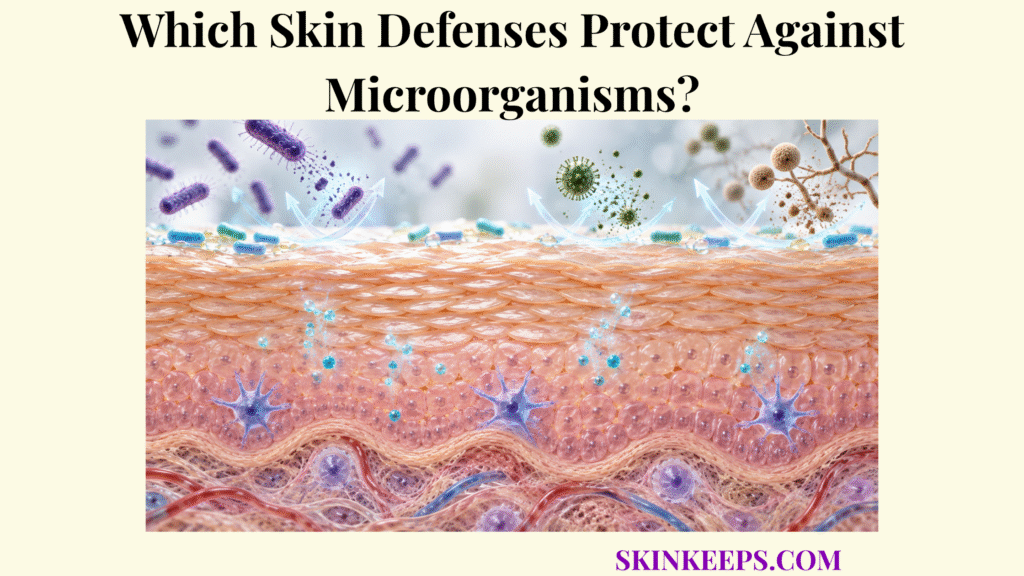
Skin protects against microorganisms through four coordinated defense layers: an intact physical barrier restricts entry, surface chemistry suppresses selected microbial growth, resident microbiota provide colonization resistance, and cutaneous immune systems recognize and eliminate threats that penetrate into living tissue. Healthy skin is therefore colonized and ecologically active rather than sterile.
Protection depends on how these layers interact at the same time. The key tasks are to prevent invasion first, constrain microbial expansion at the surface, recognize meaningful barrier breach, recruit appropriate effector mechanisms, and then switch inflammatory activity back down after the threat is controlled.
What Makes Skin Defenses the First Barrier Against Microorganisms?
Skin defenses form the body’s first antimicrobial barrier because the epidermal surface physically separates vulnerable tissues from external microbes while chemical, microbial, and immune mechanisms continuously shape which organisms can remain, multiply, or penetrate deeper. The broader skin barrier protection system therefore includes far more than germ killing.
Bacteria, fungi, viruses, bacteriophages, and microscopic eukaryotes can normally occupy the skin surface without establishing infection. Colonization means organisms are present and may reproduce without necessarily invading tissue or causing disease; infection implies tissue-level invasion or damage with a host response. Microbial presence alone is not proof of infection.
| Skin Defense | Primary Mechanism | Main Antimicrobial Outcome |
|---|---|---|
| Stratum corneum | Compact corneocytes + lipid matrix | Restricts microbial penetration into viable tissue |
| Desquamation | Controlled surface-cell shedding | Removes some attached organisms |
| Acid mantle | Acidic physicochemical environment | Limits growth of selected potential pathogens |
| Sebum-derived lipids | Antimicrobial FFAs + ecological shaping | Inhibits selected organisms and shapes sebaceous-site microbiota |
| Eccrine sweat | Dermcidin + surface chemistry | Provides constitutive antimicrobial activity |
| Host AMPs | Direct antimicrobial effects + immune modulation | Inhibits selected bacteria, fungi, and other microbes |
| Resident microbiome | Niche competition + antimicrobial metabolites | Creates colonization resistance |
| Keratinocyte PRRs | Detect conserved microbial patterns | Initiates cytokine, chemokine, and AMP responses |
| Langerhans/DC networks | Antigen sampling and presentation | Connects local exposure with adaptive immunity |
| Recruited leukocytes | Phagocytosis + antimicrobial effector mechanisms | Controls organisms invading living tissue |
| Adaptive lymphocytes | Antigen-specific response + memory | Improves targeted and recall defense |
No single row functions independently; antimicrobial defense emerges from interactions among these systems.
How Do Skin Defenses Physically Block Microorganisms From Entering?
Skin defenses physically block microorganisms primarily through the stratum corneum, where compact corneocytes and intercellular lipids create a continuous, low-permeability surface that separates microbes from viable epidermal tissue. This first structural layer is treated in more depth in the stratum corneum barrier page.
How Does the Stratum Corneum Restrict Microbial Penetration?
The stratum corneum restricts microbial penetration because microorganisms encounter multiple layers of cornified cells and extracellular lipids before they can reach metabolically active epidermal tissue. Barrier protection is strong but not absolute; wounds, fissures, maceration, inflammation, and other structural disruption can create easier access to viable tissue.
How Do Corneocytes and Intercellular Lipids Work Together Against Microorganisms?
Corneocytes provide the mechanically resistant cellular framework, while tightly organized ceramide-, cholesterol-, and fatty-acid-rich lipids seal the spaces between them and reduce pathways through which microorganisms or microbial products could penetrate. Keratin and cornified protein structures reinforce the cells, whereas the extracellular lipid matrix provides much of the low-permeability pathway between cells.
How Does Continuous Epidermal Shedding Remove Attached Microorganisms?
Controlled desquamation contributes to microbial defense by continually removing superficial corneocytes together with some microorganisms and environmental material attached to the skin surface. Shedding contributes to clearance but does not sterilize the skin or eliminate resident microbial communities.
How Do Skin Defenses Use Surface Chemistry to Limit Microbial Growth?
Skin defenses use surface chemistry by maintaining an acidic, lipid-rich, saline environment containing host antimicrobial molecules that makes the surface less favorable for selected pathogens while remaining compatible with adapted resident microbes. The broader physicochemical system is explained in the skin acid mantle page.
How Does the Skin’s Acidic Surface Limit Microbial Growth?
The mildly acidic skin surface limits the growth of selected microorganisms and supports antimicrobial and barrier processes that function differently when surface conditions shift substantially. Skin pH is not one fixed universal number; it varies with body site, age, technique, sweat, sebum, environment, cleansing, and disease state.
How Do Sebum Lipids Contribute to Skin Antimicrobial Defense?
Sebum contributes to antimicrobial defense because sebaceous lipids and their breakdown products include free fatty acids with direct activity against selected microorganisms while also shaping the nutrient environment of sebaceous skin. Sapienic and lauric acids are useful experimental examples, but sebum is not simply an antimicrobial fluid because sebaceous sites also support organisms adapted to lipid-rich niches.
How Does Sweat Add Antimicrobial Protection?
Eccrine sweat adds antimicrobial protection by transporting constitutively produced dermcidin-derived peptides and other soluble factors onto the epidermal surface. Sweat also changes moisture, salt, and local physicochemistry, so its contribution is better understood through antimicrobial molecules and surface conditions than through a simplistic washing or disinfecting effect.
How Do Antimicrobial Peptides Control Microorganisms on Skin?
Skin antimicrobial peptides inhibit selected microorganisms through direct actions such as membrane disruption while also influencing inflammatory and immune signaling. Important examples include cathelicidin LL-37, β-defensins, RNase 7, psoriasin/S100A7, and dermcidin; their expression patterns, targets, and physiological potency are not identical.
How Do Skin Defenses Use the Microbiome to Resist Harmful Microorganisms?
Skin defenses use the resident microbiome as an ecological barrier in which adapted microorganisms occupy niches, consume resources, produce inhibitory compounds, and interact with host cells in ways that can restrict colonization by competing pathogens. This ecological role is developed further under skin microbiome barrier defense.
How Do Resident Microorganisms Compete for Space and Nutrients?
Resident microorganisms create colonization resistance partly by occupying physical attachment sites and consuming nutrients that would otherwise be available to newly arriving competitors. Competition is ecological rather than a literal wall, and its effect varies across dry, moist, and sebaceous body sites.
How Can Skin Microbes Directly Antagonize Potential Pathogens?
Some resident microbial strains directly antagonize competitors by producing bacteriocins, antimicrobial peptides, proteases, organic acids, or other metabolites capable of suppressing selected organisms. Selected coagulase-negative Staphylococcus strains can inhibit S. aureus, but this function cannot be assumed for every strain within the same species.
How Does the Microbiome Communicate With Host Skin Defenses?
Resident microbiota communicate with keratinocytes and immune cells through microbial molecules and metabolites that can alter antimicrobial-peptide production, barrier function, cytokine signaling, and local immune tone. These interactions can reinforce defense, but a particular community pattern does not guarantee pathogen exclusion.
Why Should Skin Microbes Not Be Divided Simply Into “Good” and “Bad” Organisms?
Skin microorganisms cannot be divided reliably into universally good and bad categories because microbial behavior depends on strain, body site, community structure, host immunity, and barrier condition. A pathobiont is a normally tolerated resident organism capable of contributing to disease when ecological or host conditions change; the label is therefore contextual rather than absolute.
How Do Skin Defenses Recognize Microorganisms That Cross the Barrier?
Skin defenses recognize microorganisms that reach viable tissue through pattern-recognition systems in keratinocytes and immune cells that detect conserved microbial molecules and convert that recognition into antimicrobial and inflammatory signaling. This detection step is explored in the dedicated skin microorganism recognition page.
How Do Keratinocytes Detect Microbial Signals?
Keratinocytes detect microbial signals through pattern-recognition receptors including Toll-like, NOD-like, C-type lectin, and nucleic-acid sensing pathways that recognize broad microbial molecular patterns. A PAMP or MAMP is a conserved microbial pattern; a PRR is the host sensor that recognizes classes of microbial or danger signals. Keratinocytes therefore function as active sentinels, a role developed further in keratinocyte immune surveillance.
How Do Langerhans Cells and Dendritic Cells Recognize and Process Microbial Antigens?
Langerhans cells and dermal dendritic cells sample and process microbial antigens and can present antigen-derived information to T lymphocytes, linking local microbial exposure with adaptive immune responses. Langerhans cells are not the only cutaneous antigen-presenting cells; multiple dermal dendritic-cell populations contribute to this bridge between tissue sensing and adaptive immunity.
How Do Cytokines and Chemokines Convert Recognition Into a Coordinated Response?
Microbial recognition activates cytokine and chemokine programs that modify nearby cells, induce antimicrobial molecules, activate vascular recruitment pathways, and guide leukocytes toward invaded tissue. Cytokines are a broad class of immune signaling proteins, whereas chemokines are a subgroup specialized in directing cell migration. The transition from recognition to coordinated skin immune defense depends on both signal types.
How Do Skin Defenses Eliminate Microorganisms After Detection?
Skin defenses eliminate microorganisms that escape surface barriers by escalating from local antimicrobial signaling to recruited innate effector cells and, when needed, antigen-specific adaptive responses before regulatory mechanisms return the tissue toward homeostasis. Recruitment itself is explained in greater depth under skin immune-cell recruitment.
How Does Skin Immune Signaling Recruit Defensive Cells?
Keratinocyte and immune-cell cytokines activate nearby tissue and blood vessels, while chemokine gradients help recruit neutrophils, monocytes, and other leukocytes from the circulation toward microbial invasion sites. This recruitment is context-dependent and is not necessary for every microorganism merely present on the surface.
How Do Neutrophils Control Microbial Threats in Skin?
Neutrophils provide rapid antimicrobial defense through phagocytosis, granule-derived molecules, oxidative mechanisms, and additional antimicrobial programs after recruitment into infected tissue. Their importance is especially well established in selected bacterial infections such as S. aureus, but neutrophil dominance should not be generalized to every bacterial, fungal, or viral threat.
How Do Macrophages Contribute to Microbial Clearance and Tissue Recovery?
Macrophages contribute by phagocytosing microorganisms and damaged material, producing inflammatory mediators, coordinating other immune cells, and later participating in inflammatory resolution and tissue recovery. Their activities shift with local signals and cannot be reduced accurately to a rigid two-state M1/M2 binary.
How Does Adaptive Immunity Add Targeted Defense?
Adaptive immunity adds specificity when microbial antigens activate appropriate lymphocyte populations, generating targeted effector responses and memory capable of accelerating defense during later encounters with the same or related antigens. Skin-resident memory T cells can provide rapid local recall responses, although similar persistent populations can also contribute to inflammatory disease in other contexts.
How Does Skin Return Toward Immune Balance After Microbial Control?
After microbial control, inflammatory signals and leukocyte recruitment must decline while damaged material is cleared and barrier function is restored, preventing successful defense from becoming persistent tissue-damaging inflammation. Effective antimicrobial defense therefore requires both threat control and timely inflammatory resolution.
What Happens When Skin Defenses Against Microorganisms Break Down?
Skin defenses against microorganisms break down when physical exclusion, surface chemistry, microbial ecology, immune recognition, or immune regulation becomes impaired enough to allow microbial overgrowth, deeper invasion, inadequate clearance, or excessive inflammatory tissue damage. These failure modes should not be collapsed into one vague concept of “weak immunity.”
How Can Barrier Damage Make Microbial Entry Easier?
Barrier damage can increase microbial access by creating fissures or permeability defects that reduce the physical separation between surface microorganisms and viable epidermal or dermal tissue. Wounds, maceration, fissuring, and inflammatory barrier disruption are mechanistic examples rather than diagnoses.
How Can Microbiome Disruption Reduce Colonization Resistance?
Changes in microbial-community composition can reduce colonization resistance when competitive niches, inhibitory metabolites, or normal host–microbe signaling are altered. The relationship is often bidirectional, so dysbiosis should not be treated as a standalone diagnosis or as automatic proof that microbial change caused disease.
How Can Weak Immune Responses Impair Microbial Control?
Insufficient recognition, antimicrobial signaling, leukocyte recruitment, or effector-cell activity can allow microbes that enter viable tissue to persist or spread more readily. Recurrent irritation by itself does not establish immunodeficiency or identify which immune mechanism is impaired.
How Can Excessive Immune Activation Damage Skin?
Excessive or persistent antimicrobial inflammation can injure keratinocytes, disrupt barrier structure, prolong vascular and leukocyte activation, and create tissue damage even after the initiating microbial burden has decreased. More inflammation is therefore not automatically better microbial defense.
| Defense Failure | Mechanistic Change | Possible Consequence |
|---|---|---|
| Stratum-corneum disruption | Easier access to viable tissue | Greater susceptibility to penetration |
| Altered surface chemistry | Reduced selective antimicrobial pressure | Changes in microbial growth patterns |
| Reduced AMP activity | Less direct innate antimicrobial activity | Impaired control of selected organisms |
| Loss of colonization resistance | Reduced niche competition or microbial antagonism | Easier pathogen expansion |
| Poor microbial recognition | Inadequate cytokine/AMP response | Delayed local defense |
| Weak leukocyte recruitment/function | Reduced tissue-level microbial clearance | Persistent or spreading infection |
| Excessive inflammatory activation | Collateral host-tissue injury | Barrier damage and chronic inflammation |
| Failed resolution | Continued immune activation after control | Persistent tissue damage and delayed recovery |
These are biological mechanisms, not diagnoses. Similar symptoms can arise from very different microbial, inflammatory, or noninfectious causes.
What Are the Key Takeaways About Skin Defenses Against Microorganisms?
The key fact about skin defenses against microorganisms is that protection depends on layered control rather than sterilization: the physical barrier limits entry, surface chemistry and resident microbiota constrain microbial growth, and immune systems escalate only when organisms reach vulnerable tissue. Successful defense ends with resolution and restoration of barrier homeostasis, not with endless inflammation.
- Intact stratum corneum: Creates the first physical obstacle to microbial penetration.
- Corneocytes and intercellular lipids: Maintain low permeability and separate microbes from viable tissue.
- Desquamation: Removes some attached microorganisms as superficial corneocytes are shed.
- Acidic surface conditions: Make the skin environment less favorable for selected pathogens.
- Sebum lipids: Supply fatty acids and other surface lipids that can influence microbial growth.
- Sweat: Carries dermcidin and contributes to the physicochemical surface environment.
- Antimicrobial peptides: Directly inhibit selected microbes and also modify immune signaling.
- Resident microbiome: Provides colonization resistance through competition, antagonism, metabolites, and host interaction.
- Keratinocytes: Detect microbial patterns and initiate AMP, cytokine, and chemokine responses.
- Langerhans and dendritic cells: Sample antigen and connect local exposure to adaptive immunity.
- Neutrophils and macrophages: Provide tissue-level antimicrobial clearance after deeper invasion.
- Adaptive immunity: Adds antigen specificity and memory when required.
- Resolution: Stops unnecessary inflammation and helps restore barrier homeostasis after microbial control.
- Healthy defense does not mean sterile skin: It means controlled colonization and restricted invasion.
What Common Questions Do People Ask About Skin Defenses Against Microorganisms?
Common questions about skin antimicrobial defense focus on whether healthy skin is sterile, how acidity and the microbiome constrain pathogen overgrowth, and what happens when microorganisms cross the physical barrier.
Is Healthy Skin Supposed to Be Free of Microorganisms?
No. Healthy skin normally supports complex microbial communities; effective defense controls which organisms can colonize, prevents excessive pathogen expansion, and limits microbial penetration into vulnerable tissue.
Does Acidic Skin Kill All Harmful Microorganisms?
No. Acidic surface conditions can inhibit or disadvantage selected potential pathogens and support normal barrier biology, but many microorganisms are adapted to skin and pH alone does not sterilize the surface.
How Does the Skin Microbiome Protect Against Pathogens?
Resident microorganisms can protect through competition for space and nutrients, production of inhibitory molecules, alteration of pathogen behavior, and stimulation of host antimicrobial defenses.
What Happens When Bacteria Cross the Stratum Corneum?
Microbial molecules reaching viable tissue can activate keratinocyte and immune-cell recognition systems, triggering antimicrobial peptides, cytokines, chemokines, leukocyte recruitment, and—when required—adaptive immune responses.
Is More Skin Inflammation Always Better for Fighting Infection?
No. Regulated inflammation can control invading microorganisms, but excessive or persistent immune activation can injure tissue, damage the barrier, and interfere with restoration of normal skin homeostasis.
Skin Barrier Function and the Microbiome: integrated physical, chemical, microbial, innate, and adaptive barrier relationships.
Lipids and the Permeability and Antimicrobial Barriers of the Skin: stratum-corneum lipids, permeability, sebum, and antimicrobial lipid biology.
Overcoming pH Defenses on the Skin to Establish Infections: acidic surface conditions, pH variability, skin-surface composition, and microbial adaptation.
The Multiple Facets of Dermcidin in Cell Survival and Host Defense: eccrine-gland dermcidin production and constitutive antimicrobial activity in sweat.
Epithelial Antimicrobial Defence of the Skin and Intestine: cathelicidins, defensins, epithelial AMPs, and innate antimicrobial defense.
The Skin Microbiota: Balancing Risk and Reward: context-dependent resident-microbial functions, colonization resistance, and host–microbe interaction.
Cytokinocytes: The Diverse Contribution of Keratinocytes to Immune Responses in Skin: keratinocyte PRRs, cytokines, chemokines, and sentinel-cell functions.
Human Skin Dendritic Cells in Health and Disease: Langerhans cells, dermal dendritic-cell diversity, antigen sampling, and adaptive immune linkage.
Innate and Adaptive Immune Responses Against Staphylococcus aureus Skin Infections: contextual evidence for PRR signaling, neutrophil recruitment, and innate/adaptive interaction in bacterial skin infection.
Functional Heterogeneity of Human Skin-Resident Memory T Cells in Health and Disease: local recall defense, TRM heterogeneity, and the distinction between protective and inflammatory memory responses.
This page explains normal skin antimicrobial physiology and is not a diagnosis or treatment guide. Seek prompt medical evaluation for spreading redness, increasing pain or swelling, pus, red streaking, fever with skin symptoms, rapidly worsening lesions, or significant wound deterioration; urgent assessment is appropriate for severe systemic illness or rapidly progressive tissue damage.



