
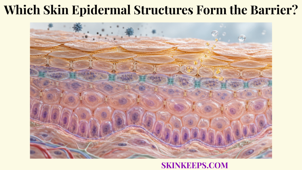
The epidermal barrier is formed primarily by the stratum corneum’s corneocytes and extracellular lipids, reinforced by granular-layer tight junctions and continuously rebuilt through keratinocyte differentiation, lipid secretion, and controlled surface shedding. No single layer, protein, or lipid performs every barrier task.
This anatomy becomes clearer when the epidermis is followed from its proliferative basal layer toward the surface: deeper cells generate replacements, differentiating layers build junctions and barrier components, mature corneocytes acquire nested protein and lipid structures, and the outer surface sheds cells without normally opening persistent gaps.
Which Skin Epidermal Layers Contribute to Barrier Formation?
Skin epidermal barrier formation depends on all major keratinocyte layers, but their roles differ: basal cells generate replacements, spinous cells strengthen cohesion and begin differentiation, granular cells prepare junctional and lipid structures, and the stratum corneum provides the main external permeability barrier. This structural hierarchy is one part of broader skin barrier protection and sits within the wider system of skin functions.
How Does the Stratum Corneum Form the Main External Permeability Barrier?
The stratum corneum forms the main external permeability barrier because flattened protein-rich corneocytes are embedded within highly ordered extracellular lipid lamellae that strongly resist passive molecular and water movement. Its architecture limits both outside-to-inside penetration and inside-to-outside water escape rather than functioning as an absolute, impermeable wall.
The dedicated stratum corneum barrier therefore reflects a two-component structural idea: resistant corneocytes provide the cellular framework, while intercellular lipids provide much of the low-permeability pathway around those cells.
How Does the Stratum Granulosum Prepare Cells and Lipids for Barrier Formation?
The stratum granulosum prepares the outer barrier by forming tight junctions, organizing terminal-differentiation proteins, and releasing lamellar-body contents that are processed into the extracellular lipid matrix of the stratum corneum. Granular keratinocytes contain keratohyalin granules, profilaggrin, developing keratin networks, and abundant lamellar bodies.
Functional tight junctions are concentrated in the upper granular layer, where they restrict paracellular passage between living keratinocytes. This deeper junctional seal reinforces barrier control beneath the stratum corneum rather than replacing it as the principal outer barrier.
How Do Deeper Viable Epidermal Layers Supply New Barrier Cells?
Deeper epidermal layers maintain barrier renewal because proliferative basal keratinocytes produce daughter cells that move outward through spinous and granular differentiation before becoming surface corneocytes. The stratum spinosum contributes strong desmosomal cohesion and advancing keratin differentiation, while the basal layer contributes renewal more than direct surface permeability resistance.
Most body sites are described with four major strata—basale, spinosum, granulosum, and corneum. A recognizable stratum lucidum is characteristic of thick skin on palms and soles and should not be treated as a universal layer.
| Epidermal layer | Main structures | Primary barrier contribution |
|---|---|---|
| Stratum basale | Proliferative keratinocytes, basement-membrane attachments | Generates new keratinocytes for continual renewal |
| Stratum spinosum | Differentiating keratinocytes, abundant desmosomes, keratin network | Maintains mechanical cohesion and advances barrier differentiation |
| Stratum granulosum | Keratohyalin granules, tight junctions, lamellar bodies | Provides deeper paracellular sealing and prepares proteins/lipids for cornification |
| Stratum lucidum | Compact transitional cornified layer in thick skin | Adds a structural layer in palms and soles; not universal |
| Stratum corneum | Corneocytes, envelopes, corneodesmosomes, lipid lamellae | Provides principal outer permeability and mechanical barrier |
Stratum lucidum is characteristic of thick skin and should not be presented as a universal epidermal layer.
How Does the Stratum Corneum Form the Skin’s Main Epidermal Barrier?
The stratum corneum forms the skin’s main epidermal barrier through repeated layers of terminally differentiated corneocytes reinforced by protein envelopes and surrounded by organized extracellular lipids. This composite construction gives the outer epidermis both mechanical durability and unusually low permeability.
How Are Flattened Corneocytes Arranged Into Protective Layers?
Corneocytes are flattened terminally differentiated cells arranged in overlapping layers roughly parallel to the skin surface, creating a mechanically resistant cellular framework through which penetrating substances must pass or move around. Their number and thickness vary by body site, friction, thick-versus-thin skin, and physiological state, so a single universal layer count is not appropriate.
Corneocytes are anucleate and lack ordinary cytoplasmic organelles, yet they are not empty debris. They are highly specialized terminal structures packed with organized keratin-rich material, envelopes, natural moisturizing components, and adhesion machinery appropriate for barrier function.
How Does Keratin Strengthen Mature Corneocytes?
Dense keratin intermediate-filament networks strengthen mature corneocytes by providing intracellular mechanical resilience within the cross-linked cornified architecture. During differentiation, filaggrin helps aggregate keratin filaments before later processing contributes to the biochemical environment of the stratum corneum.
Keratin therefore reinforces the cellular “brick,” but it is not the extracellular permeability seal. Much of the low-permeability pathway lies in the ordered lipids between corneocytes.
How Does Stratum-Corneum Organization Restrict Irritant, Chemical, and Microbial Penetration?
Stratum-corneum organization restricts penetration because potential external agents encounter both resistant corneocyte structures and a tortuous extracellular lipid pathway before reaching viable epidermal tissue. Barrier effectiveness still depends on the molecule, dose, duration and route of exposure, and the condition of the skin.
That architecture also contributes strongly to the skin water barrier by restricting passive water movement outward. It reduces penetration rather than completely excluding every chemical, allergen, or microorganism.
How Do Corneocytes and Epidermal Lipids Build the Barrier Matrix?
Corneocytes and epidermal lipids build the barrier matrix through complementary architecture: protein-rich corneocytes provide the structural units, while specialized envelopes and extracellular lipid lamellae create mechanical reinforcement and low permeability around them. The familiar brick-and-mortar analogy is useful only when its missing layers are made explicit.
How Do Corneocytes Act as the Structural “Bricks” of the Barrier?
Corneocytes act as structural “bricks” because terminal differentiation converts keratinocytes into flattened, mechanically resistant cells packed with keratin and surrounded by a cross-linked protein shell. The analogy describes their position in a layered wall, not their full molecular complexity.
How Do Ceramides, Cholesterol, and Fatty Acids Form the Intercellular “Mortar”?
Ceramides, cholesterol, and free fatty acids form organized extracellular lamellae between corneocytes that provide much of the stratum corneum’s resistance to water diffusion and penetration by external compounds. This extracellular system is the structural core of skin lipids and chemical barrier function.
These lipid classes work as a coordinated matrix rather than a “ceramides alone” barrier. Lipid subclasses, chain lengths, packing, processing, and lamellar organization all influence how effectively the extracellular pathway restricts molecular movement.
How Does the Cornified Envelope Strengthen Each Corneocyte?
The cornified envelope strengthens each corneocyte through a densely cross-linked protein shell containing proteins such as involucrin, loricrin, and small proline-rich proteins. Transglutaminase-dependent cross-linking creates a tough scaffold beneath the former plasma membrane and mechanically reinforces the mature cell.
The cornified envelope is therefore a protein-rich shell. It should not be confused with the lipid layer that coats its outside surface.
How Does the Corneocyte Lipid Envelope Connect Corneocytes With Surrounding Lipid Layers?
The corneocyte lipid envelope creates a covalently bound hydrophobic surface around the cornified envelope that helps anchor and organize the extracellular lipid lamellae surrounding each corneocyte. ω-hydroxyceramides are important components of this specialized lipid interface.
Three structures must remain separate: the cornified envelope is the cross-linked protein shell, the corneocyte lipid envelope is the covalently attached lipid layer outside it, and the intercellular lipid lamellae are free extracellular lipid sheets between neighboring corneocytes.
How Do Epidermal Cell Junctions Strengthen Barrier Integrity?
Epidermal cell junctions strengthen barrier integrity by sealing selected paracellular spaces and mechanically linking neighboring keratinocytes or corneocytes so that the multilayered epidermis remains cohesive under environmental stress. Tight junctions, desmosomes, and corneodesmosomes solve different structural problems and should not be treated as interchangeable seals.
How Do Tight Junctions Restrict Movement Between Cells in the Granular Layer?
Tight junctions in the upper granular epidermis restrict paracellular movement between neighboring keratinocytes, creating a deeper permeability seal beneath the stratum corneum. Claudin proteins, occludin, zonula-occludens proteins, and junctional adhesion molecules participate in the junctional complex.
Claudin-1 is expressed across broader epidermal regions, while complete functional sealing is concentrated in upper granular keratinocytes. Tight junctions therefore reinforce inside-out and outside-in control at a deeper level rather than serving as the main outer permeability barrier.
How Do Desmosomes Maintain Cohesion Between Viable Keratinocytes?
Desmosomes maintain epidermal mechanical cohesion by anchoring neighboring viable keratinocytes to one another and coupling those cell-cell contacts to intracellular keratin filament networks. Desmogleins and desmocollins are key desmosomal cadherins within this mechanically strong adhesion system.
Their primary contribution is resistance to mechanical stress, especially within the spinous layer. Desmosomes should not be described mainly as permeability seals.
How Do Corneodesmosomes Hold Corneocytes Together Before Controlled Shedding?
Corneodesmosomes preserve stratum-corneum cohesion by linking neighboring corneocytes until controlled proteolytic degradation near the surface allows those cells to be shed during normal desquamation. Corneodesmosin is an important component of this modified adhesive system.
Healthy barrier architecture therefore requires adhesion that is strong enough to maintain cohesion but releasable enough to permit renewal. Excessively rapid breakdown or excessive persistence can both disturb normal surface homeostasis.
| Structure | Main location | Structural type | Primary barrier function |
|---|---|---|---|
| Corneocyte | Stratum corneum | Terminal differentiated cell | Mechanical cellular framework |
| Cornified envelope | Around corneocyte interior | Cross-linked protein shell | Cell reinforcement |
| Corneocyte lipid envelope | Outside cornified envelope | Covalently bound lipid layer | Hydrophobic interface and lipid organization |
| Extracellular lipid lamellae | Between corneocytes | Organized lipid matrix | Main low-permeability pathway |
| Corneodesmosome | Between corneocytes | Adhesion complex | SC cohesion and controlled desquamation |
| Tight junction | Upper stratum granulosum | Sealing junction | Deeper paracellular permeability control |
| Desmosome | Viable epidermis; prominent in spinosum | Mechanical adhesion junction | Keratinocyte cohesion |
| Lamellar body | Differentiating granular keratinocyte | Secretory organelle | Supplies lipid precursors and processing material |
| Basal keratinocyte | Stratum basale | Proliferative epidermal cell | Continual barrier-cell renewal |
How Do Epidermal Renewal Structures Maintain the Skin Barrier?
Epidermal renewal maintains the barrier through continuous basal-cell proliferation, ordered keratinocyte differentiation, lamellar-body secretion, cornification, and controlled desquamation that replaces surface material without leaving the skin persistently exposed. A functioning barrier is therefore a dynamic production-and-loss system rather than a static wall.
How Do Basal Keratinocytes Generate New Epidermal Cells?
Basal keratinocytes divide to maintain the epidermal cell population, while selected daughter cells leave the proliferative compartment and begin upward differentiation toward the skin surface. Their contribution to keratinocyte barrier function is renewal: they supply the cells that will later become specialized corneocytes.
Turnover speed varies with anatomical site, age, disease, and measurement method, so a single universal epidermal-turnover time should not be presented as a fixed biological constant.
How Does Keratinocyte Differentiation Produce Mature Corneocytes?
Keratinocyte differentiation progressively changes cell shape, keratin expression, protein cross-linking, lipid metabolism, organelle content, and junctional structures until granular cells undergo cornification and become terminal corneocytes. The sequence moves from basal proliferation through spinous and granular differentiation into a specialized cornified state.
Cornification is not ordinary apoptosis. It is a specialized terminal-differentiation program that builds the structures required for the outer barrier while removing the nucleus and conventional organelles.
How Do Lamellar Bodies Deliver Lipids Needed for the Surface Barrier?
Lamellar bodies accumulate lipid precursors and processing components in differentiated keratinocytes and release them near the granular–cornified interface, where extracellular enzymatic processing generates mature barrier lipids. Their cargo can include glucosylceramides, phospholipids, cholesterol, lipid-processing enzymes, proteases, protease inhibitors, and other barrier-associated molecules.
The organelles themselves do not become the extracellular lipid barrier. Their contents are secreted and processed into the organized lipid system surrounding corneocytes.
How Does Controlled Desquamation Remove Old Corneocytes Without Destroying Barrier Continuity?
Controlled desquamation removes only the most superficial corneocytes as corneodesmosomes are progressively degraded, while continual production and differentiation from deeper layers maintains overall stratum-corneum continuity. Normal shedding is therefore a regulated homeostatic event rather than simple uncontrolled loss of barrier cells.
What Happens When Skin Epidermal Barrier Structures Become Disrupted?
When skin epidermal barrier structures become disrupted, permeability resistance and mechanical cohesion can fall, allowing greater water escape and easier interaction between viable skin and external irritants, allergens, chemicals, or microorganisms. Structural failure can originate in lipids, differentiation, cohesion, junctions, or more than one system at the same time.
What Happens When Intercellular Lipid Organization Is Depleted or Disturbed?
Disturbed extracellular lipid composition or lamellar organization increases the permeability of the stratum corneum because the normally ordered low-permeability pathway between corneocytes becomes less effective. This is one structural route to skin barrier disruption, but it should not be reduced to “low ceramides” alone.
Cholesterol, free fatty acids, ceramide subclasses, chain length, processing enzymes, and lipid phase organization all contribute to barrier architecture. Different defects can therefore produce similar functional consequences through different mechanisms.
How Does Loss of Corneocyte Cohesion Weaken the Barrier?
Loss of normal corneocyte cohesion can create abnormal scaling, fissuring, or structural gaps when corneodesmosome regulation or cornified architecture no longer maintains orderly attachment between surface cells. Excessive cohesion can also disturb normal desquamation, so both insufficient and excessive adhesion can impair surface homeostasis.
How Can Junction Disruption Increase Epidermal Permeability?
Disruption of granular-layer tight junctions can increase paracellular permeability beneath the stratum corneum and weaken the coordinated inside-out and outside-in epidermal barrier. Junctional changes can be primary, secondary, inflammation-associated, or disease-specific; they should not be assumed to explain every skin disorder.
Why Can Structural Barrier Damage Increase Water Loss, Irritation, and External Exposure?
Structural barrier damage can increase transepidermal water loss and external-substance penetration because disrupted lipid, cellular, or junctional pathways provide less resistance to molecular movement across the epidermis. Dryness, stinging, scaling, redness, or sensitivity do not by themselves identify which structure is defective.
What Are the Key Takeaways About the Skin’s Epidermal Barrier Architecture?
The key fact about the skin’s epidermal barrier architecture is that no single structure forms the barrier alone: corneocytes, protein and lipid envelopes, extracellular lipids, cell junctions, and continuously differentiating keratinocytes function as one renewing anatomical system.
- Stratum corneum: Provides the principal external permeability barrier.
- Corneocytes: Form the strong cellular framework of the outer barrier.
- Keratin: Provides intracellular mechanical resilience.
- Cornified envelope: Creates a highly cross-linked protein shell around mature corneocytes.
- Corneocyte lipid envelope: Creates a hydrophobic interface linking the corneocyte surface to surrounding lipid organization.
- Extracellular lipid lamellae: Ceramides, cholesterol, and free fatty acids create much of the low-permeability pathway.
- Tight junctions: Add deeper paracellular control in the upper stratum granulosum.
- Desmosomes: Maintain mechanical cohesion between viable keratinocytes.
- Corneodesmosomes: Maintain corneocyte cohesion until controlled surface shedding occurs.
- Lamellar bodies: Deliver lipid precursors and processing material needed to construct the extracellular barrier matrix.
- Basal keratinocytes: Continually replenish cells lost from the surface.
- Differentiation: Converts living keratinocytes into highly specialized barrier corneocytes.
- Desquamation: Removes old surface cells in a regulated manner without normally disrupting overall continuity.
- Barrier disruption: Structural defects can increase permeability, TEWL, and exposure of viable epidermis to external agents.
What Common Questions Do People Ask About Skin Epidermal Barrier Structures?
Common questions about skin epidermal barrier structures focus on whether the stratum corneum is the only barrier, what the brick-and-mortar model means, and how envelopes, lipids, and junctions differ.
Is the Stratum Corneum the Only Epidermal Barrier?
No. The stratum corneum provides the main outer permeability barrier, while functional tight junctions in the upper stratum granulosum add a deeper paracellular barrier beneath it.
What Are the “Bricks” and “Mortar” in the Skin Barrier?
Corneocytes are commonly described as the structural “bricks,” while organized extracellular lipids made mainly from ceramides, cholesterol, and free fatty acids form the “mortar.” The analogy is useful, but it leaves out envelopes, junctions, corneodesmosomes, enzymes, and renewal.
What Is the Difference Between the Cornified Envelope and the Corneocyte Lipid Envelope?
The cornified envelope is a cross-linked protein shell that reinforces the corneocyte. The corneocyte lipid envelope is a covalently attached hydrophobic lipid layer outside that shell that interfaces with surrounding extracellular lipids.
What Do Tight Junctions Do in the Epidermis?
Tight junctions in the upper granular layer restrict movement through spaces between living keratinocytes, adding deeper paracellular permeability control beneath the stratum corneum.
How Does the Epidermal Barrier Replace Cells Without Opening Gaps?
Basal keratinocytes continually generate new cells, differentiation supplies new corneocytes from below, and controlled desquamation removes only superficial corneocytes, so renewal and surface loss remain balanced.
Epidermal Tight Junctions in Health and Disease: dual epidermal barrier architecture, upper-granular tight-junction localization, claudins, occludin, and paracellular sealing.
Mammalian Epidermis: A Compendium of Lipid Functionality: corneocytes, corneocyte lipid envelope, ceramide/cholesterol/free-fatty-acid matrix, lamellar organization, and epidermal lipid biology.
Five Functional Aspects of the Epidermal Barrier: keratinization, desmosomes, corneodesmosomes, tight junctions, stratum-corneum architecture, and the limits of simplified barrier models.
An Update of the Defensive Barrier Function of Skin: stratum-corneum permeability function, lamellar bodies, extracellular lipid processing, barrier homeostasis, and barrier disruption.
The Role of Tight Junctions in Atopic Dermatitis: A Systematic Review: tight-junction anatomy, claudin/occludin/ZO components, and the relevance of junctional disruption to epidermal permeability.
Cellular Mechanisms of Skin Repair in Humans and Other Mammals: normal human epidermal stratification, basal proliferation, spinous/granular differentiation, thick-skin stratum lucidum, and corneocyte formation.
Cytokines and the Skin Barrier: basal-to-corneocyte differentiation, cornification, cornified-envelope formation, lipid-envelope development, and desmosome-to-corneodesmosome transition.
Epidermal Lamellar Bodies, Essential Organelles for the Skin Barrier: lamellar-body cargo, extracellular lipid delivery, corneocyte cohesion, desquamation control, and epidermal barrier homeostasis.
This page explains normal epidermal anatomy and does not diagnose a “damaged barrier” or skin disease from dryness, redness, scaling, sensitivity, or itching. Persistent severe scaling, cracking, inflammation, blistering, recurrent infection, or rapidly worsening skin changes should be assessed by a qualified healthcare professional.



