
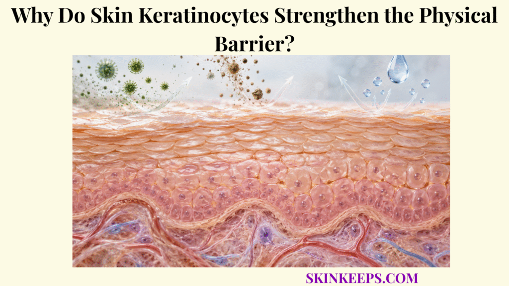
Skin keratinocytes strengthen the physical barrier by undergoing a tightly regulated differentiation program that builds resilient keratin networks, cross-linked cornified envelopes, lipid-associated corneocytes, and the extracellular lipid matrix required for stratum-corneum integrity.
Keratinocytes are not merely cells positioned beneath the barrier; they manufacture most of its structural components. Basal-cell renewal, keratin remodeling, envelope construction, lipid secretion, cornification, and controlled desquamation continuously rebuild a surface that must remain mechanically strong and selectively low in permeability.
What Role Do Skin Keratinocytes Play in Building the Physical Barrier?
Skin keratinocytes build the physical barrier by dividing in the basal epidermis and then undergoing progressive differentiation as daughter cells move through the spinous and granular layers toward the stratum corneum. This cell-to-surface sequence places keratinocytes at the center of the broader skin barrier protection system.
Where Do Skin Keratinocytes Originate Within the Epidermis?
Barrier-forming keratinocytes arise from proliferative cells in the stratum basale, where cell division maintains the epidermal population and supplies daughter cells for outward differentiation. The basal cells remain attached to the basement-membrane zone while selected daughters leave the proliferative compartment, a spatial relationship described in the wider map of skin epidermal barrier structures.
How Do Skin Keratinocytes Change as They Move Toward the Surface?
As keratinocytes move outward, they progressively stop proliferating and change their keratin expression, protein composition, lipid metabolism, junctions, cell shape, and organelles to prepare for cornification. Basal cells are dominated by K5/K14, suprabasal differentiation brings K1/K10 to prominence, and granular cells accumulate keratohyalin proteins, lamellar bodies, and the machinery needed for envelope formation.
How Does Skin Keratinocyte Differentiation Create the Protective Outer Layers?
Keratinocyte differentiation creates the protective outer layers by converting living epidermal cells into flattened corneocytes reinforced by protein envelopes and embedded within a hydrophobic extracellular lipid matrix. The resulting cells become the cellular framework of the stratum corneum barrier, but their final barrier properties depend on both intracellular structure and the lipids organized around them.
| Differentiation stage | Characteristic keratinocyte change | Major barrier output |
|---|---|---|
| Stratum basale | Proliferation; K5/K14 network | Supplies replacement cells and basal mechanical resilience |
| Stratum spinosum | K1/K10 expression; enlarged keratin network; strong desmosomal cohesion | Increases structural resistance and advances differentiation |
| Stratum granulosum | Profilaggrin/loricrin expression; transglutaminases; abundant lamellar bodies | Prepares cornified protein shell and permeability lipids |
| Cornification | Organelle removal, keratin compaction, protein cross-linking | Produces resistant corneocyte |
| Stratum corneum | Corneocytes embedded in extracellular lipid lamellae | Creates final mechanical and permeability barrier |
Barrier formation is a continuous differentiation program rather than one isolated step at the skin surface.
How Do Skin Keratinocytes Produce Keratin for Barrier Strength?
Skin keratinocytes strengthen the epidermis by assembling intermediate-filament networks whose keratin composition changes during differentiation and whose mechanical resilience allows epidermal cells to withstand deformation and friction. Keratin therefore reinforces cells mechanically, but it is not the extracellular material that seals the spaces between surface corneocytes.
How Do Basal K5 and K14 Strengthen Skin Keratinocytes?
Basal keratinocytes predominantly pair K5 and K14 into intermediate-filament networks that distribute mechanical stress through the cytoplasm and connect structurally with epidermal adhesion systems. Human disorders caused by severe K5/K14 disruption demonstrate how loss of this architecture can make basal keratinocytes fragile under frictional stress, but mechanical fragility should not be equated automatically with the same degree of permeability failure.
Why Do Differentiating Skin Keratinocytes Switch Toward K1 and K10?
As keratinocytes leave the basal layer, expression shifts toward suprabasal keratins such as K1 and K10 as part of the broader differentiation program that produces increasingly specialized barrier cells. The shift is not simply “more keratin”; it reflects a change in cytoskeletal identity, cell architecture, and the type of mechanical work required in suprabasal layers.
How Does Keratin Organization Prepare Skin Keratinocytes to Become Corneocytes?
In upper epidermal layers, keratin filaments become increasingly organized and are bundled in part through filaggrin-associated processes as granular keratinocytes transform into mechanically resistant corneocytes. Profilaggrin stored in keratohyalin granules is processed to filaggrin, which promotes keratin-filament aggregation during terminal differentiation; its later contribution to natural moisturizing factor is a separate downstream topic.
How Do Skin Keratinocytes Form the Cornified Envelope?
Skin keratinocytes form the cornified envelope by producing specialized structural proteins and covalently cross-linking them during terminal differentiation into a tough, insoluble shell around the future corneocyte. This process converts a living membrane-bound cell into a structure able to tolerate repeated surface stress.
Which Proteins Do Differentiating Skin Keratinocytes Use to Build the Cornified Envelope?
Differentiating keratinocytes synthesize envelope components including involucrin, loricrin, and small proline-rich proteins that become progressively incorporated into the developing cornified envelope. No single protein forms the entire envelope; it is a layered, multi-protein structure assembled in a defined sequence.
How Do Transglutaminases Cross-Link the Cornified Envelope?
Keratinocyte transglutaminases—especially transglutaminase 1—create highly stable covalent cross-links between envelope proteins, producing the insoluble protein shell characteristic of mature corneocytes. Cross-linking here means chemically joining structural proteins into a resistant network rather than merely packing them closer together.
How Does the Cornified Envelope Improve Resistance to Friction and External Stress?
The cornified envelope improves physical resistance by surrounding the keratin-rich corneocyte with a cross-linked protein shell that distributes and resists mechanical deformation at the skin surface. It provides mechanical reinforcement but does not alone create the entire low-permeability lipid seal.
Outside this protein-rich shell, specialized keratinocyte-derived lipids form the corneocyte lipid envelope, a distinct hydrophobic interface that helps organize the extracellular lipid lamellae. The cornified protein envelope, corneocyte lipid envelope, and free extracellular lipid matrix should be treated as separate structures.
How Do Skin Keratinocytes Support the Lipid Barrier Between Cells?
Skin keratinocytes build the extracellular permeability barrier by synthesizing lipid precursors, packaging them with processing enzymes inside lamellar bodies, and releasing those materials at the granular–cornified interface where they are converted into organized barrier lipids. This means keratinocytes manufacture the future cellular “brick” and the lipid “mortar” in parallel.
How Do Skin Keratinocytes Form and Load Lamellar Bodies?
Differentiating keratinocytes form lamellar bodies containing lipid precursors, cholesterol, and enzymes required to generate and organize the mature stratum-corneum lipid matrix. Their cargo can include glucosylceramides, sphingomyelin, phospholipids, cholesterol, and lipid-processing enzymes, but the organelles themselves do not become the final lipid lamellae.
How Do Lamellar Bodies Release Keratinocyte-Derived Barrier Material?
Upper granular keratinocytes release lamellar-body contents at the interface with the stratum corneum, placing lipid constituents, precursors, and processing enzymes into the extracellular space where the mature barrier matrix forms. Extracellular processing then converts precursor pools into the lipid species needed for the final surface organization.
How Do Ceramides, Cholesterol, and Fatty Acids Organize Around Corneocytes?
After secretion and extracellular processing, ceramides, cholesterol, and free fatty acids organize into densely packed lamellar structures between corneocytes, creating much of the stratum corneum’s resistance to water movement and penetration by external compounds. These are the central components discussed in skin barrier lipids, while the organization between cells is treated in greater depth under skin intercellular lipids.
This keratinocyte-derived lipid matrix also underpins the skin water barrier, because ordered extracellular lipids restrict both outside-to-inside penetration and inside-to-outside water movement.
How Do Skin Keratinocytes Maintain the Physical Barrier Through Renewal?
Skin keratinocytes maintain the physical barrier through continuous renewal in which basal cells generate replacements, differentiating cells rebuild barrier structures, new corneocytes enter the stratum corneum, and controlled desquamation removes older surface cells. The epidermis therefore renews the barrier without normally exposing a persistent gap to the environment.
How Do Basal Skin Keratinocytes Continually Supply Replacement Cells?
Basal keratinocyte proliferation maintains a reservoir of epidermal cells while selected daughter cells leave the basal compartment and enter the differentiation pathway. There is no single universal turnover duration because renewal rates vary by body site, age, inflammation, disease, and measurement method.
How Does Upward Skin Keratinocyte Differentiation Replace the Surface Barrier?
As replacement keratinocytes progress outward, they reproduce the keratin networks, cornified proteins, lamellar-body contents, and other structures needed to replace barrier material continuously lost from the surface. New barrier material is therefore generated before older surface material is shed.
How Does Corneocyte Formation Maintain Surface Protection?
Terminal cornification continually adds newly formed corneocytes to the lower stratum corneum, preserving the layered cellular framework as older corneocytes move toward the surface. Cornification is a specialized terminal-differentiation program involving organelle loss, protein cross-linking, keratin reorganization, and envelope formation rather than ordinary apoptosis.
How Does Controlled Shedding Preserve Barrier Continuity?
Controlled desquamation progressively weakens corneodesmosomal adhesion only in superficial stratum-corneum layers, allowing old corneocytes to detach while newly formed cells below preserve overall barrier continuity. Normal shedding is therefore a maintenance mechanism, not a form of barrier breakdown.
What Happens When Skin Keratinocyte Barrier Functions Are Disrupted?
When skin keratinocyte barrier functions are disrupted, the result depends on which part of differentiation fails: cytoskeletal defects can increase mechanical fragility, defective cornification can weaken corneocytes, lipid-processing defects can increase permeability, and abnormal cohesion can disturb surface integrity. The mechanistic patterns below are part of the wider biology of skin barrier disruption, not diagnoses by themselves.
How Can Abnormal Skin Keratinocyte Differentiation Weaken the Stratum Corneum?
Abnormal differentiation can weaken the stratum corneum when keratinocytes fail to generate properly organized corneocytes, differentiation proteins, envelopes, or surface architecture. Dryness, scaling, or cracking cannot identify the molecular defect because many inflammatory, genetic, environmental, and mechanical conditions can alter epidermal differentiation.
How Can Keratin Defects Reduce Mechanical Barrier Strength?
Keratin defects can reduce mechanical barrier strength by weakening intermediate-filament networks, making keratinocytes less resistant to friction and deformation. K5/K14 defects provide strong human evidence for this form of mechanical fragility, but a weak cytoskeleton does not automatically predict an equivalent increase in transepidermal water loss.
How Can Defective Keratinocyte Lipid Processing Increase Permeability?
Defective keratinocyte lipid synthesis, lamellar-body transport, secretion, or extracellular lipid processing can weaken the permeability barrier by disrupting the composition and organization of extracellular ceramide-, cholesterol-, and fatty-acid-rich lamellae. Because the lipid matrix is the main continuous diffusion pathway through the stratum corneum, changes in its organization can alter both water retention and substance penetration.
How Can Poor Keratinocyte Cohesion Weaken Barrier Integrity?
Abnormal cell adhesion or corneodesmosomal regulation can weaken barrier integrity by allowing inappropriate separation of epidermal cells or premature corneocyte shedding. The opposite problem can also occur: excessive persistence of corneocyte cohesion can impair normal desquamation, so more adhesion is not automatically better.
Why Can Keratinocyte Barrier Failure Increase Water Loss and External Exposure?
Keratinocyte-derived barrier failure can increase transepidermal water loss and external-substance penetration when cornified architecture or extracellular lipid organization no longer provides normal resistance to molecular movement. Dry or stinging skin alone does not prove keratinocyte dysfunction because those symptoms are nonspecific.
| Keratinocyte function | Normal barrier role | Effect if disrupted |
|---|---|---|
| Basal proliferation | Supplies replacement epidermal cells | Poor renewal or abnormal epidermal architecture |
| K5/K14 filament network | Mechanical resilience of basal cells | Greater cell fragility under mechanical stress |
| K1/K10 suprabasal differentiation | Builds specialized suprabasal cytoskeleton | Abnormal differentiation and mechanical architecture |
| Filaggrin-mediated keratin organization | Compacts keratin network during cornification | Altered corneocyte structure and hydration biology |
| Cornified-envelope formation | Reinforces mature corneocytes | Structurally weaker or abnormal cornification |
| Corneocyte lipid-envelope formation | Links corneocyte surface with surrounding lipid architecture | Impaired corneocyte–lipid interface |
| Lamellar-body secretion | Delivers lipid precursors and enzymes | Defective extracellular lipid formation |
| Ceramide/cholesterol/FFA organization | Produces low-permeability extracellular matrix | Increased water and substance permeability |
| Corneodesmosomal regulation | Balances cohesion with shedding | Premature separation or abnormal retention of corneocytes |
| Coordinated differentiation | Integrates all barrier outputs | Mechanical and/or permeability failure |
Keratinocyte dysfunction is not one single barrier defect. Different molecular failures can predominantly affect mechanical resilience, permeability, adhesion, differentiation, or several systems at once.
What Are the Key Takeaways About Skin Keratinocytes and Physical Barrier Strength?
The key fact about skin keratinocytes and physical barrier strength is that keratinocytes construct the barrier through coordinated differentiation: they build resilient cytoskeletons, reinforce future corneocytes with protein envelopes, generate the lipids that seal intercellular spaces, and continually replace the barrier as surface cells are shed.
- Basal skin keratinocytes continually generate the cells needed for epidermal renewal.
- K5/K14 strengthen the intermediate-filament network of basal keratinocytes.
- K1/K10 become prominent as keratinocytes enter suprabasal differentiation.
- Keratin filaments provide mechanical resilience but are not the extracellular permeability seal.
- Filaggrin-related processing helps organize keratin bundles during terminal differentiation.
- Involucrin, loricrin, and other proteins contribute to cornified-envelope construction.
- Transglutaminases covalently cross-link envelope proteins into a resistant shell.
- The cornified envelope reinforces the future corneocyte mechanically.
- The corneocyte lipid envelope is a distinct outer lipid-associated structure and is not the protein envelope.
- Lamellar bodies carry lipid precursors and processing enzymes toward the stratum-corneum interface.
- Ceramides, cholesterol, and free fatty acids form much of the final extracellular permeability matrix.
- Cornification converts differentiated keratinocytes into specialized barrier corneocytes.
- Controlled desquamation removes superficial corneocytes while deeper replacement cells preserve continuity.
- Keratinocyte defects can affect barrier functions differently—mechanical strength, permeability, cohesion, renewal, or several systems at once.
What Common Questions Do People Ask About Skin Keratinocytes and the Physical Barrier?
Common questions about skin keratinocytes focus on whether keratin alone makes the barrier, how keratinocytes become corneocytes, what lamellar bodies do, and why abnormal differentiation can weaken skin.
Are Skin Keratinocytes the Same as Corneocytes?
No. Keratinocytes are living epidermal cells at earlier differentiation stages, while corneocytes are their terminally differentiated, flattened surface descendants within the stratum corneum.
Does Keratin Alone Create the Skin’s Physical Barrier?
No. Keratin provides important intracellular mechanical strength, but the complete physical barrier also depends on cornified envelopes, corneocyte cohesion, extracellular lipids, and continual keratinocyte renewal.
Why Do Skin Keratinocytes Change Which Keratins They Produce?
Keratin expression changes with differentiation because basal and suprabasal epidermal cells require different cytoskeletal programs; basal cells predominantly express K5/K14, while differentiating suprabasal cells shift toward proteins including K1/K10.
What Do Lamellar Bodies Do in Skin Keratinocytes?
Lamellar bodies transport lipid constituents, precursors, processing enzymes, and other barrier-associated material toward the granular–cornified interface, where their contents contribute to formation of the extracellular permeability barrier.
Can Abnormal Skin Keratinocyte Differentiation Weaken Both Strength and Water Control?
Yes. Depending on the affected process, abnormal differentiation can weaken cellular mechanical resilience, cornified structure, lipid-barrier organization, cohesion, or several systems together, potentially altering both physical strength and permeability.
Epidermal Differentiation in Barrier Maintenance and Wound Healing: K5/K14 and K1/K10 expression, filaggrin, cornified-envelope assembly, transglutaminase cross-linking, lamellar-body lipids, and conversion to corneocytes.
Dynamics and Epigenetics of the Epidermal Differentiation Complex: basal-to-granular differentiation, the terminal differentiation program, cornified-envelope gene regulation, and coordinated barrier construction.
Epidermal Lamellar Bodies, Essential Organelles for the Skin Barrier: lamellar-body anatomy, secretory cargo, differentiation-linked production, lipid metabolism enzymes, and barrier homeostasis.
The Skin Barrier: An Extraordinary Interface With an Exceptional Lipid Organization: ceramides, cholesterol, free fatty acids, extracellular lamellae, lipid organization, and permeability control.
Barrier Functions of the Skin: stratum-corneum physical barrier, protein–lipid integration, water homeostasis, and mechanical protection.
The Role of Ceramides in the Disruption of the Cutaneous Permeability Barrier: ceramide diversity, extracellular-lipid organization, and permeability consequences of altered lipid composition.
Epidermolysis Bullosa Simplex: A Paradigm for Disorders of Tissue Fragility: human K5/K14 evidence for basal-keratinocyte mechanical fragility under frictional stress.
An Update of the Defensive Barrier Function of Skin: keratinocyte differentiation, involucrin, loricrin, filaggrin, transglutaminase 1, lamellar-body contribution, and permeability-barrier biology.
This page explains normal keratinocyte and barrier biology and does not diagnose a skin disorder or prescribe treatment. Seek medical evaluation for recurrent unexplained blistering, severe persistent scaling or fissuring, spreading redness, pus, fever, significant skin detachment, or rapidly worsening skin breakdown.



