
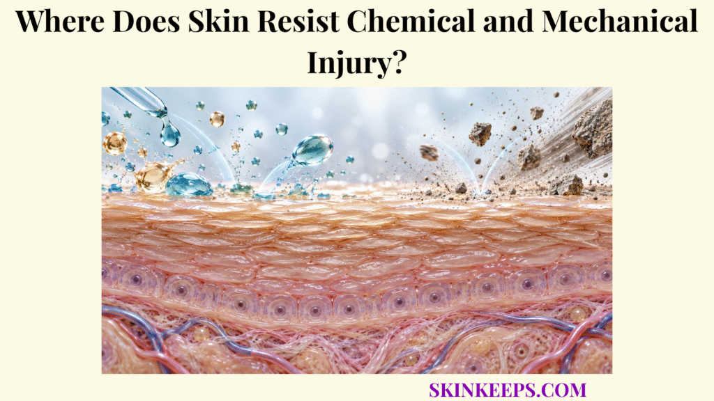
Skin resists chemical and mechanical injury at multiple structural levels: the stratum corneum provides the main barrier to chemical penetration and superficial exposure, while cohesive epidermal cells and the collagen-rich dermis provide progressively deeper resistance to friction, shear, stretch, and deformation.
Chemical permeability, surface mechanics, dermal strength, adaptation, and failure therefore have to be considered separately. No skin barrier provides unlimited resistance, and protection changes with exposure intensity, duration, repetition, hydration, anatomical site, and the condition of the tissue being stressed.
Where Does Skin Resistance Begin Against Injury?
Skin resistance begins at the epidermal surface, especially the stratum corneum, but the structures that resist chemical penetration are not identical to those that bear deeper mechanical loads.
The broad skin barrier protection system combines physical, chemical, microbial, immune, and repair functions. Within that system, skin epidermal barrier structures create the first interface with both external chemicals and mechanical contact, while deeper connective tissues become increasingly important as forces penetrate below the surface.
Where Does Skin Resistance to Chemicals Begin?
Skin resistance to most passive external chemical penetration begins in the stratum corneum, where compact corneocytes and organized extracellular lipids create the principal permeability barrier.
Corneocytes provide a protein-rich structural path, but the extracellular lipid lamellae form the only continuous route through the stratum corneum. Molecules attempting to cross intact skin must repeatedly partition into and diffuse through this heterogeneous corneocyte–lipid architecture. The result is strong resistance to many substances rather than absolute impermeability.
Where Does Skin Resistance to Mechanical Stress Begin?
Mechanical resistance begins at the cornified surface but extends through cohesive viable epidermis into the collagen-rich dermis, because abrasion, shear, stretch, and tensile loading stress different tissue structures.
The outer cornified layer provides a durable contact surface. Living keratinocytes below it are mechanically coupled through junctional systems and keratin filaments, while the dermis supplies the fibrous extracellular matrix that carries much of whole-skin tensile loading. Mechanical protection is therefore a multilayer property rather than a function of surface thickness alone.
Why Must Chemical and Mechanical Skin Resistance Be Distinguished?
Chemical resistance is governed mainly by permeability and chemical–tissue interactions, whereas mechanical resistance depends on material strength, cohesion, deformation, load direction, magnitude, and repetition.
A substance can cross the skin without immediately producing visible surface injury, and another can injure surface tissue before large amounts penetrate deeply. Mechanical stress likewise can fail in different ways: superficial abrasion, cracking, intraepidermal shear separation, or deeper tensile and pressure-related damage are not the same process.
How Does Skin Resistance Limit Chemical Injury?
Skin resistance limits chemical injury primarily by slowing the partitioning and diffusion of external substances through the stratum corneum, although the effectiveness of that barrier varies substantially with the chemical, exposure conditions, and skin state.
The stratum corneum barrier is usually the principal rate-limiting structure for passive percutaneous transport. Its extracellular ceramide-, cholesterol-, and free-fatty-acid-rich matrix is central to skin lipids and chemical penetration, but neither lipid composition nor layer thickness can be interpreted as a universal chemical-safety threshold.
How Do Corneocytes and Lipids Limit Chemical Penetration?
Corneocytes create a structurally resistant pathway while extracellular ceramide-, cholesterol-, and free-fatty-acid-rich lamellae provide the continuous low-permeability matrix through which many chemicals must diffuse.
The arrangement is often simplified as cellular “bricks” surrounded by a lipid matrix, but real transport is more complex. Molecules may favor lipid-rich or more polar microenvironments differently, and appendageal routes can contribute for selected compounds. Even so, the intact stratum corneum remains the dominant passive barrier for most external chemical exposure.
Which Chemical Properties Affect Skin Penetration?
Chemical penetration varies with molecular size, lipid–water partitioning, ionization, solubility, concentration, and interactions between the chemical, its vehicle, and stratum-corneum components.
Smaller molecules with suitable lipid–water partitioning often penetrate intact skin more readily, but no single molecular-weight threshold defines chemical safety. The frequently cited roughly 500-Da observation comes from drug-delivery literature and is an empirical permeability heuristic, not a boundary above which chemicals become incapable of crossing skin.
Which Exposure Conditions Alter Chemical Penetration?
The amount of chemical crossing skin depends not only on the molecule itself but also on concentration, contact duration, exposed area, temperature, hydration, occlusion, rubbing, anatomical site, and barrier integrity.
Hydration and occlusion can alter stratum-corneum organization and increase transport for some compounds. Damaged skin can shorten or disorganize diffusion pathways, but the size of any increase remains chemical-specific. Volatility, removal, reactivity, and the applied vehicle can also change the effective dose remaining available at the surface.
Why Does the Skin Not Resist Every Chemical Equally?
The skin cannot resist every chemical equally because different substances partition into lipids and proteins differently, diffuse at different rates, and may themselves extract lipids, denature proteins, damage cells, or chemically react with tissue.
Penetration potential and local injury potential are related but not synonymous. A solvent may disrupt barrier lipids and increase permeability; an irritant may damage membranes or proteins; and a strongly corrosive exposure can destroy tissue locally without first requiring extensive systemic absorption. Surfactant effects likewise depend on chemistry, formulation, concentration, exposure time, frequency, and the pre-existing barrier state.
| Barrier / Exposure Factor | Chemical-Defense Role | Limitation |
|---|---|---|
| Corneocytes | Add structural and protein-rich diffusion resistance | Chemicals can still partition into or transit cellular pathways |
| Intercellular lipids | Main continuous low-permeability pathway | Lipophilic chemicals and solvents may interact strongly with the matrix |
| Stratum-corneum thickness | Can lengthen the diffusion path at some sites | Thickness alone does not predict penetration |
| Lipid organization | Restricts molecular diffusion | Selected solvents and surfactants can disturb organization |
| Molecular size | Larger molecules generally diffuse less readily | Not an absolute safety cutoff |
| Lipophilicity / ionization | Influence partitioning into the stratum corneum and deeper aqueous layers | No single property predicts total absorption |
| Concentration | Can increase the driving force for transport | Local injury may occur before deep penetration |
| Contact duration | Increases opportunity for absorption | Effect depends on volatility, removal, reaction, and dose |
| Occlusion / hydration | Can increase permeability | Magnitude varies by compound and skin site |
| Barrier damage | Removes or disorganizes resistance | Increase in penetration is compound-specific |
How Does Skin Resistance Reduce Mechanical Injury?
Skin resistance reduces mechanical injury by combining a keratinized surface, strong cell–cell adhesion, compliant epidermal tissue, and layered architecture that distributes minor frictional and deformational stresses before they produce structural failure.
The epidermis is more than a passive cover. Keratinocyte physical-barrier function depends on differentiation, keratin architecture, and junctional cohesion that allow the tissue to resist and redistribute ordinary deformation without every surface contact becoming a tear.
How Do Keratinized Corneocytes Resist Surface Stress?
Keratin-rich corneocytes resist minor surface stress by providing a tough outer layer whose cornified envelopes and cellular architecture tolerate repeated low-level contact better than living epidermal cells would alone.
The stratum corneum is therefore important for abrasion resistance and minor surface deformation, but it does not carry most whole-skin tensile load. Its role is better understood as a mechanically durable interface that protects living tissue while retaining enough flexibility to move with the body.
How Does Epidermal Cell Cohesion Resist Mechanical Deformation?
Desmosomal adhesion and keratin filament networks mechanically couple neighboring keratinocytes, allowing stresses to be distributed through the epidermal sheet rather than concentrated within isolated cells.
Keratin filaments reinforce the intracellular cytoskeleton, while desmosomes transmit force between cells. This network can deform within limits, but repeated or sufficiently large shear cycles can produce fatigue and separation in the viable epidermis.
How Do Friction and Shear Differ in Skin Injury?
Friction acts at the skin–surface interface, whereas shear deformation occurs within tissue when adjacent layers or underlying structures move relative to one another.
In the current mechanical model of foot friction blisters, a high-friction interface provides traction while underlying bone movement deforms soft tissue. Repetition creates mechanical fatigue and an intraepidermal tear, commonly associated with the stratum spinosum. The blister is therefore not simply the result of an object scraping away the stratum corneum.
Why Does Skin Hydration Affect Mechanical Resistance?
Stratum-corneum hydration changes its flexibility and frictional behavior: excessive dryness can promote cracking, while prolonged overhydration or maceration can soften tissue and reduce mechanical integrity.
A mechanically durable surface requires an appropriate material state rather than maximum dryness or maximum hydration. Dry, inflexible corneum can concentrate strain around cracks, while prolonged wetting can soften the epidermal interface and increase susceptibility to localized damage.
How Do Deeper Skin Structures Strengthen Mechanical Resistance?
Deeper skin structures strengthen mechanical resistance primarily through the collagen-rich dermal extracellular matrix, which bears tensile loads while elastic fibers and ground substance allow controlled deformation and recovery.
The detailed connective-tissue contribution is developed in the child mechanism page on skin keratin and connective-tissue resistance. At overview level, whole-skin strength is best treated as an integrated response in which superficial layers protect against contact while the dermal network increasingly bears extension, tension, and load redistribution.
How Does Dermal Collagen Provide Tensile Strength?
Dermal collagen provides much of skin’s tensile strength because densely organized fibrillar collagen networks progressively recruit under stretch and resist excessive extension.
Type I collagen is the dominant fibrillar collagen in adult dermis, with type III contributing to the network. Collagen fibers are wavy at low load and progressively straighten and align as strain increases. Orientation, cross-linking, and fiber recruitment make skin mechanically nonlinear and anisotropic, so resistance differs with the direction of applied force.
How Does Elastin Contribute to Flexibility and Recoil?
Elastic fibers permit skin to deform under relatively low loads and contribute to recoil toward its previous configuration after the force is removed.
Elastin should not be treated as the sole source of “flexibility.” The mechanical response emerges from elastic fibers interacting with collagen, hydrated matrix molecules, cells, and the geometry of the tissue. Elastic recoil is one component of a larger viscoelastic system.
How Does the Dermal Extracellular Matrix Distribute Mechanical Stress?
The dermal extracellular matrix distributes mechanical stress through an integrated collagen–elastic-fiber network embedded in hydrated proteoglycan-rich ground substance, producing nonlinear and viscoelastic mechanical behavior.
Viscoelasticity means that the response depends partly on time: skin can creep under sustained load and relax stress after deformation. These behaviors help distribute everyday forces, but they also mean that magnitude, rate, direction, and duration of loading all influence whether deformation remains reversible.
| Skin Structure | Dominant Mechanical Property | Protective Role |
|---|---|---|
| Stratum corneum | Surface toughness / abrasion resistance | Protects living epidermis from minor surface stress |
| Keratin filaments | Cellular resilience | Reduce keratinocyte rupture during deformation |
| Desmosomes | Cell–cell cohesion | Transfer forces across epidermal cells |
| Viable epidermis | Layered deformation resistance | Tolerates limited shear and stretch before failure |
| Basement-membrane zone | Epidermal–dermal attachment | Transfers force between major tissue layers |
| Dermal collagen | Tensile strength and stiffness | Resists excessive extension and tearing |
| Elastic fibers | Extensibility and recoil | Contribute to reversible deformation |
| Proteoglycan-rich ground substance | Viscoelastic damping / hydration | Contributes to time-dependent load distribution |
Mechanical properties arise from interaction among layers, not from one isolated protein.
How Does Skin Resistance Adapt to Repeated Stress?
Skin resistance can adapt to repeated non-destructive mechanical stress through increased epidermal production and local hyperkeratosis, but the adaptation is site- and load-dependent and can still be overwhelmed.
This is a local mechanical response, not evidence that skin can be trained to tolerate hazardous chemicals. Repeated irritant exposure has complex and exposure-specific effects and should not be presented as a reliable form of protective “hardening.”
How Does Repeated Friction or Pressure Increase Epidermal Thickness?
Chronic repetitive friction or pressure can stimulate keratinocyte proliferation and cornification, increasing stratum-corneum thickness in repeatedly loaded areas.
This mechanical hyperkeratosis changes the local architecture of the surface so that the same load is applied across a thicker cornified layer. The response can be useful within ordinary physiological loading but varies with site, force pattern, footwear or tools, tissue hydration, and individual skin biology.
How Does Callus Formation Change Local Skin Resistance?
A callus is a localized hyperkeratotic response to chronic mechanical loading that increases surface thickness at frequently stressed sites such as the hands or feet.
A callus can provide useful local protection, yet more callus does not always mean more protection. Excessive hyperkeratosis may increase local stiffness, alter pressure distribution, or fissure when the thickened surface becomes dry or deformed. Callused skin can still blister if internal shear is sufficient.
How Does Epidermal Renewal Replace Mechanically Damaged Surface Cells?
Basal keratinocyte proliferation and differentiation continually replace superficial cells lost or damaged during ordinary mechanical exposure, maintaining barrier continuity when injury remains within regenerative capacity.
Renewal moves newly formed cells outward as they differentiate, ultimately replacing shed corneocytes. This routine turnover is part of barrier maintenance; it is not the same as the inflammatory and reparative response required after a deeper wound.
Why Does Mechanical Adaptation Have Limits?
Mechanical adaptation has limits because sufficiently large, prolonged, or repetitive pressure, shear, or friction can exceed tissue tolerance regardless of surface thickening.
A thickened surface does not eliminate the possibility of blistering, fissuring, abrasion, or deeper pressure-related injury. Adaptation changes local material properties, but it does not remove the finite failure thresholds of living epidermis, dermis, vasculature, and underlying soft tissue.
What Weakens Skin Resistance to Chemical and Mechanical Injury?
Skin resistance weakens when barrier architecture, hydration, cellular cohesion, or dermal tissue integrity is altered, allowing chemical penetration or mechanical deformation to occur more readily.
How Does Barrier Disruption Weaken Chemical Resistance?
Barrier disruption weakens chemical resistance by creating less organized or shorter diffusion pathways through the stratum corneum, which can increase penetration of selected substances.
Disrupted lipids, removed corneocytes, inflammation, erosions, or fissures can all change the effective barrier. The detailed process of skin barrier disruption matters because the magnitude of increased penetration depends on both the type of damage and the chemical being applied.
How Can Dryness Weaken Mechanical Skin Resistance?
Excessive stratum-corneum dryness reduces flexibility and can promote scaling or fissuring, creating structural stress concentrations that make the surface less tolerant of deformation.
The skin water barrier therefore has a mechanical consequence as well as a water-conservation role. At the opposite extreme, prolonged overhydration can soften the corneum and reduce mechanical integrity, so neither maximal dryness nor maximal wetness is a durability goal.
How Can Solvents or Harsh Cleansing Weaken Skin Resistance?
Repeated solvent or harsh-surfactant exposure can weaken resistance by extracting or reorganizing lipids, interacting with epidermal proteins, and repeatedly activating barrier-disruption and inflammatory responses.
These effects are formulation- and exposure-dependent. Acetone, detergent systems, alcohols, oils, and other solvents do not have identical mechanisms or potency, and normal cleansing should not be described as inherently destructive. Dose, frequency, duration, chemistry, and baseline barrier condition all modify the outcome.
How Do Friction, Pressure, and Shear Weaken Skin Resistance?
Excessive friction, pressure, and repeated shear can mechanically fatigue epidermal or deeper tissue structures until deformation exceeds their capacity for reversible recovery.
High friction can increase traction and therefore the amount of internal shear generated during repeated motion. Sustained pressure can deform tissue and, when sufficiently intense or prolonged, compromise deeper structures and perfusion. These mechanisms are distinct even when they occur together.
How Does Inflammation Change Skin Resistance?
Inflammation can weaken skin resistance by altering epidermal differentiation, lipid organization, hydration, cell adhesion, and tissue mechanics, although the exact effect depends on the underlying condition.
Barrier weakness alone cannot identify a particular inflammatory disorder. The same functional consequence can arise from different biological causes, so this physiology should not be used to diagnose irritant dermatitis, allergic dermatitis, psoriasis, eczema, or another condition from appearance or symptoms alone.
What Happens When Skin Resistance Is Overwhelmed?
When skin resistance is overwhelmed, injury reflects the type, intensity, and duration of exposure: chemicals can disrupt or destroy barrier tissue, while mechanical loads can produce fissures, abrasions, intraepidermal blisters, or deeper tissue damage.
What Happens When Chemical Exposure Overwhelms Skin Resistance?
Chemical exposure can overwhelm skin resistance through direct irritant injury, lipid extraction, protein damage, cellular toxicity, or corrosive tissue destruction depending on the substance, concentration, duration, and barrier condition.
Mild or repeated irritation, acute irritant injury, and chemical corrosion should not be collapsed into one mechanism. Some exposures primarily disturb barrier lipids and inflammatory signaling, whereas corrosive substances can cause rapid, deeper tissue destruction. Visible severity may also lag behind the initial exposure for some substances.
What Happens When Shear Overwhelms Epidermal Resistance?
Repeated shear deformation can produce mechanical fatigue and intraepidermal separation, which can fill with fluid to form a friction blister.
For classic foot friction blisters, research places the mechanical tear within the epidermis, often in the stratum spinosum. Friction is important because it provides traction between the external surface and skin, while the repeated internal shear created by relative motion produces the tissue failure.
How Do Abrasions and Fissures Differ From Blisters?
Abrasions involve superficial mechanical tissue loss, fissures are cracks that commonly develop when inflexible or disrupted skin separates under stress, and friction blisters involve intraepidermal shear separation.
These terms describe different structural failures. An abrasion removes tissue from the surface; a fissure opens along a crack; a blister creates a plane of separation within the epidermis. They can coexist in a damaged area, but they should not be used interchangeably.
What Happens When Mechanical Injury Extends Into Deeper Tissue?
When loading exceeds deeper tissue tolerance, damage can extend beyond epidermal failure into dermal structures or, under prolonged pressure and impaired perfusion, involve still deeper tissues.
That deeper injury is outside the scope of a barrier-mechanism overview, but it reinforces the central rule: visible surface resistance does not guarantee protection of the tissues beneath it. The relevant failure mechanism depends on load magnitude, duration, repetition, direction, anatomy, and perfusion.
| Exposure | Primary Stressed System | Possible Failure Pattern |
|---|---|---|
| Repeated mild irritant | Stratum-corneum lipids / proteins | Barrier disruption + inflammation |
| Lipid-extracting solvent | Intercellular lipid matrix | Increased permeability + dryness / irritation |
| Strong corrosive chemical | Epidermis / deeper tissue | Acute tissue destruction |
| Repeated shear | Viable epidermis | Intraepidermal blister |
| Surface abrasion | Stratum corneum / epidermis | Superficial tissue loss |
| Excessive dryness + deformation | Stratum corneum | Fissuring / cracking |
| Chronic pressure / friction | Epidermal adaptation | Hyperkeratosis / callus |
| Excessive or prolonged mechanical load | Epidermis / dermis / deeper tissue | Structural injury beyond adaptive capacity |
The same exposure can produce different outcomes depending on dose, duration, anatomical location, tissue hydration, and barrier condition.
What Are the Key Takeaways About Skin Resistance?
The key fact about skin resistance is that chemical and mechanical protection rely on different but overlapping structures: the stratum corneum controls most passive chemical penetration, while keratinized epidermis, cellular cohesion, and dermal connective tissue progressively resist mechanical deformation.
Chemical protection therefore depends on both the barrier and the substance approaching it, while mechanical protection depends on how multiple tissue layers respond to force. Renewal and hyperkeratotic adaptation can preserve function under ordinary stress, but neither mechanism creates unlimited protection.
- Skin resistance begins at the stratum corneum, but deeper layers contribute increasingly to mechanical protection.
- Chemical resistance is mainly a permeability function of the stratum corneum.
- Corneocytes and extracellular lipids create the main passive chemical diffusion barrier.
- No chemical is resisted equally: molecular properties, concentration, vehicle, duration, site, hydration, and barrier condition matter.
- Chemical penetration and chemical injury are not identical: chemicals can damage surface tissue without extensive systemic absorption.
- Keratinized epidermis provides surface mechanical protection.
- Keratin filaments and cell junctions help living epidermal cells tolerate deformation.
- Friction and shear are different: friction can facilitate traction, while repeated shear deformation can cause intraepidermal blistering.
- Dermal collagen provides much of skin’s tensile strength.
- Elastic fibers contribute extensibility and recoil.
- Skin mechanics are nonlinear and direction-dependent.
- Repeated mechanical loading can cause adaptive hyperkeratosis or callus formation.
- Adaptation has limits: excessive stress can still cause blistering, fissuring, abrasion, or deeper injury.
- Excessive dryness can reduce flexibility, while excessive moisture or maceration can weaken mechanical integrity.
- Barrier disruption can increase chemical penetration and reduce resistance to further exposure.
- No skin barrier provides unlimited chemical or mechanical protection.
What Common Questions Do People Ask About Skin Resistance to Chemical and Mechanical Injury?
Common questions about skin resistance focus on whether the stratum corneum blocks all chemicals, which skin layer provides mechanical strength, why calluses form, and how friction actually causes blisters.
Can the Stratum Corneum Block Every Chemical?
No. The stratum corneum strongly limits passive penetration of many substances, but chemicals differ greatly in molecular properties and reactivity, and penetration also changes with dose, vehicle, exposure time, skin site, hydration, and barrier integrity.
Which Skin Layer Provides the Most Mechanical Strength?
The dermis provides much of whole-skin tensile strength because of its dense collagen-rich extracellular matrix, while the epidermis and stratum corneum provide important surface toughness, cohesion, and resistance to abrasion or shear.
Does Thicker Skin Always Provide Better Chemical Protection?
No. Greater stratum-corneum thickness can lengthen the diffusion pathway, but chemical penetration also depends on lipid organization, molecular properties, exposure conditions, anatomical site, hydration, and whether the chemical itself disrupts the barrier.
Why Does Repeated Friction Create a Callus in Some Areas but a Blister in Others?
Repeated moderate loading can stimulate adaptive hyperkeratosis over time, while sufficiently large or repetitive shear deformation can mechanically fatigue the viable epidermis and cause intraepidermal tearing before protective adaptation is sufficient.
Can Skin Become Fully Resistant to Repeated Chemicals or Mechanical Stress?
No. Skin can renew and adapt to selected mechanical stresses, but every barrier has finite structural and biochemical capacity, and sufficiently intense, prolonged, repeated, or chemically aggressive exposure can overwhelm it.
Sources & Evidence
Research grounding
Chedik L, et al. An Update of Skin Permeability Data Based on a Systematic Review of Recent Research. Scientific Data. 2024. Used for xenobiotic permeability, experimental variability, and the importance of skin integrity and exposure conditions.
Moore TC, et al. Using Molecular Simulation to Understand the Skin Barrier. Current Opinion in Colloid & Interface Science. 2022. Used for stratum-corneum architecture, corneocytes, lipid lamellae, and the continuous extracellular lipid pathway.
Nielsen JB, Benfeldt E, Holmgaard R. Penetration Through the Skin Barrier. Current Problems in Dermatology. 2016. Used for skin-site, integrity, molecular-size, solubility, ionization, logP, and vehicle effects on percutaneous penetration.
Patel K, Nixon R. Irritant Contact Dermatitis — a Review. Current Dermatology Reports. 2022. Used for irritant, solvent, detergent, wet-work, dose, repetition, and barrier-disruption context without generalizing all formulations.
Limbert G. Mathematical and Computational Modelling of Skin Biophysics: a Review. Proceedings of the Royal Society A. 2017. Used for nonlinear skin mechanics, anisotropy, constitutive behavior, and structural contributions to load bearing.
Corr DT, Hart DA. Biomechanics of Scar Tissue and Uninjured Skin. Advances in Wound Care. 2013. Used for viscoelasticity, direction-dependent behavior, tensile resistance, and load dissipation in uninjured skin.
Rushton R. Friction Blisters of the Feet: A New Paradigm to Explain Causation. Journal of Athletic Training. 2024. Used for the friction-versus-shear distinction, repeated shear deformation, mechanical fatigue, and intraepidermal blister formation.
Chaturvedi P, et al. Quantifying Skin Sensitivity Caused by Mechanical Insults: A Review. Skin Research and Technology. 2022. Used for stratum-corneum hydration, dryness, prolonged moisture, mechanical integrity, and localized susceptibility to mechanical damage.
Medical note: This page explains normal protective physiology and does not diagnose or treat chemical or mechanical injury. For significant chemical exposure, follow the product SDS or emergency instructions and seek poison-control or professional guidance; urgent assessment is appropriate for corrosive exposure, severe pain, extensive blistering or tissue damage, eye/face exposure, systemic symptoms, or deep mechanical injury.



