
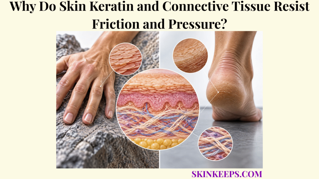
Skin keratin and connective tissue resist mechanical injury through different but connected layers: keratin filament networks and epidermal adhesion resist surface deformation and shear, while collagen-rich dermal connective tissue bears deeper tensile loads and elastic fibers support reversible deformation.
Mechanical protection therefore changes with depth and with the type, magnitude, direction, duration, and repetition of force. Friction, shear, pressure, stretching, hydration, and tissue condition all modify the response, and neither epidermal keratinization nor connective tissue makes skin mechanically indestructible.
Where Does Skin Keratin and Connective Tissue Resistance Begin?
Skin keratin and connective tissue resistance begins at the keratinized epidermal surface and extends through mechanically coupled keratinocytes into the collagen- and elastin-rich dermis, with each depth resisting a different component of mechanical load.
How Does Skin Keratin Strengthen the Epidermis?
Skin keratin strengthens the epidermis by assembling into intermediate-filament networks that allow keratinocytes to deform substantially while maintaining cellular integrity under mechanical stress. These filaments are not simply rigid rods: they redistribute stress, tolerate large deformation, and become mechanically more resistant as strain rises.
Keratin expression also changes with differentiation. Basal keratinocytes primarily express K5/K14, whereas suprabasal differentiated keratinocytes shift toward K1/K10, creating mechanically specialized cytoskeletal systems across epidermal depth.
How Do Epidermal Cells Share Mechanical Loads?
Desmosomes anchor neighboring keratinocytes to their keratin networks, creating a supracellular scaffold that distributes force across the epidermal sheet rather than leaving isolated cells to bear loads independently. Desmogleins and desmocollins form adhesive components, while plaque proteins including desmoplakin connect those junctions to keratin intermediate filaments.
Keratin intermediate filament: a cytoskeletal filament assembled from keratin proteins that gives epithelial cells high mechanical resilience.
Desmosome: a strong cell–cell adhesion junction that mechanically couples neighboring keratinocytes to their keratin filament networks.
How Does Skin Connective Tissue Provide Deeper Stability?
Dermal connective tissue provides deeper stability through a collagen-rich extracellular matrix that resists excessive extension while elastic fibers and hydrated ground substance permit controlled deformation and recoil. This depth-dependent organization places the topic within broader skin resistance to chemical and mechanical injury and the wider system of skin barrier protection.
How Does Skin Keratin Resist Friction?
Skin keratin resists friction-related mechanical stress by strengthening corneocytes and living keratinocytes, while keratin-linked adhesion systems preserve epidermal cohesion as tangential forces are transmitted through the surface.
How Do Keratin-Filled Corneocytes Resist Surface Stress?
Keratin-rich corneocytes create a tough outer interface that tolerates minor abrasion and repetitive low-level contact before living epidermal cells are directly exposed. The stratum corneum barrier is mechanically useful because densely keratinized cells, cornified envelopes, and corneodesmosomal cohesion create a durable but hydration-dependent surface.
This outer layer is not a rigid plate and it does not carry most whole-skin tensile load. Its flexibility and frictional behavior change with water content, while deeper mechanical behavior depends increasingly on viable epidermis and dermis.
How Do Keratin Filaments Resist Deformation in Living Keratinocytes?
Keratin intermediate filaments resist deformation by forming resilient intracellular networks capable of stretching, redistributing stress, and becoming mechanically more resistant as strain increases. Human genetic evidence linking keratin defects to epithelial fragility reinforces the importance of this cytoskeletal system for normal mechanical integrity.
How Do Desmosomes Preserve Epidermal Cohesion During Friction?
Desmosomes transmit mechanical force between adjacent keratinocytes by linking cell–cell adhesion complexes to keratin intermediate filaments, preventing minor local forces from separating cells immediately. Epidermal tissue strength therefore emerges from both intracellular resilience and intercellular force transfer.
Why Are Friction and Shear Not the Same Mechanical Stress?
Friction acts at the interface between skin and an external surface, whereas shear describes tangential deformation within skin and deeper tissue; high friction can increase the shear transmitted into the epidermis without being the tissue tear itself.
Friction: resistance to relative movement at the interface between skin and another surface.
Shear: tangential stress or deformation that makes adjacent tissue regions move or distort relative to one another.
How Does Skin Connective Tissue Resist Pressure?
Skin connective tissue resists pressure by deforming and distributing mechanical loads through a collagen-rich, elastic, hydrated extracellular matrix, but sustained compression can still overwhelm tissue and damage cells or impair perfusion.
How Does Dermal Collagen Provide Tensile Strength During Pressure and Deformation?
Dermal collagen provides tensile resistance as pressure deforms the skin laterally and stretches connective-tissue fibers, with increasing collagen-fiber recruitment making tissue progressively stiffer at larger strains. Type I collagen dominates the fibrillar dermal network, while type III collagen contributes to the broader architecture.
Pressure itself is normal force distributed over an area, but compression creates complex internal tension, shear, and distortion. Collagen therefore should not be described as simply “absorbing” pressure; it mainly resists tensile components of the resulting tissue deformation.
How Do Elastic Fibers Allow Deformation and Recoil?
Elastic fibers allow dermal tissue to extend at lower loads, store elastic energy, and recoil after ordinary deformation is removed. Elastin contributes strongly to reversible extensibility, but it does not bear most tensile load and is not the sole determinant of skin flexibility.
How Does the Dermal Extracellular Matrix Distribute Loads?
Collagen, elastic fibers, proteoglycans, glycosaminoglycans, and tissue water function as a composite viscoelastic matrix that redistributes and dissipates mechanical energy over space and time. Viscoelastic means that skin shows both elastic recovery and time-dependent deformation, so the response depends on force magnitude, loading rate, duration, and previous loading.
Skin is also anisotropic: collagen-fiber architecture differs by direction, so identical pulling forces can produce different deformation depending on orientation.
Why Can Pressure Still Damage Connective Tissue?
Connective tissue resistance is finite, and sufficiently intense or prolonged compression can produce damaging internal deformation, stress concentrations, vascular compromise, and cell injury despite an intact skin surface. Deep damage may begin before obvious superficial breakdown, especially where loading is concentrated over deeper structures.
| Skin structure | Main mechanical property | Protective role |
|---|---|---|
| Keratin intermediate filaments | High resilience / strain tolerance | Reduces keratinocyte mechanical failure |
| Desmosome–keratin scaffold | Strong intercellular cohesion | Distributes force across epidermal cells |
| Cornified corneocytes | Surface toughness | Resists minor abrasion and surface stress |
| Stratum corneum | Hydration-dependent stiffness/flexibility | Provides the first mechanical contact interface |
| Basement-membrane attachment system | Layer anchoring | Couples epidermis to connective tissue |
| Dermal collagen | Tensile strength + strain stiffening | Resists excessive stretching and tissue deformation |
| Elastic fibers | Extensibility + recoil | Supports reversible deformation |
| Proteoglycan-rich ground substance | Viscoelasticity / hydration | Contributes to time-dependent load distribution |
Mechanical protection is a multilayer property: no single protein carries all forms of force.
How Do Skin Keratin and Connective Tissue Work Together?
Skin keratin and connective tissue work together by forming a mechanically continuous system in which the epidermis handles the external interface and early shear, while dermal connective tissue redistributes deeper tension, stretch, and compression.
How Does the Epidermis Handle the First Mechanical Load?
The epidermis receives the external load first, using its cornified surface, keratin cytoskeleton, and strong cell adhesion to resist surface abrasion and limit local deformation. The organization of these skin epidermal barrier structures allows a thin tissue layer to act as a mechanically coherent sheet rather than a loose stack of cells.
How Is Mechanical Force Transferred Toward the Dermis?
Mechanical forces pass through epidermal cell–cell and cell–matrix attachment systems into the basement-membrane zone and underlying connective tissue, allowing multiple layers to participate in load distribution. Desmosomes transfer force laterally among keratinocytes, while hemidesmosomal and basement-membrane attachments couple the epidermis to deeper tissue.
How Does the Dermis Resist Forces the Epidermis Cannot Carry Alone?
As deformation reaches deeper tissue, dermal collagen and elastic-fiber networks bear increasing mechanical load and distribute strain across a larger tissue volume. The epidermis and dermis therefore have distinct but mechanically coupled jobs: one handles the first interface and early shear, while the other carries much of whole-skin tensile deformation.
Surface Friction Pathway
Skin–object contact → interface friction / traction → stratum corneum + keratinized epidermis → keratin IF + desmosomal cohesion → limited epidermal deformation.
If repetitive shear stays within tolerance, tissue remains intact. If shear exceeds tolerance, epidermal fatigue can progress to blistering or abrasion.
Pressure Pathway
Normal mechanical load → surface compression → epidermal + dermal deformation → collagen recruitment + elastic deformation + ECM load distribution.
If magnitude or duration exceeds tolerance, cell deformation, vascular compromise, and deeper tissue injury can develop.
Friction, shear, and pressure travel through different mechanical pathways but converge on the same finite multilayer tissue system.
How Do Skin Keratin and Connective Tissue Adapt to Repeated Stress?
Skin keratin and connective tissue adapt to ordinary repeated mechanical stress through epidermal renewal and localized hyperkeratosis, while healthy dermal connective tissue repeatedly deforms and recovers within its normal viscoelastic range.
How Does Repeated Friction Increase Keratinization?
Repeated mild-to-moderate mechanical loading can stimulate keratinocyte proliferation and differentiation, increasing stratum-corneum thickness at repeatedly stressed sites. This mechanical hyperkeratosis reflects an epidermal response rather than a simple accumulation of inert keratin.
The adaptive process overlaps with normal keratinocyte physical-barrier function, because basal-cell renewal and differentiation continuously replace mechanically worn surface cells.
Why Do Pressure-Prone Areas Develop Calluses?
Calluses develop when localized repetitive friction or pressure produces epidermal hyperplasia and hyperkeratosis, creating a thicker cornified surface over mechanically stressed areas such as palms, soles, and pressure-bearing sites.
A callus can increase local surface thickness and alter mechanical tolerance, but it is not universally beneficial. Excessive hyperkeratosis can become stiff, uncomfortable, fissure-prone, or associated with altered pressure distribution.
How Does Connective Tissue Tolerate Repeated Normal Deformation?
Healthy connective tissue tolerates physiological repetition through viscoelastic deformation, collagen-fiber recruitment, and elastic recoil without requiring permanent structural remodeling after every load cycle. Chronic overload is different: it can drive inflammatory signaling, matrix remodeling, fibrosis, or tissue damage rather than simple strengthening.
Why Does Mechanical Adaptation Have Limits?
Adaptation has limits because sufficiently large, prolonged, or repetitive forces can exceed epidermal fatigue resistance or deeper connective-tissue tolerance even when surface thickening has occurred. A thickened surface does not eliminate the possibility of blistering, fissuring, abrasion, or deeper pressure-related injury.
What Weakens Skin Keratin and Connective Tissue Resistance?
Skin keratin and connective tissue resistance weakens when epidermal flexibility, cellular cohesion, collagen architecture, elastic recoil, or tissue hydration is disturbed, reducing the amount of mechanical deformation the skin can tolerate before injury.
How Does Excessive Dryness Weaken Skin Keratin Resistance?
Excessive dryness reduces stratum-corneum flexibility and can promote scaling or fissuring, making the surface less able to deform smoothly under repeated mechanical loading. This is one reason the skin water barrier and surface mechanics are functionally linked.
How Does Excessive Moisture Weaken Mechanical Resistance?
Prolonged overhydration or maceration softens the stratum corneum, alters its frictional properties, and can increase susceptibility to superficial mechanical damage. Normal hydration is not harmful; the problem is excessive or sustained water exposure that changes material behavior.
How Can Inflammation Weaken Epidermal Mechanical Resistance?
Inflammation can alter keratinocyte differentiation, junctional organization, barrier lipids, hydration, and cellular cohesion, making epidermal mechanical behavior less predictable and sometimes more fragile. Broader structural failure belongs to the topic of skin barrier disruption rather than to normal mechanics alone.
How Can Collagen or Elastic-Fiber Disorganization Reduce Deeper Resistance?
Disorganization or damage of collagen and elastic-fiber networks can alter stiffness, tensile strength, recoil, and load distribution, reducing normal dermal mechanical performance. Skin flexibility alone cannot diagnose a collagen or elastin disorder.
Why Does Existing Tissue Injury Increase Mechanical Vulnerability?
Previously damaged or scarred tissue can distribute mechanical loads differently from normal skin, creating local stress concentrations and changing tolerance to future friction, stretch, or pressure. The exact effect depends on tissue depth, scar structure, hydration, and loading direction.
What Happens When Skin Keratin and Connective Tissue Are Overloaded?
When skin keratin and connective tissue are overloaded, injury depends on the force pattern: repetitive shear can separate epidermal layers, abrasion can remove surface tissue, fissuring can split inflexible skin, and sustained compression can damage deeper soft tissue.
How Does Friction and Repeated Shear Produce a Blister?
A friction blister develops when high interface friction permits repeated shear deformation within the epidermis until mechanical fatigue produces an intraepidermal tear that subsequently fills with fluid. Classic foot-blister studies place this failure commonly within the stratum spinosum, where repeated shear cycles progressively exceed tissue tolerance.
How Does Surface Mechanical Overload Cause Abrasion?
Abrasion occurs when mechanical contact directly disrupts or removes superficial epidermal tissue faster than the surface can withstand the applied stress. This is distinct from a blister, which is an intraepidermal separation rather than simple surface tissue loss.
How Does Dry or Thickened Skin Develop Fissures?
Fissures can develop when relatively inflexible, dry, or hyperkeratotic surface tissue is repeatedly deformed until mechanical stress concentrates and produces a linear crack. Mechanical stress is one contributor, but fissures are not always caused by mechanics alone.
How Can Pressure Cause Deeper Tissue Injury?
Prolonged or intense pressure can produce damaging internal tissue deformation and, when combined with shear, can increase stress concentrations and impair local perfusion in deeper tissues even before surface breakdown becomes visible. There is no single universal safe pressure threshold because tolerance depends on force distribution, duration, anatomy, tissue state, and individual susceptibility.
What Happens After Skin Mechanical Resistance Fails?
Once mechanical loading produces structural tissue injury, inflammatory and repair mechanisms replace normal resistance as the dominant biological response. That transition belongs to skin wound repair; the present page stops at the mechanical failure boundary rather than duplicating wound-healing phases.
| Mechanical stress | Main structure stressed | Potential failure pattern |
|---|---|---|
| Mild surface contact | Stratum corneum | Usually tolerated |
| Repeated surface abrasion | Cornified surface | Erosion / abrasion |
| High-friction repetitive shear | Viable epidermis | Intraepidermal blister |
| Dryness + repetitive bending | Stratum corneum | Fissuring |
| Chronic moderate pressure/friction | Epidermal differentiation system | Hyperkeratosis / callus |
| Excessive focal pressure | Dermis / subcutis | Tissue deformation |
| Pressure + shear | Skin + deeper soft tissue | Amplified internal strain / stress |
| Prolonged sustained load | Deep soft tissue / vasculature | Cell damage, perfusion impairment, pressure injury |
| Previous scar + new loading | Altered ECM | Local stress concentration / reduced adaptability |
Mechanical outcome depends on magnitude, direction, duration, repetition, anatomical site, tissue hydration, and pre-existing tissue condition.
This page explains normal mechanical physiology rather than treatment. Recurrent unexplained blistering, repeated skin breakdown, persistent painful fissures, suspected pressure injury, non-healing abrasions, or signs of infection warrant professional assessment.
What Are the Key Takeaways About Skin Keratin and Connective Tissue?
The key fact about skin keratin and connective tissue is that mechanical resistance is layered: keratin and epidermal adhesion protect cells against surface deformation and shear, while collagen-rich connective tissue and elastic fibers distribute deeper loads and permit controlled deformation.
Friction, shear, pressure, hydration, load direction, repetition, and tissue condition determine whether a force remains within normal tolerance, produces adaptive thickening, or progresses to structural failure. Mechanical adaptation changes tolerance; it does not make skin injury-proof.
- Skin keratin is not a simple hard shell: keratin intermediate filaments give keratinocytes mechanical resilience.
- Basal and suprabasal keratinocytes use different keratin networks as the epidermis differentiates.
- Desmosomes connect keratin networks between neighboring keratinocytes, allowing force to spread across the epidermis.
- Keratin-filled corneocytes and cornified envelopes provide surface toughness against minor abrasion.
- Friction is an interface force, while shear is internal tangential deformation.
- Friction blisters are primarily repetitive shear-fatigue injuries, not simple surface burns.
- Dermal collagen provides much of whole-skin tensile resistance.
- Collagen fibers progressively recruit as strain increases, producing nonlinear strain stiffening.
- Skin is anisotropic: mechanical resistance depends partly on loading direction.
- Elastic fibers contribute extensibility and recoil, but do not carry all mechanical load.
- Proteoglycans, water, collagen, and elastic fibers together create viscoelastic tissue behavior.
- Pressure produces tissue compression and deformation, not merely a surface force.
- Pressure combined with shear can damage deeper tissue even before major surface breakdown is visible.
- Skin hydration changes friction and mechanical properties.
- Excessive dryness can promote fissuring, while prolonged maceration can reduce mechanical integrity.
- Repeated moderate loading can cause hyperkeratosis and callus formation.
- Callus changes local mechanical tolerance but does not provide unlimited protection.
- When mechanical resistance fails, injury may appear as abrasion, fissure, blister, or deeper pressure-related tissue damage.
What Common Questions Do People Ask About Skin Keratin and Connective Tissue?
Common questions about skin keratin and connective tissue focus on which structure provides strength, whether friction directly causes blisters, how collagen differs from elastin, and why repeated pressure sometimes creates callus but sometimes causes injury.
Does Skin Keratin Make the Epidermis Hard?
Keratin makes epidermal cells mechanically resilient rather than simply hard; intermediate-filament networks allow keratinocytes to tolerate deformation, while keratin-rich corneocytes provide additional surface toughness.
Does Friction Directly Cause Skin Blisters?
Friction is important because it increases traction between skin and an external surface, but a classic friction blister forms when repeated internal shear deformation mechanically fatigues and tears the viable epidermis.
Is Collagen or Elastin More Important for Skin Strength?
Collagen provides most dermal tensile-load resistance and progressive strain stiffening, while elastic fibers are particularly important for reversible extensibility and recoil; normal skin mechanics depend on both plus the surrounding extracellular matrix.
Does a Callus Make Skin Fully Resistant to Friction and Pressure?
No. Callus formation increases local cornified thickness after repeated mechanical loading, but sufficiently intense or repetitive shear, friction, pressure, or dryness can still produce blistering, fissuring, abrasion, or deeper injury.
Can Pressure Damage Tissue Even When the Skin Surface Looks Intact?
Yes. Sustained pressure and shear can create damaging deformation and stress concentrations in deeper soft tissue before visible epidermal breakdown develops, particularly over bony prominences or beneath rigid devices.
Sources & Evidence
Research grounding
Types I and II Keratin Intermediate Filaments — keratin intermediate-filament structure, differentiation-dependent expression, cellular integrity, and mechanical stress resistance.
Desmosomes and Intermediate Filaments: Their Consequences for Tissue Mechanics — desmosome–keratin coupling, intercellular cohesion, and force distribution across epithelial tissue.
Skin Microstructure Is a Key Contributor to Its Friction Behaviour — stratum-corneum mechanics, hydration-dependent friction, and surface microstructure.
Friction Blisters of the Feet: A Critical Assessment of Current Prevention Strategies — friction versus shear, repetitive shear deformation, mechanical fatigue, and intraepidermal blister mechanics.
Mathematical and Computational Modelling of Skin Biophysics: A Review — whole-skin nonlinear mechanics, collagen architecture, anisotropy, and strain-dependent behavior.
Mechanical Properties and Functions of Elastin: An Overview — elastic-fiber extensibility, elastic-energy storage, recoil, and interaction with the extracellular matrix.
Our Contemporary Understanding of the Aetiology of Pressure Ulcers/Pressure Injuries — sustained soft-tissue deformation, pressure–shear interaction, vascular compromise, and deeper damage beneath intact skin.
Corns and Calluses Resulting from Mechanical Hyperkeratosis — hyperkeratosis and callus formation as responses to chronic mechanical pressure or friction.
Medical note: This page is educational and does not diagnose mechanical skin injury or prescribe treatment. Seek timely assessment for worsening pain, spreading redness, drainage, recurrent unexplained blistering, non-healing skin breakdown, or suspected pressure injury; urgent care may be needed for severe crush injury, deep wounds, extensive skin loss, or systemic illness.



