
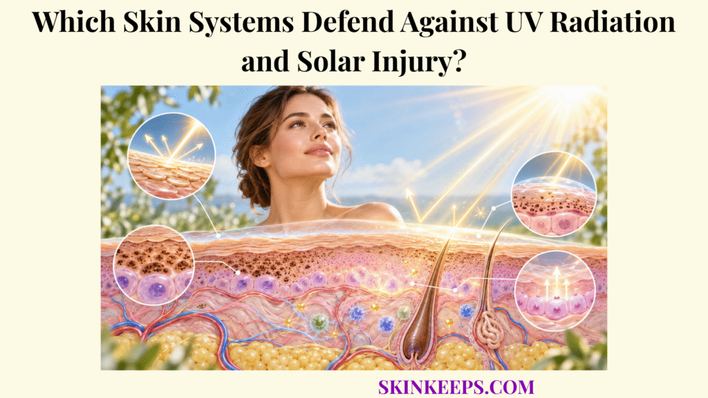
Skin defends against UV radiation and solar injury through several overlapping systems: the epidermis reduces penetration, melanin absorbs and redistributes UV energy, antioxidants limit oxidative stress, DNA-repair systems correct specific lesions, cell-cycle checkpoints delay division, apoptosis removes severely damaged cells, and cutaneous immune responses help manage injured tissue. UVA and UVB challenge skin at structural, molecular, cellular, and immunological levels, and both can produce injury through partly overlapping pathways.
This page explains which natural skin systems respond at each stage—from incoming radiation to molecular damage and tissue injury—and where those defenses reach their limits. These mechanisms reduce damage; they do not make excessive or cumulative ultraviolet exposure safe.
What UV Threats Must the Skin’s Protective Systems Defend Against?
The skin’s protective systems must defend against both UVA and UVB radiation because each can damage skin cells, DNA, proteins, lipids, and extracellular tissue through different but overlapping photochemical pathways. UV defense is one of the major physiological skin functions, but it operates across several layers rather than through one protective molecule.
UVA spans approximately 320–400 nm and penetrates more deeply than UVB, reaching the dermis more efficiently. UVB spans approximately 280–320 nm and is absorbed more strongly in the epidermis, where it is especially effective at producing direct DNA photoproducts. Wavelength influences penetration and molecular targets, but neither range should be treated as biologically harmless.
UVA strongly promotes reactive oxygen species, or ROS, which can oxidize lipids, proteins, DNA, and signaling molecules. In the dermis, oxidative signaling can alter fibroblasts, increase matrix-degrading enzymes, and damage collagen and elastin. UVA can also contribute to mutagenic DNA lesions rather than acting only as an “aging” wavelength.
UVB is highly effective at directly exciting DNA bases and generating cyclobutane pyrimidine dimers, or CPDs, and 6-4 photoproducts. It also contributes to ROS production, inflammation, erythema, and mutagenesis. Repeated exposure to either UVA or UVB can therefore add oxidative, genomic, inflammatory, and structural injury faster than normal repair and tissue turnover can completely resolve it.
How Does UVA Penetrate Deeper Skin Layers and Contribute to Oxidative Stress and Photoaging?
UVA penetrates more deeply through the epidermis into the dermis, where it strongly promotes reactive oxygen species formation, oxidative injury, matrix-degrading signaling, and long-term photoaging. Fibroblasts and extracellular-matrix proteins are therefore exposed to a greater proportion of UVA than of UVB.
UVA-driven oxidative stress can oxidize membrane lipids, proteins, and DNA while activating matrix metalloproteinases that degrade collagen and alter dermal matrix organization. Elastin can also become structurally abnormal during chronic photodamage. These processes help explain photoaging, but UVA also contributes to mutagenic DNA injury and skin-cancer risk.
How Does UVB Primarily Affect the Epidermis and Directly Damage DNA?
UVB is absorbed strongly within the epidermis and directly excites DNA bases, efficiently producing cyclobutane pyrimidine dimers and 6-4 photoproducts that can become mutations if they are not correctly repaired before replication. Keratinocytes are therefore major targets of UVB photochemistry.
Direct DNA injury is not the only UVB effect. UVB also generates reactive oxygen species, activates inflammatory pathways, contributes to erythema, and can influence upper dermal tissue. Its dominant pattern is stronger epidermal absorption and direct photoproduct formation, not an absolute restriction to the epidermis.
How Does Repeated UV Exposure Create Cumulative Cellular and Tissue Injury?
Repeated UV exposure creates cumulative injury when successive episodes of DNA damage, oxidative stress, inflammatory signaling, and extracellular-matrix degradation occur faster or more extensively than normal repair and turnover can completely resolve them. A DNA lesion can be removed, a damaged protein can be replaced, and an injured cell can die, but those systems are finite rather than perfectly efficient.
Over time, unrepaired mutations can persist in descendant cells while repeated oxidative signaling alters dermal matrix structure. The result is a rising burden of photoaging and carcinogenic risk rather than a single universal “safe cumulative dose.”
| UV type | Relative skin penetration | Major biological effects | Key natural responses |
|---|---|---|---|
| UVA | Penetrates through the epidermis and more efficiently reaches the dermis. | Strong ROS production, oxidative DNA/protein/lipid injury, fibroblast and matrix damage, photoaging; also contributes to mutagenic DNA damage. | Melanin, antioxidant systems, DNA-damage responses, checkpoints, and apoptosis. |
| UVB | Absorbed predominantly within the epidermis. | Direct DNA photoproducts, erythema, inflammatory signaling, mutagenesis; also contributes to oxidative stress. | Epidermal interception, melanin, DNA repair, p53-associated signaling, apoptosis, and inflammatory responses. |
UVA and UVB effects overlap; the table describes dominant patterns rather than exclusive biological effects.
How Does the Epidermal Defense System Reduce UV Penetration?
The epidermal defense system reduces UV penetration through superficial absorption and scattering, keratinized tissue, epidermal pigmentation, and adaptive thickening that increases the amount of tissue incoming radiation must cross before reaching deeper vulnerable cells. The broader skin epidermis UV response includes both passive interception and active cellular adaptation.
How Does the Stratum Corneum Absorb and Scatter Part of Incoming UV Radiation?
The stratum corneum provides partial UV interception because its keratinized cells and molecular components absorb and scatter a fraction of incoming radiation before that energy reaches living epidermal and dermal tissue. This effect reduces exposure but does not stop all UVA or UVB at the surface.
Keratin, urocanic-acid-related chemistry, and the optical properties of the outer epidermis all influence how radiation is transmitted. The stratum corneum should therefore be understood as one attenuation layer rather than a sunscreen equivalent.
How Does Epidermal Thickness Influence UV Penetration?
Greater epidermal thickness can reduce the fraction of UV energy reaching deeper cells because radiation encounters more absorbing and scattering tissue before passing through the epidermis. Tissue path length and optical density therefore influence attenuation without creating a universal percentage of protection.
How Can Repeated UV Exposure Trigger Epidermal Thickening as an Adaptive Response?
UV injury can stimulate keratinocyte proliferation and epidermal hyperplasia, producing a thicker epidermis that provides greater resistance to subsequent UV penetration but does not make further exposure safe. This adaptive epidermal thickening is a response to exposure-related stress and injury.
UV-induced thickening is an adaptive response to UV exposure; it is not a reason to intentionally expose skin to ultraviolet radiation.
How Does the Melanin Protection System Shield Skin Cells From UV Damage?
The melanin protection system shields skin cells when melanocytes synthesize pigment inside melanosomes and transfer those pigment-containing organelles to keratinocytes, where melanin absorbs and redistributes UV energy and can concentrate around nuclei to reduce genomic exposure. This mechanism is the basis of skin melanin UV absorption, but it provides partial rather than complete photoprotection.
How Do Melanocytes Produce Melanin Inside Melanosomes?
Melanocytes synthesize melanin inside specialized organelles called melanosomes, where melanogenic enzymes convert tyrosine-derived substrates into eumelanin or pheomelanin pigments. Tyrosinase initiates key steps in the pathway, while melanosomes mature as pigment accumulates.
Eumelanin generally provides more effective photoprotection than pheomelanin because their chemical and photophysical properties differ. The relevant point for UV defense is not the full melanogenesis pathway but the production of pigment capable of absorbing and redistributing part of incoming radiation.
How Is Melanin Transferred From Melanocytes to Keratinocytes?
Melanocytes extend dendritic processes toward neighboring keratinocytes and transfer melanosomes containing pigment into those epidermal cells, distributing melanin across the epidermal melanin unit. UV-related signaling can increase this pigmentation response through pathways involving p53, POMC-derived α-MSH, and melanocortin-1 receptor signaling.
How Do Melanin-Containing Structures Help Shield Keratinocyte Nuclei From UV Radiation?
Transferred melanosomes can accumulate in a supranuclear distribution within keratinocytes, positioning pigment between incoming UV radiation and nuclear DNA and thereby reducing part of the genomic UV burden. This spatial organization contributes to melanin DNA protection by placing an absorbing pigment cap above vulnerable genetic material.
Why Does Melanin Reduce UV Damage Without Providing Complete Protection?
Melanin reduces UV injury by absorbing and dispersing radiation and limiting some oxidative and DNA damage, but endogenous pigmentation cannot stop all UVA or UVB from reaching living cells or prevent all mutations and photoaging. Pigmentation changes risk; it does not eliminate risk.
All skin tones can sustain UV injury. A tanning response indicates UV-triggered biological adaptation and associated cellular stress, so it should not be presented as a protective treatment or a safe strategy for building resistance.
How Do Cellular UV-Defense Systems Limit Oxidative Stress and DNA Damage?
Cellular UV-defense systems limit damage by neutralizing reactive oxygen species, recognizing molecular lesions, repairing repairable DNA damage, delaying cell division through checkpoints, and eliminating severely damaged cells through apoptosis when safe recovery is unlikely. These post-penetration mechanisms include skin antioxidant defenses and specialized DNA-damage responses.
How Do Antioxidant Defenses Help Neutralize UV-Generated Reactive Oxygen Species?
Skin antioxidant defenses limit UV-generated oxidative stress through coordinated enzymatic and non-enzymatic systems that neutralize reactive oxygen species before those molecules extensively damage DNA, proteins, lipids, and cellular membranes. Enzymatic defenses include superoxide dismutase, catalase, glutathione peroxidase, and glutathione reductase.
Non-enzymatic defenses include glutathione, tocopherol, ascorbate, ubiquinol, and other redox-active molecules. These systems maintain redox balance but have finite capacity; a sufficiently large UV-driven ROS load can shift tissue toward oxidative stress.
How Do DNA-Repair Mechanisms Correct Certain Forms of UV-Induced DNA Damage?
DNA-repair systems correct many UV-induced lesions by recognizing damaged DNA and removing or replacing abnormal nucleotides before those lesions become fixed mutations during replication. Nucleotide excision repair, or NER, is a major pathway for bulky UV photolesions such as CPDs and 6-4 photoproducts.
Damage-recognition proteins including XPC and DDB2 help initiate NER, after which the damaged DNA segment is excised and resynthesized. Base excision repair, or BER, handles selected oxidative base lesions such as 8-oxoG through proteins including OGG1. This skin DNA repair machinery is essential but not perfectly efficient.
Xeroderma pigmentosum demonstrates why intact UV-DNA-repair pathways matter: defects in nucleotide excision repair produce extreme UV sensitivity and a markedly increased risk of UV-related skin cancers. The condition is useful here as a biological teaching example rather than as a diagnostic topic.
How Do Cell-Cycle Checkpoints Prevent Damaged Cells From Dividing Immediately?
Cell-cycle checkpoints delay division after UV damage so cells have additional time to assess and repair DNA before duplicating potentially mutagenic lesions. p53 and p21 participate in this response by slowing progression through cell-cycle checkpoints, including G1 and G2 control.
Checkpoint arrest is not the same as DNA repair. Arrest creates time and changes the cellular decision environment; repair enzymes still have to recognize and process the lesion, and other checkpoint proteins also contribute beyond p53.
How Can Severely Damaged Keratinocytes Undergo Programmed Cell Death?
Severely UV-damaged keratinocytes can activate apoptosis when cellular damage exceeds a recoverable threshold, removing potentially dangerous cells rather than allowing them to survive and continue dividing with extensive genomic injury. Histologically, apoptotic keratinocytes after UV exposure are often described as sunburn cells.
p53-associated signaling can promote apoptosis, but UV-triggered cell death also involves death receptors, mitochondrial pathways, caspases, and other mechanisms. Apoptosis reduces the survival of some highly damaged cells; it does not guarantee that every potentially abnormal cell is eliminated.
How Does the Cutaneous Immune System Control UV-Related Injury?
The cutaneous immune system responds to UV-related injury when stressed keratinocytes and resident immune cells release signaling molecules that promote inflammation, recruit or regulate immune activity, and participate in surveillance of damaged tissue, although excessive UV exposure can simultaneously suppress important immune functions. This response overlaps with the broader skin immune defense network.
How Do Keratinocytes Release Signaling Molecules After UV Injury?
UV-stressed keratinocytes act as immune-signaling cells by releasing cytokines, chemokines, and other mediators that alert nearby tissue, influence vascular and inflammatory responses, and communicate with resident immune cells. Mediators can include IL-1, TNF, prostaglandins, and other stress-related signals.
How Does Inflammation Help Respond to Damaged Skin Cells?
Controlled inflammation helps damaged skin respond to UV injury by increasing signaling, altering local blood flow, recruiting defensive cells, and coordinating removal or processing of damaged tissue. Acute inflammation can therefore support damage control, while chronic or excessive inflammatory signaling can contribute to additional tissue injury and photoaging.
How Do Immune-Surveillance Mechanisms Help Identify Abnormal or Severely Damaged Cells?
Cutaneous immune surveillance helps identify cells expressing abnormal or stress-associated signals and contributes to the broader control of potentially dangerous cellular changes within skin. Dendritic cells, T cells, innate immune cells, and keratinocyte-derived signals all participate in this layered surveillance system.
Immune surveillance is not a perfect filter. Mutated cells can escape recognition or survive in altered tissue environments, which is why immunity represents one cancer-prevention layer rather than an absolute safeguard.
How Can Excessive UV Exposure Alter Normal Skin Immune Responses?
Excessive UV exposure can suppress aspects of cutaneous immunity by altering antigen-presenting cells, cytokine networks, and regulatory immune pathways, potentially weakening normal immune surveillance even while UV injury simultaneously triggers inflammation. Changes in Langerhans-cell function and regulatory T-cell pathways are among the mechanisms described in UV-induced immunosuppression.
UV exposure can produce inflammation and immunosuppression at the same time through different biological pathways; these effects are not contradictory.
What Happens When the Skin’s UV-Protection Mechanisms Are Overwhelmed?
Skin UV-protection mechanisms become insufficient when radiation produces more molecular and cellular injury than epidermal interception, pigmentation, antioxidant control, DNA repair, checkpoint responses, apoptosis, and immune surveillance can successfully contain. Natural photoprotection is therefore a damage-reduction network, not a guarantee against sunburn, photoaging, mutation accumulation, or cancer.
How Does Excessive UV Exposure Produce Erythema and Sunburn?
Sunburn develops when UV-induced molecular injury triggers inflammatory mediators and vascular responses that produce erythema, pain, heat, and—in more severe injury—epidermal cell death. UVB strongly contributes to erythema, but the clinical response reflects interacting DNA damage, inflammatory signals, vasodilation, and apoptotic keratinocytes.
Sunburn is evidence of tissue injury rather than a harmless temporary cosmetic response. Significant blistering, extensive burns, severe pain, systemic illness, dehydration symptoms, or confusion may warrant prompt medical care.
How Does Accumulated Oxidative Damage Contribute to Photoaging?
Accumulated UV-driven oxidative stress contributes to photoaging by activating matrix-degrading pathways, damaging dermal fibroblasts, disrupting collagen and elastin organization, and progressively altering skin structure and pigmentation. UVA-associated ROS and matrix metalloproteinase activation are major contributors to dermal matrix degradation.
How Can Unrepaired DNA Damage Produce Mutations?
Unrepaired UV DNA lesions can become permanent mutations when damaged DNA is replicated or inaccurately repaired, allowing altered genetic information to persist in descendant skin cells. A DNA lesion is therefore not automatically a mutation: many lesions are repaired before replication, while a mutation represents a lasting sequence change.
Why Can Repeated Solar Injury Increase the Risk of Precancerous Changes and Skin Cancer?
Repeated solar injury raises skin-cancer risk because cumulative DNA mutations, altered growth-control genes, oxidative injury, clonal expansion, and changes in immune surveillance can progressively weaken the safeguards that normally constrain abnormal cells. TP53 mutations and other growth-control changes can be selected during chronic UV exposure, but no single UV-induced mutation makes cancer inevitable.
Actinic keratosis is a UV-associated precancerous keratinocyte lesion, and some lesions can progress to squamous cell carcinoma. Basal cell carcinoma, squamous cell carcinoma, and melanoma also have important relationships with ultraviolet exposure, but suspicious lesions require professional examination rather than diagnosis from an article.
A persistent rough or scaly sun-exposed patch, a non-healing lesion, recurrent bleeding, a changing mole or pigmented lesion, a new persistent growth, or a lesion that repeatedly crusts or ulcerates should be evaluated by a dermatologist.
| Defense system | Primary mechanism | Main threat controlled | Important limitation |
|---|---|---|---|
| Epidermal interception | Keratinized tissue and epidermal thickness absorb/scatter part of incoming radiation. | Reduces deeper UV penetration. | Does not block all UVA or UVB. |
| Melanin system | Melanosomes absorb/scatter UV and can shield keratinocyte nuclei. | Reduces cellular and DNA UV burden. | Natural pigmentation provides incomplete protection. |
| Antioxidant system | Enzymatic and non-enzymatic antioxidants neutralize ROS. | Limits oxidative damage. | High UV loads can overwhelm antioxidant capacity. |
| DNA-repair system | NER and BER recognize and remove appropriate lesions. | Limits mutation fixation. | Repair is not perfectly efficient. |
| Cell-cycle checkpoints | Damage signals delay cell division. | Reduces immediate replication of damaged DNA. | Checkpoints can fail or be bypassed. |
| Apoptosis | Severely damaged cells undergo programmed death. | Removes some highly damaged keratinocytes. | Not every abnormal cell is eliminated. |
| Immune defense | Inflammatory signaling and surveillance respond to tissue injury and abnormal cells. | Supports damage control. | UV can itself suppress aspects of immunity. |
What Are the Key Takeaways About the Skin’s Natural UV Defenses?
The key fact about the skin’s natural UV defenses is that photoprotection depends on multiple systems acting at different stages—from reducing incoming radiation to repairing or removing damaged cells—and none of those systems provides complete protection from excessive or cumulative UV exposure.
- UV threat: UVA and UVB produce overlapping but different patterns of penetration and molecular injury.
- Epidermal defense: The epidermis absorbs and scatters part of incoming UV, and UV exposure can stimulate adaptive epidermal thickening.
- Melanin defense: Melanocytes produce melanin in melanosomes and transfer pigment to keratinocytes, where it reduces UV injury and can protect nuclear DNA.
- Antioxidant defense: Enzymatic and non-enzymatic antioxidants reduce UV-generated oxidative stress.
- DNA repair: Nucleotide excision repair and other pathways remove many UV-induced DNA lesions before mutations become fixed.
- Cell-cycle control: Checkpoints can pause damaged cells before division.
- Apoptosis: Severely damaged keratinocytes can undergo programmed cell death.
- Immune defense: Skin immune signaling responds to injured tissue, but heavy UV exposure can also suppress aspects of immune surveillance.
- Defense limitation: Natural photoprotection cannot completely prevent sunburn, photoaging, mutation accumulation, precancerous change, or skin cancer when UV exposure exceeds defensive capacity.
What Common Questions Do People Ask About Skin UV Defense?
Common questions about skin UV defense focus on whether melanin is enough protection, how UVA differs from UVB, what happens to damaged DNA, and whether tanning strengthens the skin safely.
Which Is More Dangerous to Skin, UVA or UVB?
Neither UVA nor UVB should be considered harmless. UVB is especially efficient at producing direct epidermal DNA photolesions and sunburn, while UVA penetrates more deeply and strongly contributes to oxidative damage and photoaging; both contribute to skin-cancer risk.
Does Melanin Completely Block UV Radiation?
No. Melanin absorbs and disperses part of incoming ultraviolet energy and reduces cellular damage, but significant UVA and UVB exposure can still reach living skin cells and produce DNA damage in every skin tone.
Can Skin Repair UV-Damaged DNA?
Yes. Skin cells use DNA-repair pathways such as nucleotide excision repair to remove many UV-induced lesions, but repair is not perfectly efficient, so some damage can persist or become permanent mutations.
Does Tanning Make Skin Safe From Future UV Exposure?
No. Tanning is an adaptive pigmentation response triggered by UV exposure and associated cellular damage; the additional pigment provides only limited protection and does not make later UV exposure safe.
What Happens If a UV-Damaged Skin Cell Cannot Be Repaired?
A heavily damaged skin cell may remain arrested, undergo apoptosis, or survive with persistent DNA alterations. Mutations that escape repair and growth-control mechanisms can contribute to abnormal clonal expansion over repeated exposures, but one surviving mutation does not automatically produce cancer.
Journal of Cosmetic Dermatology / PubMed — The impact of ultraviolet radiation on skin photoaging — review of in vitro studies: UVA/UVB tissue effects, reactive oxygen species, fibroblast and matrix injury, collagen fragmentation, and photoaging.
International Journal of Molecular Sciences / PMC — UV Radiation and the Skin: UV-induced inflammation, epidermal thickening, p53-associated damage responses, cell-cycle arrest, DNA repair, apoptosis, and sunburn cells.
Journal of Investigative Dermatology / PubMed — UV signaling pathways within the skin: p53/POMC/α-MSH signaling, melanin synthesis, melanosome transfer, and supranuclear pigment localization in keratinocytes.
Redox Report / PubMed — Oxidative stress and antioxidant strategies in dermatology: superoxide dismutase, catalase, glutathione peroxidase, glutathione, tocopherol, ascorbate, and endogenous skin antioxidant systems.
Biomedicines / PubMed — UV Radiation in DNA Damage and Repair Involving DNA-Photolyases and Cryptochromes: UVA/UVB DNA injury and nucleotide-excision-repair biology.
Photodermatology, Photoimmunology & Photomedicine / PubMed — Molecular mechanisms of UV-induced apoptosis: UV-triggered keratinocyte apoptosis, p53-associated pathways, death receptors, mitochondria, and sunburn cells.
Nature Reviews Immunology / PMC — Immune surveillance in the skin: mechanisms and clinical consequences: cutaneous immune surveillance, dendritic-cell trafficking, T-cell responses, and inflammatory recruitment.
Journal of Dermatological Science / PMC — Photocarcinogenesis of the skin: Current status and future trends: UV-induced DNA damage, oxidative stress, inflammation, immunosuppression, Langerhans-cell changes, and photocarcinogenesis.
American Academy of Dermatology — Sunscreen FAQs: both UVA and UVB are harmful, UV radiation and tanning damage skin, and broad-spectrum sun protection reduces short- and long-term UV injury.
American Academy of Dermatology — Actinic keratosis: Overview: chronic UV damage, precancerous keratinocyte changes, and the need for professional evaluation of suspicious sun-damaged lesions.
Medical note: This article explains normal skin photobiology for educational purposes and does not diagnose sunburn severity, actinic keratosis, melanoma, basal cell carcinoma, or squamous cell carcinoma. A persistent rough or scaly sun-exposed patch, a non-healing lesion, recurrent bleeding, a changing mole or pigmented lesion, a new persistent growth, or a lesion that repeatedly crusts or ulcerates should be evaluated by a dermatologist. Significant blistering sunburn, extensive burns, severe pain, dehydration symptoms, confusion, or systemic illness may require prompt medical care.



