
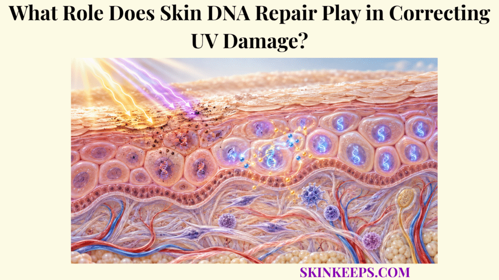
Skin DNA repair corrects UV damage by recognizing abnormal DNA structures, removing lesion-containing DNA, rebuilding the missing sequence from the intact complementary strand, and sealing the repaired region before persistent lesions can interfere with transcription or become fixed as mutations during replication.
The principal pathway for major direct UV photoproducts is nucleotide excision repair (NER), supported by checkpoint systems that can slow unsafe cell-cycle progression. Repair is highly effective but finite and does not guarantee correction of every lesion after strong or repeated UV exposure.
How Does Skin DNA Repair Detect UV-Induced Damage?
Skin DNA repair detects UV-induced damage through complementary surveillance systems that recognize abnormal DNA structure throughout the genome or respond when lesions stall transcription in actively expressed genes.
Which UV Lesions Activate Skin DNA Repair?
The principal direct UV lesions handled by NER are cyclobutane pyrimidine dimers (CPDs) and 6-4 photoproducts (6-4PPs) created between neighboring pyrimidine bases. UVB is particularly efficient at forming these lesions, although UVA and UVB have overlapping genotoxic effects. This downstream repair step sits within the broader skin UV defense hierarchy.
How Does Global-Genome Skin DNA Repair Find UV Lesions?
Global-genome NER surveys chromosomal DNA for lesion-associated structural abnormalities, with XPC–RAD23B serving as a major recognition complex. This genome-wide surveillance operates after upstream systems described in skin defense against solar injury have failed to prevent a lesion from forming.
Why Does Skin DNA Repair Need DDB2 for Many CPDs?
CPDs distort the helix relatively weakly and are inefficiently recognized by XPC alone, so UV-DDB containing DDB1 and DDB2 helps identify these lesions in chromatin and facilitates recruitment of XPC. By contrast, 6-4PPs distort DNA more strongly and are generally recognized more efficiently by XPC-dependent GG-NER.
How Does Transcription-Coupled Skin DNA Repair Detect Damage?
Transcription-coupled NER begins when RNA polymerase II stalls at a transcription-blocking lesion, triggering CSA-, CSB-, UVSSA-, and related machinery that prioritizes the affected transcribed strand. TC-NER responds to transcription blockage rather than searching for one specific lesion type.
How Is a Suspected UV Lesion Verified?
After initial recognition, TFIIH opens the local DNA region and helps verify whether the DNA structure is compatible with an NER substrate. XPB contributes ATPase/translocase activity, XPD contributes ATPase/helicase-dependent scanning and verification, and XPA with RPA helps organize and stabilize the pre-incision complex.
Photoprotection lowers lesion formation; DNA repair corrects lesions that nevertheless form. Upstream protection includes skin melanin UV absorption and skin melanin distribution and photoprotection.
How Does Skin DNA Repair Remove UV-Damaged DNA?
Skin DNA repair removes UV-damaged DNA by opening a local repair bubble around the lesion and using the structure-specific nucleases XPF–ERCC1 and XPG to incise the damaged strand on opposite sides of the photolesion.
How Does TFIIH Open DNA Around the UV Lesion?
XPB- and XPD-dependent TFIIH activities locally open and inspect DNA around the suspected lesion, allowing downstream NER proteins to assemble around the damaged strand. The opening is local; NER does not unwind an entire chromosome.
How Do XPA and RPA Prepare DNA for Excision?
XPA helps verify and organize the pre-incision complex, while RPA binds exposed single-stranded DNA, stabilizes the undamaged strand, and helps orient the incision machinery. Neither XPA nor RPA is the nuclease that cuts the damaged strand.
Which Enzyme Cuts Before the UV Lesion?
The XPF–ERCC1 complex makes the incision on the 5′ side of the lesion. XPF contains the catalytic nuclease activity, while ERCC1 provides important structural and targeting interactions within the repair complex.
Which Enzyme Cuts After the UV Lesion?
XPG makes the corresponding incision on the 3′ side. XPG is recruited before its catalytic incision is triggered, and current models support a coordinated sequence in which XPF–ERCC1 usually makes the first productive cut and repair synthesis can begin before the XPG incision is completed.
How Much DNA Does Skin DNA Repair Remove?
Human NER removes a short segment surrounding the lesion—on the order of a few dozen nucleotides rather than only the chemically altered bases. Many human NER models report an excised oligonucleotide of roughly 24–32 nucleotides, but the exact number is not a universal reader-facing repair deadline or fixed measurement for every context.
How Does Skin DNA Repair Restore the DNA Sequence?
Skin DNA repair restores the DNA sequence by using the intact complementary strand as a template for DNA polymerase-mediated gap filling, followed by DNA ligase sealing of the remaining backbone nick.
How Do DNA Polymerases Rebuild the Excised Region?
Repair polymerases copy sequence information from the undamaged complementary strand and insert replacement nucleotides across the excision gap. Human NER can use polymerases δ, ε, or κ with supporting factors such as PCNA, RFC, and RPA, with usage influenced by cell state and repair context.
Why Is the Complementary DNA Strand Essential for Skin DNA Repair?
The intact strand preserves the base-pairing information needed to reconstruct the removed sequence accurately. Repair synthesis is therefore template-directed; polymerases do not retrieve a stored copy of the sequence from elsewhere in the cell.
How Does DNA Ligase Finish Skin DNA Repair?
After gap synthesis, DNA ligase seals the remaining nick in the sugar-phosphate backbone and restores strand continuity. Depending on cellular state and repair context, ligation can involve DNA ligase I or DNA ligase IIIα with XRCC1.
Does Completing NER Mean Every Effect of UV Is Reversed?
No. NER restores the targeted DNA region, but the same UV exposure can also produce oxidative, protein, lipid, mitochondrial, and inflammatory injury that requires separate responses. The distinction is especially important because skin antioxidant systems reduce oxidative lesion formation but do not replace DNA repair after a lesion exists.
How Does Skin DNA Repair Work With Cell-Cycle Control?
Skin DNA repair works with cell-cycle control because checkpoints can slow replication or division when UV lesions remain, reducing the chance that damaged DNA is copied before repair systems have processed it.
How Does ATR–CHK1 Respond to UV-Damaged DNA?
UV lesions that stall replication can generate RPA-coated single-stranded DNA intermediates that strongly activate ATR–CHK1 signaling. This pathway helps stabilize replication stress and delays inappropriate progression; ATR is signaling machinery, not the nuclease that excises a CPD.
How Does p53–p21 Support Skin DNA Repair?
UV-associated DNA-damage signaling can stabilize p53 and increase p21, which inhibits selected cyclin-dependent kinase activity and contributes to cell-cycle slowing while repair operates. The wider decision network—including checkpoint, survival, and apoptosis programs—belongs to the keratinocyte response to UV-induced cellular damage.
Why Does Delaying DNA Replication Matter?
Delaying replication gives repair pathways additional opportunity to remove mutagenic lesions before replication machinery encounters them. Checkpoint control can therefore lower the chance of lesion bypass without physically removing the lesion itself.
What Happens After Successful Skin DNA Repair?
When genomic integrity is sufficiently restored, checkpoint signaling can subside and the cell may resume transcription, replication, or normal cell-cycle progression according to its physiological state. Not every repaired differentiated keratinocyte is expected to re-enter proliferation.
Is Cell-Cycle Arrest Itself DNA Repair?
No. A checkpoint buys time and regulates cell-cycle progression; the repair machinery performs the molecular correction. The complete timing of these post-UV events is covered by the skin epidermal UV response.
| DNA problem | Repair / control response | Protective outcome | Important limitation |
|---|---|---|---|
| 6-4PP / strongly distorted UV lesion | XPC-driven GG-NER recognition | Efficient NER recruitment | Repair speed varies |
| CPD | DDB2-assisted GG-NER recognition | Improves CPD detection | CPDs can persist longer |
| Stalled transcription | TC-NER | Prioritizes active-gene repair | Does not replace GG-NER |
| Verified photolesion | XPF–ERCC1 + XPG | Removes lesion-containing DNA | Requires assembled NER complex |
| Excision gap | Pol δ / ε / κ + PCNA / RFC | Reconstructs missing DNA | Template strand must remain intact |
| Remaining nick | DNA ligase | Restores strand continuity | Finishes only the local repair event |
| Replication stress | ATR–CHK1 | Delays unsafe progression | Does not excise the lesion |
| p53 activation | p21 / checkpoint response | Supports repair opportunity | Not the sole checkpoint pathway |
| Selected oxidative DNA lesion | BER | Corrects oxidized bases | Mechanistically separate from NER |
| Severe unresolved damage | Apoptosis | Removes unsafe damaged cell | Cell is lost rather than repaired |
DNA repair, checkpoint control, lesion tolerance, and apoptosis solve different problems and should not be described as interchangeable responses.
How Does Skin DNA Repair Limit Mutation Accumulation?
Skin DNA repair limits mutation accumulation by removing UV-induced lesions before replication converts lesion-associated miscoding or bypass events into stable sequence changes.
How Can an Unrepaired UV Lesion Become a Mutation?
When replication machinery encounters a persistent UV lesion, replication may stall or use translesion polymerases. Bypass can be relatively accurate or error-prone depending on the lesion, polymerase, and sequence context; if an incorrect base is incorporated and subsequently copied, a permanent mutation may become fixed.
Why Are CPDs Important for UV Mutation Risk?
CPDs are abundant UV photolesions and can persist longer than 6-4PPs in many contexts, giving unrepaired CPDs greater opportunity to interfere with replication and contribute to characteristic UV-associated mutation patterns. No single repair time applies across all cells, genomic sites, or exposures.
What Are UV-Signature Mutations?
UV-associated mutational patterns include characteristic substitutions at dipyrimidine sites, such as C-to-T changes and CC-to-TT tandem substitutions. These are population-level genomic patterns and should not be treated as deterministic proof that every such mutation in every context arose from UV.
How Does Skin DNA Repair Support Long-Term Epidermal Stability?
By repeatedly removing lesions before they become permanent sequence changes, DNA repair reduces the mutational burden inherited by surviving epidermal cell lineages. Upstream skin melanin and UV-induced DNA protection reduces the lesion burden that repair must process in the first place.
Does Skin DNA Repair Prevent Every UV-Induced Mutation?
No. Repair efficiency is finite, and some lesions can persist or be bypassed before correction, especially when exposure is strong, repetitive, or repair capacity is impaired. Repair lowers mutation probability rather than removing already fixed mutations.
What Happens When Skin DNA Repair Cannot Correct UV Damage?
When skin DNA repair cannot correct UV damage completely, lesions can persist, interfere with transcription or replication, activate prolonged checkpoints, trigger apoptosis, or survive long enough to contribute to permanent mutation accumulation.
What Happens When UV Lesions Persist in Keratinocyte DNA?
Persistent UV lesions can block RNA polymerase or replication machinery, maintaining transcriptional and replication stress until the lesion is repaired, bypassed, or the affected cell changes fate. Translesion synthesis can permit replication past selected lesions but does not physically remove them.
When Does Apoptosis Replace Skin DNA Repair?
If accumulated genomic and cellular damage becomes incompatible with safe survival, keratinocytes can activate apoptosis rather than retain a severely damaged cell. UV-induced apoptotic keratinocytes are often termed sunburn cells.
Does Apoptosis Correct the Damaged DNA?
No. Apoptosis removes the damaged cell from the viable epidermal population; it does not reconstruct that cell’s genome. Repair and cell removal are distinct biological outcomes.
What Happens if a Damaged Cell Survives With Mutations?
A surviving keratinocyte with relevant permanent mutations can transmit those alterations to descendant cells, allowing altered clones to expand under continued tissue selection. Persistent mutations increase biological risk but do not mean that an individual cell will inevitably become malignant.
Why Does Repeated UV Exposure Challenge Skin DNA Repair?
Repeated UV exposure continually creates new CPDs, 6-4PPs, and oxidative lesions, so repair systems must process recurrent injury while earlier molecular and cellular stress may still be resolving. Endogenous repair is a response to genotoxic injury, not a reason to deliberately create UV damage.
What Does Xeroderma Pigmentosum Show About Skin DNA Repair?
Xeroderma pigmentosum provides strong human genetic evidence for the importance of UV-damage processing: inherited defects affecting NER components, or POLH in XP-V, can produce extreme UV sensitivity and markedly increased susceptibility to UV-associated skin malignancy. This example demonstrates biological importance and is not a self-diagnostic guide.
What Are the Key Takeaways About Skin DNA Repair?
The key fact about skin DNA repair is that cells correct major UV photolesions by finding damaged DNA, opening and verifying the affected region, removing a short lesion-containing segment, rebuilding it from the intact complementary strand, and coordinating the process with checkpoints that reduce premature replication of unresolved damage.
NER is a restoration system rather than a UV-blocking system. GG-NER and TC-NER differ mainly in how they recognize damage, while both converge on shared opening, incision, synthesis, and ligation machinery; repair reduces mutation fixation but cannot guarantee complete correction after excessive or repeated UV exposure.
- Skin DNA repair acts after a DNA lesion has already formed.
- CPDs and 6-4PPs are two major direct UV-induced DNA photolesions.
- Nucleotide excision repair is the principal human pathway for removing CPDs and 6-4PPs.
- Global-genome NER surveys DNA broadly.
- Transcription-coupled NER prioritizes lesions that stall active transcription.
- XPC is a major global-genome damage-recognition factor.
- DDB2 is especially important for efficient recognition of poorly distorting CPDs in chromatin.
- CPDs and 6-4PPs do not have identical recognition or repair kinetics.
- TFIIH opens and verifies the lesion-containing DNA region.
- XPA and RPA help organize and stabilize the pre-incision repair complex.
- XPF–ERCC1 makes the 5′ incision.
- XPG makes the 3′ incision.
- NER removes a short lesion-containing DNA segment rather than only the damaged bases.
- DNA polymerases rebuild the excision gap using the intact complementary strand as template.
- DNA ligase seals the final nick in the repaired strand.
- Checkpoints delay unsafe cell-cycle progression but do not perform molecular repair.
- ATR–CHK1 is especially important during UV-associated replication stress.
- p53–p21 can contribute to cell-cycle control after UV damage.
- A DNA lesion is not automatically a permanent mutation.
- Successful repair reduces the opportunity for mutations to become fixed during replication.
- Translesion synthesis allows lesion bypass but does not remove the lesion.
- Selected oxidative lesions such as 8-oxoG are handled mainly by BER rather than ordinary NER.
- Antioxidants reduce lesion formation but do not replace DNA repair.
- Severely damaged keratinocytes can undergo apoptosis when safe repair is insufficient.
- Apoptosis removes the cell rather than repairing its DNA.
- Skin DNA repair is highly protective but cannot correct every lesion generated by excessive or repeated UV exposure.
What Common Questions Do People Ask About Skin DNA Repair?
Common questions about skin DNA repair focus on which UV lesions are repaired, how damaged DNA is removed, whether repair restores the original sequence, why cell division pauses, and what happens when repair fails.
Which UV-Induced DNA Lesions Does Skin DNA Repair Correct?
Nucleotide excision repair primarily removes bulky UV photolesions such as cyclobutane pyrimidine dimers and 6-4 photoproducts, while other pathways such as base excision repair correct selected oxidative DNA lesions.
Does Skin DNA Repair Cut Out Only the Damaged DNA Bases?
No. Nucleotide excision repair makes incisions on both sides of the lesion and removes a short DNA segment containing the damage, after which polymerases rebuild the missing region from the intact complementary strand.
Can Skin DNA Repair Restore the Original DNA Sequence?
Yes, when repair proceeds accurately and the opposite DNA strand remains intact, polymerases can reconstruct the missing sequence using complementary base pairing before ligase seals the repaired strand.
Why Does Cell Division Slow While Skin DNA Repair Is Active?
DNA-damage checkpoints can delay replication or cell-cycle progression so repair systems have more opportunity to remove lesions before damaged DNA is copied or bypassed.
What Happens if Skin DNA Repair Cannot Remove All UV Damage?
Persistent lesions may continue disrupting transcription or replication, contribute to mutation fixation if bypassed incorrectly, or trigger apoptosis when the cell's damage becomes too severe for safe survival.
Sources & Evidence
1. Mechanisms of UV-Induced Mutations and Skin Cancer
2. Nucleotide Excision Repair: From Molecular Defects to Neurological Abnormalities
3. Mechanism of Action of Nucleotide Excision Repair Machinery
4. Mechanism and Regulation of DNA Damage Recognition in Nucleotide Excision Repair
5. Nucleotide Excision Repair in Humans
6. p53 and the Ultraviolet Radiation-Induced Skin Response
Medical/Educational Disclaimer
This page explains normal DNA-repair biology and is not medical or genetic advice. Seek professional evaluation for unusual severe photosensitivity, persistent or changing sun-exposed lesions, or suspected inherited photosensitivity.



