
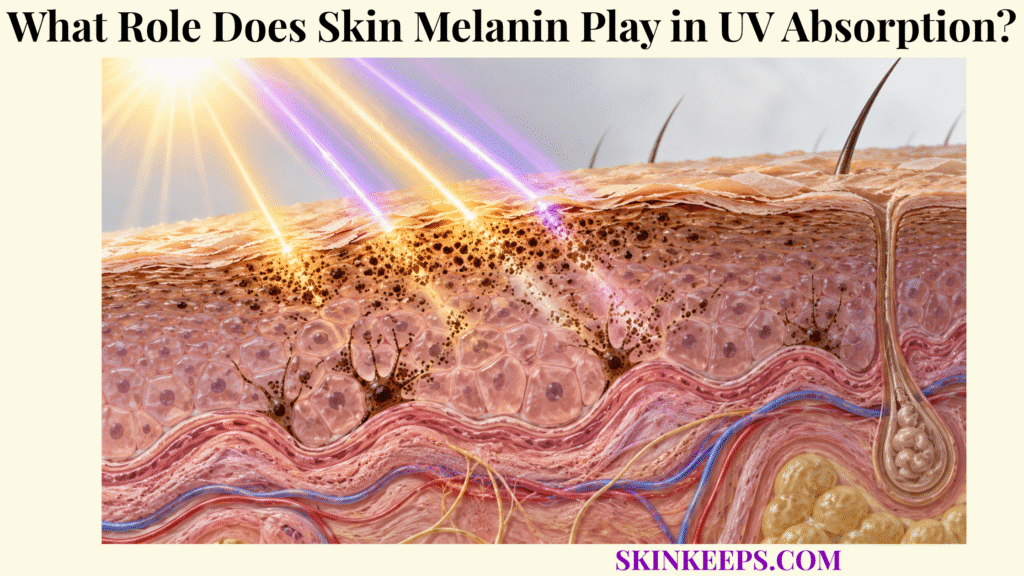
Skin melanin reduces ultraviolet exposure by absorbing and attenuating part of incoming UVA and UVB inside epidermal melanosomes, especially after those pigment-containing organelles are transferred from melanocytes to surrounding keratinocytes. Basal melanocytes manufacture melanin, melanosomes package it, and keratinocyte distribution places pigment where it can intercept radiation before more of that energy reaches vulnerable cellular targets.
This page follows that pathway from pigment production to optical absorption, supranuclear DNA shielding, UV-induced pigmentation, and the different photochemical behavior of eumelanin and pheomelanin. Melanin lowers effective ultraviolet exposure; it does not make epidermal cells UV-proof, and tanning does not convert prior UV injury into complete future protection.
Where Does Skin Melanin Provide UV Protection?
Skin melanin provides UV protection mainly within the epidermis, where basal melanocytes synthesize pigment inside melanosomes and transfer those organelles to surrounding keratinocytes that distribute melanin across UV-exposed tissue. This pigment system operates as one component of broader skin UV defense, rather than as an isolated optical coating.
Where Do Melanocytes Produce Skin Melanin?
Melanocytes located mainly along the epidermal basal layer synthesize melanin inside specialized membrane-bound organelles called melanosomes. Their dendritic shape allows one melanocyte to interact with multiple neighboring keratinocytes, creating a functional epidermal melanin unit without requiring melanocytes themselves to cover most of the skin surface.
Melanin synthesis depends on melanogenic machinery that includes tyrosinase and related proteins such as TYRP1 and DCT/TYRP2. These enzymes help convert tyrosine-derived intermediates into pigment as melanosomes mature, but the photoprotective outcome depends on subsequent pigment transfer and distribution as well as synthesis.
How Is Skin Melanin Packaged Inside Melanosomes?
Melanosomes provide the specialized biochemical compartment in which melanogenic enzymes generate and accumulate eumelanin- and pheomelanin-containing pigment. Progressive organelle maturation organizes the pigment before mature melanosomes are moved toward melanocyte dendrites for transfer.
Melanocyte pigment-producing epidermal cell.
Melanosome organelle in which melanin is synthesized, stored, matured, and transported.
Melanin family of biological pigments that includes eumelanin and pheomelanin.
How Does Skin Melanin Reach Keratinocytes?
Mature pigment-containing melanosomes move through melanocyte dendrites and are transferred to neighboring keratinocytes, distributing pigment far beyond the relatively small population of melanocytes themselves. Several transfer models have experimental support, including exocytosis followed by uptake, cytophagocytosis-like mechanisms, and transfer of melanin-containing material; no single mechanism should be treated as universally exclusive.
Why Does Melanosome Distribution Matter for Skin Melanin Protection?
Melanosome distribution determines where pigment intercepts incoming light, so pigment amount, intracellular positioning, dispersion, and persistence all influence the photoprotection produced by the same broad melanocyte system. Constitutive pigmentation therefore reflects melanin production and melanosome biology much more strongly than large differences in melanocyte number.
| Structure / Process | Main Location | Photoprotective Role |
|---|---|---|
| Melanocyte | Basal epidermis | Synthesizes melanin and produces melanosomes |
| Melanosome | Inside melanocyte, then keratinocyte | Packages and carries melanin |
| Melanocyte dendrites | Extending among keratinocytes | Facilitate pigment distribution |
| Keratinocyte | Across viable epidermis | Receives and distributes melanosomes |
| Supranuclear melanosome cap | Around or above keratinocyte nucleus | Reduces UV delivery toward nuclear DNA |
| Epidermal melanin | Distributed through keratinocytes | Absorbs and scatters part of incident radiation |
Constitutive pigmentation depends heavily on melanin production, pigment chemistry, melanosome characteristics, transfer, distribution, and persistence rather than simply on large differences in melanocyte number.
How Does Skin Melanin Absorb UV Radiation?
Skin melanin absorbs a broad range of ultraviolet wavelengths, reducing the fraction of incident UVA and UVB that continues through the epidermis toward proteins, membranes, and nuclear DNA. Absorption is wavelength-dependent rather than uniform, and melanin also contributes a smaller optical-scattering component.
Which UV Radiation Can Skin Melanin Absorb?
Melanin absorbs broadly across the ultraviolet spectrum and into visible wavelengths, with generally stronger optical absorption toward shorter wavelengths. UVA spans roughly 320–400 nm and UVB roughly 280–320 nm; both can be attenuated by epidermal melanin, although their depth distribution and dominant biological effects differ.
UVB is especially efficient at generating direct DNA photoproducts such as cyclobutane pyrimidine dimers and 6-4 photoproducts. UVA penetrates more deeply on average and strongly contributes to reactive oxygen species and oxidative injury, but UVA can also contribute to DNA photolesions, so the two bands should not be reduced to an absolute “ROS versus DNA” split.
What Happens to UV Energy After Skin Melanin Absorbs It?
Eumelanin can deactivate absorbed electronic excitation extremely rapidly through non-radiative processes, reducing the opportunity for that energy to drive damaging photochemical reactions and ultimately dissipating much of it as molecular motion and thermal energy. Non-radiative relaxation means that excited electronic energy is redistributed without being re-emitted as visible light.
This is more precise than saying that melanin simply “turns sunlight into heat.” The protective step involves ultrafast electronic relaxation and molecular energy redistribution before the absorbed energy can participate in more reactive photochemistry.
Does Skin Melanin Only Absorb UV Radiation?
No. Melanin and melanosome-containing tissue can also scatter incident radiation, changing photon direction and adding optical attenuation beyond molecular absorption alone. Because this page focuses on UV absorption, scattering is best treated as a secondary pathway rather than the main mechanism.
Why Does Skin Melanin Not Block All UV Radiation?
Melanin provides partial attenuation rather than complete optical exclusion, so a fraction of incident ultraviolet radiation can still reach cellular chromophores and produce molecular damage. The effective protection depends on pigment amount, type, melanosome distribution, wavelength, intensity, and exposure duration.
How Does Skin Melanin Protect Cellular DNA?
Skin melanin protects cellular DNA by concentrating pigment-containing melanosomes around keratinocyte nuclei, where their position and optical absorption reduce the amount of ultraviolet radiation delivered toward nuclear genetic material. The detailed consequences of this mechanism belong to the Better-Owner page on skin melanin and UV-induced DNA protection.
How Do Melanosomes Form Supranuclear Caps Around Keratinocyte DNA?
Transferred melanosomes can accumulate in perinuclear and supranuclear positions within keratinocytes, forming pigment-rich caps between incident light and the cell nucleus. This “microparasol” geometry matters because pigment located directly in the optical path toward DNA can intercept radiation more effectively than pigment positioned elsewhere.
The broader importance of intracellular pigment arrangement is explored in skin melanin distribution and photoprotection, where melanosome dispersion, clustering, degradation, and persistence are the primary mechanisms.
How Does Supranuclear Skin Melanin Reduce DNA Exposure?
Supranuclear pigment reduces effective UV delivery toward nuclear DNA by absorbing and redirecting part of the radiation before it reaches DNA chromophores. Experimental and human evidence supports lower photolesion burden with greater physiological pigmentation, but the melanin cap is not a complete sunscreen and cannot exclude every photon.
Does More Skin Melanin Mean No UV-Induced DNA Damage?
No. Greater constitutive pigmentation generally reduces UV-induced DNA damage and can shift the depth distribution of photolesions toward more superficial epidermal layers, but CPDs, oxidative DNA injury, and other lesions can still form. Reduced damage is biologically meaningful, yet it is not equivalent to zero damage.
How Does Skin Melanin Respond to UV Exposure?
Skin melanin responds to ultraviolet exposure through rapid pigment changes and slower melanogenic signaling that increases melanin production and melanosome distribution after UV-induced cellular stress. The broader skin epidermal UV response includes many additional cellular defenses beyond pigmentation.
How Does UV-Damaged Skin Signal Melanocytes to Produce More Melanin?
UV-induced DNA stress in keratinocytes can stabilize p53, increasing POMC-derived melanocortin signaling such as α-MSH; α-MSH activates MC1R on melanocytes, which drives cAMP–CREB–MITF signaling and increases melanogenic machinery. This pathway connects the keratinocyte response to UV damage with a delayed increase in pigment synthesis and transfer.
UV stress → p53 → POMC → α-MSH → MC1R → cAMP / CREB → MITF → increased melanogenesis and melanosome transfer.
How Does Skin Melanin Change After UVA Exposure?
UVA can rapidly darken existing pigment through oxidation and redistribution mechanisms, producing immediate or persistent pigment darkening without requiring the same degree of new melanin synthesis that characterizes delayed UVB-associated tanning. Visible darkening therefore does not automatically indicate a large increase in biologically protective pigment.
How Does Skin Melanin Change After UVB Exposure?
UVB more strongly drives delayed melanogenesis by activating DNA-damage signaling, melanogenic enzyme expression, new melanin synthesis, and increased melanosome transfer to keratinocytes. This delayed response develops after the initiating exposure has already created cellular stress.
Does Tanning Make Skin Safe From Further UV Exposure?
No. UV-induced tanning provides limited additional photoprotection and is itself triggered by UV exposure that can already have produced DNA and cellular damage. Human studies show little meaningful DNA photoprotection from UVA-induced tanning and only modest protection from UVB-associated tanning, so deliberate tanning should not be treated as a photoprotective strategy.
How Do Skin Melanin Types Differ in UV Protection?
Skin melanin types differ substantially in photoprotection because eumelanin absorbs and dissipates ultraviolet energy more efficiently, whereas pheomelanin provides weaker optical protection and has greater potential for pro-oxidant photochemistry. Pigment chemistry therefore matters in addition to total pigment quantity.
How Does Eumelanin Protect Against UV Radiation?
Eumelanin is a dark brown-black pigment with broad optical absorption, rapid non-radiative energy dissipation, and radical-scavenging properties that make it the more photoprotective major human melanin type. It is relatively photostable, but it should not be described as perfectly inert or as a complete UV blocker.
How Does Pheomelanin Differ From Eumelanin?
Pheomelanin is a yellow-red sulfur-containing pigment that provides less effective UV photoprotection and can generate reactive oxygen species during photochemical degradation under experimental conditions. The accurate boundary is greater pro-oxidant potential—not the claim that pheomelanin is inherently “toxic pigment.”
Why Does Skin Melanin Amount Alone Not Determine Photoprotection?
Photoprotection depends not only on total melanin quantity but also on the eumelanin–pheomelanin balance, melanosome size, maturation, intracellular distribution, persistence, and the epidermal depth through which pigment remains. These variables form continuous individual phenotypes and should not be reduced to rigid racial categories.
| Pigment / Feature | Main Characteristics | UV-Protective Meaning |
|---|---|---|
| Eumelanin | Brown-black; broad absorption; relatively photostable | Stronger UV attenuation and energy dissipation |
| Pheomelanin | Yellow-red; sulfur-containing | Weaker photoprotection; greater pro-oxidant potential |
| Greater total epidermal melanin | More pigment available for absorption | Usually lowers UV penetration and photolesion burden |
| Larger / more persistent melanosomes | Greater pigment packaging and epidermal persistence | Can extend the depth and duration of pigment shielding |
| Dispersed melanosomes | Wider intracellular pigment distribution | Can increase effective optical coverage |
| Supranuclear positioning | Pigment concentrated around the nucleus | Improves DNA-directed shielding |
| Efficient melanosome transfer | Wider pigment distribution among keratinocytes | Expands protection beyond melanocyte cell bodies |
These characteristics vary continuously among individuals and should not be reduced to rigid racial categories or a single skin-tone label.
What Limits Skin Melanin UV Protection?
Skin melanin UV protection is limited because natural pigmentation can attenuate only part of incident radiation, while sufficiently intense, prolonged, or cumulative UVA and UVB exposure can still generate DNA photolesions, oxidative stress, inflammation, and broader solar injury.
Why Can UV Still Damage Highly Pigmented Skin?
Highly pigmented epidermis generally attenuates more incident UV than lightly pigmented epidermis, but it remains optically permeable enough for some ultraviolet photons to reach DNA, proteins, membranes, and other cellular chromophores. Greater natural pigmentation changes risk and response; it does not create immunity to UV injury.
How Can UVA Cause Damage Despite Skin Melanin?
UVA can penetrate deeply through the epidermis and promote reactive oxygen species, oxidative molecular damage, and photoaging pathways even when substantial melanin is present. UVA can also contribute to CPDs, so it should not be described as causing DNA damage only indirectly.
Management of ROS after photons escape pigment attenuation belongs primarily to skin antioxidant systems.
How Can UVB Cause Damage Despite Skin Melanin?
UVB that escapes melanin attenuation can be absorbed directly by DNA and generate lesions such as cyclobutane pyrimidine dimers and 6-4 photoproducts. Lesions that are not prevented by pigment must then be handled by systems such as skin DNA repair.
Why Is Melanin Only One Part of Solar Defense?
Residual UV injury requires additional defenses, including antioxidant systems, DNA repair, cell-cycle control, apoptosis, immune regulation, and broader epidermal responses, because pigmentation alone cannot neutralize every photon or molecular consequence. Those layers are integrated within skin defense against solar injury.
Should Natural Pigmentation Replace External UV Protection?
No. Natural pigmentation lowers several UV responses but does not eliminate sunburn, DNA damage, photoaging, pigmentary effects, or skin-cancer risk. Public-health guidance recommends UV protection across skin tones, and absence of visible erythema should not be interpreted as absence of molecular UV injury.
What Are the Key Takeaways About Skin Melanin and UV Absorption?
The key fact about skin melanin and UV absorption is that melanocyte-derived pigment becomes broadly photoprotective only after melanosomes distribute it through epidermal keratinocytes, where pigment amount, chemistry, positioning, and persistence determine how much ultraviolet radiation can be absorbed before reaching vulnerable cellular targets.
Eumelanin is especially effective at absorbing and safely dissipating UV excitation, while supranuclear melanosome positioning reduces UV delivery toward DNA. Yet all natural pigmentation has finite capacity: tanning begins after UV stress, residual radiation still produces photolesions and oxidative injury, and melanin functions as one component of a larger solar-defense network.
- Skin melanin is synthesized by basal epidermal melanocytes.
- Melanin is produced and packaged inside melanosomes.
- Melanosomes are transferred to surrounding keratinocytes, expanding pigment protection through the epidermis.
- Visible pigmentation is determined largely by melanin and melanosome biology, not simply by melanocyte number.
- Melanin absorbs part of UVA and UVB radiation.
- Eumelanin can dissipate absorbed excitation through rapid non-radiative pathways.
- Melanin can also contribute to optical scattering.
- Supranuclear melanosome caps reduce UV delivery toward keratinocyte DNA.
- Greater pigmentation generally reduces UV-induced DNA photolesions but does not eliminate them.
- UV exposure activates melanogenic signaling, including the p53–POMC–α-MSH–MC1R pathway.
- UVA and UVB induce different pigmentation responses.
- Tanning is a response to UV-induced stress and damage, not a harmless way to build protection.
- Eumelanin provides greater photoprotection than pheomelanin.
- Pheomelanin has weaker protection and greater pro-oxidant potential.
- Melanin amount, type, melanosome characteristics, and intracellular distribution all influence photoprotection.
- All constitutive pigmentation levels remain susceptible to UV injury.
- Melanin reduces UV exposure; it cannot completely prevent solar injury.
What Common Questions Do People Ask About Skin Melanin and UV Absorption?
Common questions about skin melanin focus on where pigment comes from, whether darker pigmentation blocks ultraviolet radiation, why eumelanin protects better than pheomelanin, and whether tanning creates meaningful UV protection.
Does Skin Melanin Absorb Both UVA and UVB?
Yes. Melanin has broad optical absorption that includes both UVA and UVB wavelengths, although absorption strength varies with wavelength and pigment chemistry, and some ultraviolet radiation still penetrates the epidermis.
Do People With Darker Skin Have More Melanocytes?
Not necessarily. Melanocyte density is broadly comparable across constitutive pigmentation groups at equivalent sites; major differences arise from melanin production, melanin type, melanosome size, number, distribution, transfer, and degradation.
How Do Melanosomes Protect Keratinocyte DNA?
Transferred melanosomes can accumulate around keratinocyte nuclei as supranuclear pigment caps, where their optical absorption and positioning reduce the amount of ultraviolet radiation reaching nuclear DNA.
Is Eumelanin More Protective Than Pheomelanin?
Yes. Eumelanin generally provides stronger broadband UV absorption and safer energy dissipation, while pheomelanin provides weaker photoprotection and has greater potential to generate reactive oxygen species under photochemical stress.
Does Tanning Make Skin Fully Protected From UV Damage?
No. UV-induced tanning provides only limited additional protection, and the UV exposure that triggers tanning can already cause DNA and cellular damage, so a tan should not be treated as complete or safe photoprotection.
Sources & Evidence
Mechanisms Regulating Melanogenesis — Melanocyte location, melanosome synthesis and transfer, pigmentation diversity, melanogenesis, and UVA/UVB-associated pigment responses.
The Photoprotection Mechanism in the Black–Brown Pigment Eumelanin — Eumelanin UV absorption and ultrafast non-radiative relaxation of absorbed excitation.
The Protective Role of Melanin Against UV Damage in Human Skin — Broadband melanin absorption, eumelanin–pheomelanin differences, antioxidant behavior, and limits of endogenous photoprotection.
Significance of Melanin Distribution in the Epidermis for Protection Against UV Light — Perinuclear pigment distribution, melanosome localization, and why physiological intracellular distribution matters for keratinocyte photoprotection.
The Impact of Skin Colour on Human Photobiological Responses — Human pigmentation phenotypes, reduced but persistent DNA photodamage, UVA/UVB photobiology, and natural-protection limits.
MITF and UV Responses in Skin: From Pigmentation to Addiction — p53–POMC–α-MSH–MC1R–cAMP–MITF signaling and pigment chemistry in the UV-tanning response.
Photobiological Implications of Melanin Photoprotection After UVB-Induced Tanning of Human Skin but Not UVA-Induced Tanning — Human evidence distinguishing UVA-associated tanning from modest UVB-associated photoprotection.
American Academy of Dermatology — The Latest in Sun Protection — Current guidance that UV protection remains relevant across skin tones and that tanning reflects skin damage rather than a safe protective strategy.
Medical note: This page is educational and does not diagnose pigment disorders, melanoma, photosensitivity, or sunburn severity. Seek professional assessment for a rapidly changing, bleeding, or non-healing pigmented lesion, persistent unexplained pigment change, unusual photosensitivity, or significant blistering; urgent care may be appropriate for severe widespread burns, dehydration, or systemic illness after major UV exposure.



