
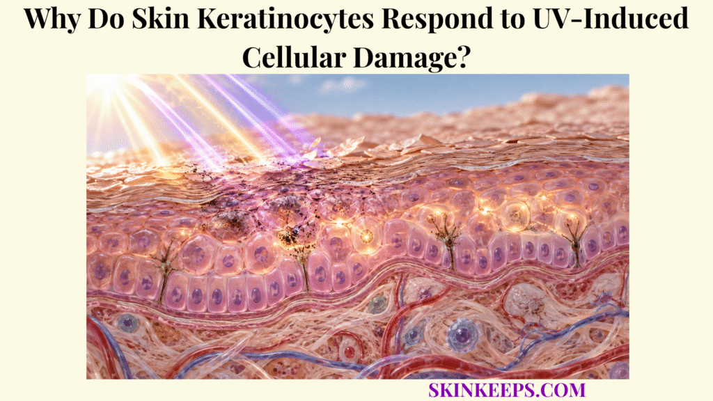
Skin keratinocytes respond to UV-induced cellular damage because ultraviolet radiation can injure their DNA, disturb redox balance, and alter cellular signaling, forcing each affected cell to coordinate repair, temporary cell-cycle control, communication with neighboring cells, survival, or apoptosis.
Because keratinocytes form most of the epidermis, these decisions protect both individual genomes and tissue integrity. Their defenses reduce UV injury but cannot guarantee complete repair after excessive or repeated exposure, so detection, repair, checkpoints, apoptosis, and cross-cell signaling must be understood as complementary rather than interchangeable processes.
How Do Skin Keratinocytes Detect UV-Induced Cellular Damage?
Skin keratinocytes detect UV-induced cellular damage through overlapping systems that recognize DNA photolesions, replication or transcription stress, reactive oxygen species, and UV-altered receptor or redox signaling.
What Damage Does UV Create Inside Skin Keratinocytes?
UV can produce direct DNA photolesions, reactive oxygen species, oxidized biomolecules, and replication or transcription stress inside keratinocytes. UVB is particularly efficient at forming cyclobutane pyrimidine dimers (CPDs) and 6-4 photoproducts (6-4PPs), while UVA strongly contributes to oxidative stress; the mechanisms overlap rather than forming an absolute UVA-versus-UVB split. This cell-level response operates within the broader skin UV defense network.
How Do Skin Keratinocytes Recognize UV-Damaged DNA?
Keratinocytes recognize UV-damaged DNA through lesion-recognition and DNA-damage-response systems. DDB2 and XPC participate in global-genome nucleotide excision repair recognition, while lesions that stall transcription or replication can generate downstream stress signals, including ATR–CHK1 checkpoint activity. p53 acts downstream as an important response regulator; it does not directly recognize CPDs.
How Do Skin Keratinocytes Detect Oxidative Stress?
UV-generated reactive species alter redox-sensitive proteins, membranes, kinases, and transcriptional regulators, allowing keratinocytes to translate oxidative imbalance into stress-response signaling. ROS generation is prominent with UVA but is not exclusive to UVA exposure.
Why Is Skin Keratinocyte UV Detection Not One Single Sensor?
UV damages several molecular systems simultaneously, so keratinocytes integrate information from DNA lesions, oxidative chemistry, membrane changes, stalled transcription, and replication stress rather than relying on one universal UV sensor. The broader timing of those events belongs to the skin epidermal UV response framework.
How Do Skin Keratinocytes Activate Damage-Control Signals?
Skin keratinocytes activate damage-control signals by engaging DNA-damage checkpoints, stress kinases, inflammatory transcription programs, and survival pathways that determine how the injured cell and surrounding epidermis respond.
How Does p53 Control Skin Keratinocyte UV Responses?
UV-induced DNA stress stabilizes p53, which can increase expression of genes involved in cell-cycle control, repair-associated responses, apoptosis, and paracrine pigmentation signaling. p21 and GADD45-related programs are important examples, but p53 is one regulator within a larger network rather than the sole UV-response pathway.
How Do MAPK Pathways Respond to UV-Damaged Keratinocytes?
UV exposure can activate ERK, JNK, and p38 signaling, which influence AP-1, NF-κB, stress transcription, inflammation, survival, differentiation, and cell-death decisions. MAPK activation is context-dependent and is not automatically protective.
How Do Skin Keratinocytes Release Inflammatory Signals?
UV-stressed keratinocytes can release mediators such as IL-1α, IL-6, TNF-α, prostaglandins, and chemokines. These signals coordinate local inflammation and neighboring-cell behavior, but excessive or persistent inflammatory signaling can also amplify tissue injury.
Why Can Survival Signals Be Both Protective and Risky?
EGFR and PI3K/AKT-associated survival signals can preserve cells whose injury remains repairable, but survival becomes biologically disadvantageous if a keratinocyte carrying persistent genomic damage escapes removal and later proliferates. Redox control is therefore complementary to, rather than a substitute for, skin antioxidant systems.
How Do Skin Keratinocytes Repair UV-Damaged DNA?
Skin keratinocytes repair UV-damaged DNA mainly through lesion-specific repair systems: nucleotide excision repair removes major UV photoproducts, while base excision repair corrects selected oxidative DNA lesions.
How Does Nucleotide Excision Repair Protect Skin Keratinocytes?
Nucleotide excision repair recognizes bulky UV-induced distortions, verifies and unwinds the damaged region, removes a short DNA segment containing the lesion, and resynthesizes the missing sequence from the undamaged strand. In humans, CPDs and 6-4PPs are major NER substrates; detailed proteins and kinetics belong to skin DNA repair.
What Is the Difference Between Global-Genome and Transcription-Coupled Repair?
Global-genome NER surveys DNA broadly, with DDB2 and XPC contributing to recognition, while transcription-coupled NER prioritizes lesions that stall RNA polymerase in actively transcribed genes. Both pathways converge on shared excision and resynthesis machinery.
How Do Skin Keratinocytes Repair Oxidative DNA Damage?
Selected oxidized DNA bases are repaired through base excision repair, in which lesion-specific glycosylases identify abnormal bases before the site is processed and resynthesized. OGG1 is a representative enzyme for 8-oxoG-related repair. This is distinct from antioxidant prevention: antioxidants limit reactive chemistry, whereas BER corrects selected lesions that have already formed.
Do Skin Keratinocytes Repair Every UV Lesion Successfully?
No. Repair efficiency varies with lesion type, genomic location, cellular condition, exposure burden, and repair capacity, so some lesions can persist. A DNA lesion is not automatically a mutation; mutation fixation requires persistence, inaccurate bypass or misrepair, or replication before correction.
How Do Skin Keratinocytes Stop Damaged Cells From Dividing?
Skin keratinocytes stop or slow damaged-cell division by activating DNA-damage checkpoints that inhibit cell-cycle progression while repair systems assess and process genomic injury.
How Does p53–p21 Signaling Pause Skin Keratinocyte Division?
p53 can increase p21/CDKN1A, which inhibits cyclin-dependent kinase activity and can reduce progression from G1 into DNA synthesis when genomic damage remains unresolved. p21 contributes to checkpoint control; it does not remove DNA lesions.
How Does ATR–CHK1 Control Replication Stress?
ATR–CHK1 signaling responds strongly to UV-associated replication stress and RPA-coated single-stranded DNA, helping stabilize stalled replication and delay inappropriate progression through S phase and later checkpoints. ATR is a signaling kinase, not a CPD-repair enzyme.
Why Does Cell-Cycle Delay Protect Skin Keratinocytes?
Delaying replication reduces the chance that unresolved lesions will be copied or bypassed before repair mechanisms have had sufficient opportunity to act. Checkpoint control therefore creates time and limits unsafe progression rather than performing repair itself.
What Happens if Skin Keratinocyte Checkpoints Fail?
Checkpoint failure increases the chance that damaged DNA enters replication or mitosis, creating additional opportunities for mutation fixation, genomic instability, or abnormal clonal survival. This raises biological risk but does not mean cancer develops automatically.
| Damage / Stress | Keratinocyte Action | Protective Result | Limitation |
|---|---|---|---|
| CPD / 6-4PP | NER | Removes bulky UV photolesions | Repair may be incomplete |
| Oxidized DNA base | BER | Restores selected damaged bases | Continued ROS can create new damage |
| Replication stress | ATR–CHK1 checkpoint | Stabilizes and slows replication | Does not remove the lesion itself |
| p53 activation | p21 + repair/apoptosis programs | Reduces unsafe proliferation | Outcome depends on cell context |
| ROS | Antioxidant/redox response | Limits further oxidative injury | Does not repair all DNA lesions |
| Moderate repairable damage | Survival + repair | Preserves viable keratinocyte | Persistent damage can remain |
| Severe genomic damage | Apoptosis | Removes highly damaged cell | Excess apoptosis contributes to sunburn |
| UV stress | POMC/α-MSH + paracrine signals | Coordinates melanocyte response | Protection is delayed |
| Cytokine release | Immune/local communication | Coordinates inflammatory response | Excess inflammation can add injury |
Keratinocyte damage control is distributed across repair, checkpoint, redox, survival, apoptotic, pigment-signaling, and immune-communication systems; no single pathway guarantees a safe outcome.
How Do Skin Keratinocytes Remove Severely Damaged Cells?
Skin keratinocytes remove severely damaged cells through apoptosis when genomic or cellular injury exceeds a level compatible with safe continued survival.
What Is a UV-Induced Sunburn Cell?
A sunburn cell is an apoptotic epidermal keratinocyte generated after sufficiently damaging UV exposure. It is a histologic and cellular phenomenon, not a label for every visibly red cell in sunburned skin.
How Does the Mitochondrial Apoptosis Pathway Remove Damaged Keratinocytes?
Severe stress can shift BCL-2-family signaling toward BAX/BAK-dependent mitochondrial permeabilization, cytochrome-c release, and downstream caspase activation. This removes the cell rather than restoring its damaged genome.
Can Death-Receptor Signaling Also Trigger Keratinocyte Apoptosis?
Yes. UV can engage death-receptor systems such as Fas and TNF-family pathways, which can contribute to caspase activation alongside mitochondrial mechanisms. The relative contribution depends on cell state and experimental context.
Is All Skin Keratinocyte UV Apoptosis Controlled by p53?
No. p53 is an important regulator, but keratinocyte differentiation state and parallel death pathways allow some UV-induced apoptosis to occur through partially p53-independent mechanisms.
Why Is Apoptosis Protective if It Kills Skin Keratinocytes?
Apoptosis can protect the epidermis at the tissue level by preventing severely damaged keratinocytes from surviving and replicating potentially dangerous genomic alterations. More apoptosis is not automatically better, because excessive cell loss contributes to sunburn, inflammation, and barrier disruption.
How Do Skin Keratinocytes Coordinate UV Defense With Other Skin Cells?
Skin keratinocytes coordinate UV defense by releasing paracrine and inflammatory signals that alter melanocyte pigmentation, immune-cell behavior, neighboring keratinocyte responses, and broader epidermal adaptation.
How Do Skin Keratinocytes Signal Melanocytes After UV Damage?
UV-induced p53 activation in keratinocytes can increase POMC-derived α-MSH, which activates MC1R on melanocytes and promotes cAMP–MITF-dependent melanogenesis. That delayed pigmentation contributes to later skin melanin and UV-induced DNA protection.
Are α-MSH Signals the Only Way Skin Keratinocytes Regulate Melanocytes?
No. Keratinocytes can also alter melanocyte activity through endothelin-1, stem-cell factor, inflammatory mediators, and other paracrine signals. The p53–POMC–α-MSH–MC1R pathway is a central model, not the only route.
How Does Melanin Return Protection to Skin Keratinocytes?
Melanocytes produce and transfer pigment-containing material toward keratinocytes, where pigment location and persistence affect functional protection. The spatial mechanism is covered in greater depth by skin melanin distribution and photoprotection.
How Do Skin Keratinocytes Communicate With Immune Cells After UV Exposure?
Keratinocyte-derived cytokines and chemokines can influence leukocyte recruitment, Langerhans-cell function, inflammatory signaling, and local immune regulation after UV injury. This cross-talk connects keratinocyte stress to the broader skin immune defense system.
Does UV Always Strengthen Skin Immune Defense?
No. UV exposure can simultaneously trigger inflammatory signals and immunoregulatory or immunosuppressive responses, so keratinocyte–immune communication should not be described as simple immune activation.
What Happens When Skin Keratinocyte UV Responses Are Overwhelmed?
Skin keratinocyte UV responses become overwhelmed when UV-induced photolesions, oxidative stress, and repeated cellular injury exceed the combined capacity of repair, checkpoints, redox control, apoptosis, and tissue-level coordination.
What Happens When DNA Damage Persists in Skin Keratinocytes?
Persistent lesions can interfere with transcription and replication, and lesions that survive until DNA synthesis can increase the opportunity for permanent mutations to become fixed. A lesion remains molecular damage; it becomes a mutation only if the sequence change is fixed.
What Happens When Oxidative Stress Exceeds Keratinocyte Defenses?
Excess reactive species can continue damaging DNA, proteins, lipids, and mitochondria while maintaining redox-sensitive inflammatory and stress pathways. This is why endogenous redox systems remain necessary even when checkpoints and repair are functioning.
How Can Repeated UV Injury Contribute to Photoaging?
Repeated keratinocyte stress can maintain inflammatory and oxidative signaling that contributes to chronic epidermal dysfunction and communicates with dermal remodeling pathways involved in photoaging. Detailed collagen, elastin, and matrix-remodeling biology belongs elsewhere.
How Can Failed Keratinocyte Control Contribute to Abnormal Cell Growth?
If keratinocytes carrying relevant genomic alterations escape repair, checkpoint control, and apoptosis, continued survival and clonal expansion can contribute over time to precancerous and malignant evolution. This is a mechanistic risk pathway, not an individual cancer prediction, and keratinocyte control remains only one layer of skin defense against solar injury.
Can Skin Keratinocyte Damage Occur Without Visible Sunburn?
Yes. Molecular DNA and oxidative injury can occur without obvious erythema, so absence of visible sunburn does not show that keratinocyte damage-control systems prevented all injury.
What Are the Key Takeaways About Skin Keratinocyte UV Responses?
The key fact about skin keratinocyte UV responses is that keratinocytes do more than endure ultraviolet injury: they actively assess damage, coordinate repair and checkpoints, communicate with neighboring cells, and eliminate cells whose damage becomes unsafe to retain.
- Skin keratinocytes are major cellular targets of epidermal UV exposure.
- UVB is especially efficient at creating CPDs and 6-4PPs.
- UVA strongly promotes oxidative stress, but UVA and UVB damage pathways overlap.
- Keratinocytes do not rely on one universal UV-damage sensor.
- NER proteins, replication-stress pathways, redox signaling, and receptor pathways all contribute to damage recognition.
- p53 is a central downstream response regulator, not the UV photon sensor itself.
- ATR–CHK1 contributes strongly to UV-associated replication-stress checkpoints.
- p21 can slow cell-cycle progression after DNA damage.
- Checkpoints do not repair DNA; they create time and control while repair operates.
- NER repairs major UV photoproducts such as CPDs and 6-4PPs.
- BER repairs selected oxidative DNA lesions.
- Not every DNA lesion is repaired successfully.
- A DNA lesion does not automatically become a permanent mutation.
- MAPK, EGFR, and other stress pathways influence survival, inflammation, and cell-fate decisions.
- Keratinocytes release cytokines and chemokines after UV stress.
- Apoptosis removes severely damaged keratinocytes rather than repairing them.
- UV-induced keratinocyte apoptosis can be p53-dependent or partly p53-independent.
- Sunburn cells are apoptotic keratinocytes.
- Keratinocyte p53–POMC–α-MSH signaling can stimulate melanocyte melanogenesis.
- Keratinocytes also communicate with immune cells after UV injury.
- UV immune effects include both inflammation and immunoregulation/suppression.
- Persistent repair/checkpoint failure increases mutation and abnormal-clonal-growth opportunity.
- Keratinocyte defense reduces UV injury but cannot eliminate damage from excessive or repeated exposure.
What Common Questions Do People Ask About Skin Keratinocyte UV Responses?
Common questions about skin keratinocyte UV responses focus on how cells recognize UV injury, whether damaged DNA can always be repaired, why cell division stops, why some cells undergo apoptosis, and how keratinocytes signal melanocytes and immune cells.
How Do Skin Keratinocytes Know Their DNA Has Been Damaged by UV?
Keratinocytes use lesion-recognition and DNA-damage-response systems that detect abnormal DNA structures, stalled transcription or replication, and related stress signals; p53 then acts downstream as an important response regulator rather than as the primary UV-lesion sensor.
Can Skin Keratinocytes Repair All UV-Induced DNA Damage?
No. Nucleotide excision repair and other pathways remove many UV lesions, but efficiency varies with lesion type, genomic location, cellular condition, exposure burden, and repair capacity, so some damage can persist.
Why Do Skin Keratinocytes Stop Dividing After UV Damage?
DNA-damage checkpoints slow cell-cycle progression so damaged DNA is less likely to be replicated before repair systems have processed the lesions, with pathways such as p53–p21 and ATR–CHK1 contributing to this control.
Why Do Some UV-Damaged Skin Keratinocytes Undergo Apoptosis?
Apoptosis removes cells whose damage is sufficiently severe that continued survival could threaten genomic integrity; this response can involve p53-dependent and p53-independent death pathways.
How Do Skin Keratinocytes Help Other Skin Cells Respond to UV?
UV-stressed keratinocytes release paracrine and inflammatory signals that can increase melanocyte pigmentation, alter neighboring keratinocyte behavior, and coordinate immune-cell activity throughout the local epidermal environment.
Sources & Evidence
S1 — UV Radiation and the Skin
https://pmc.ncbi.nlm.nih.gov/articles/PMC3709783/
Broad UVA/UVB epidermal effects, cytokines, p53, cell-cycle arrest, sunburn-cell apoptosis, and delayed epidermal responses.
S2 — Autophagy in UV Damage Response
https://pmc.ncbi.nlm.nih.gov/articles/PMC5466513/
p53/p21 control, GG-NER versus TC-NER, XPC/DDB2 recognition, and checkpoint context.
S3 — Focus on UV-Induced DNA Damage and Repair—Disease Relevance and Protective Strategies
https://pmc.ncbi.nlm.nih.gov/articles/PMC7582305/
ATR–CHK signaling, p53/p21 checkpoint control, apoptosis, and UV DNA-damage-response architecture.
S4 — Molecular Mechanisms of UV-Induced Apoptosis and Its Effects on Skin Residential Cells
https://pmc.ncbi.nlm.nih.gov/articles/PMC3634415/
Keratinocyte apoptosis, mitochondrial and death-receptor pathways, and p53-dependent versus partly p53-independent responses.
S5 — Participation of Keratinocyte- and Fibroblast-Derived Factors in Melanocyte Homeostasis, the Response to UV, and Pigmentary Disorders
https://pmc.ncbi.nlm.nih.gov/articles/PMC8906239/
Keratinocyte cytokines and paracrine melanocyte signals including endothelin-1 and melanocortin-related pathways.
S6 — UV Signaling Pathways Within the Skin
https://pmc.ncbi.nlm.nih.gov/articles/PMC4102648/
p53→POMC→α-MSH→MC1R→cAMP/MITF signaling and delayed pigment photoprotection.
S7 — DNA Damage, Apoptosis and Langerhans Cells—Activators of UV-Induced Immune Tolerance
https://pmc.ncbi.nlm.nih.gov/articles/PMC2718731/
Keratinocyte damage, apoptosis, Langerhans-cell interactions, and UV-induced immune regulation/tolerance.
S8 — American Academy of Dermatology — How to Prevent Skin Cancer
https://www.aad.org/public/diseases/skin-cancer/how-to-prevent
Current safety guidance that tanning reflects UV injury and deliberate tanning should be avoided.
Medical/Educational Disclaimer
This page is educational and does not diagnose or treat UV-related disease. Seek medical evaluation for concerning or changing lesions, unusual photosensitivity, or severe sun reactions. Keratinocyte damage-response pathways do not make deliberate UV exposure safe.



