
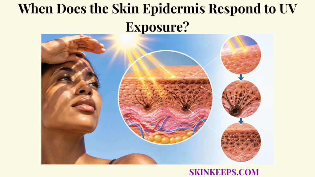
The skin epidermis begins responding to UV exposure as soon as ultraviolet energy produces molecular excitation, DNA photolesions, oxidative stress, or receptor-level stress signals, long before visible tanning or redness develops. The first events occur at the level of molecules and signaling networks rather than at the level of visible skin color.
The response then unfolds in stages: early kinase and inflammatory signaling, checkpoint control, lesion-specific repair or damaged-cell removal, delayed pigment production and transfer, and—after repeated exposure—epidermal thickening. These processes can reduce later injury, but they cannot undo all damage from the exposure that initiated them.
When Does the Skin Epidermis First Detect UV Exposure?
The skin epidermis first registers UV exposure when UVA and UVB interact with epidermal chromophores and cellular molecules, producing direct photochemical changes, reactive species, and stress signals that can begin within moments of irradiation. This is the true starting point of the broader skin UV defense response.
How Do UVA and UVB Reach the Skin Epidermis?
Both UVA and UVB reach the epidermis, but they differ in how strongly they are absorbed and which pathways they emphasize. UVB is absorbed more strongly in superficial tissue and is especially efficient at producing direct DNA photoproducts such as cyclobutane pyrimidine dimers and 6-4 photoproducts. UVA penetrates more deeply on average and strongly contributes to photosensitization, reactive oxygen species, and oxidative damage. These are tendencies rather than exclusive categories: UVA can also contribute to DNA photolesions, and UVB can also generate oxidative stress.
What First Detects UV Stress in the Skin Epidermis?
The earliest epidermal “detection” occurs when DNA and other endogenous chromophores absorb UV or when photosensitization generates reactive oxygen and nitrogen species that alter proteins, lipids, receptors, and signaling pathways. Keratinocytes therefore do not depend on one universal UV receptor. Functional detection begins through direct photochemistry, redox disturbance, membrane changes, and receptor-level stress signaling.
UV photolesion a structural DNA change created by ultraviolet exposure, such as a CPD or 6-4 photoproduct.
Checkpoint a cellular control system that slows or pauses cell-cycle progression when DNA damage is detected.
Photosensitization absorption of radiation by a cellular molecule followed by transfer of energy or electrons that can generate reactive chemistry.
How Quickly Can the Skin Epidermis Begin Signaling After UV Exposure?
Some receptor and kinase responses begin within minutes, whereas transcriptional programs such as p53-dependent checkpoint, inflammatory, and repair responses become more prominent over the following hours. Experimental human skin exposed to solar-simulated light has shown very early ERK activation, while p53 and COX-2 rise later over hours and apoptotic keratinocytes become more prominent by roughly the following day. These measurements illustrate sequence, not universal clinical time cutoffs.
How Does the Skin Epidermis Respond Immediately to UV Exposure?
The skin epidermis responds immediately to UV through photochemical injury, oxidative stress, membrane and kinase signaling, followed by early inflammatory, survival, checkpoint, and protective transcriptional responses. Damage and defense therefore begin in the same exposure window rather than occurring as neatly separated events.
How Does UV Create Immediate Stress in the Skin Epidermis?
UVB can rapidly generate DNA photoproducts, while UVA and UVB can generate reactive species that oxidize cellular molecules and activate stress pathways. Because photons can alter DNA or generate reactive chemistry at the moment of absorption, molecular injury can begin before erythema, tanning, peeling, or other visible changes appear.
How Do Keratinocytes Signal After UV Stress?
UV-stressed keratinocytes activate receptor and kinase networks including EGFR and MAPK pathways and release cytokines, prostaglandins, and other mediators. These signals coordinate inflammation, survival, repair, intercellular communication, and later adaptation. Inflammation is neither purely protective nor purely harmful: it participates in tissue communication while excessive inflammatory signaling can add to injury.
How Do Cellular Checkpoints Control UV-Damaged Keratinocytes?
DNA-damage checkpoints can slow cell-cycle progression, giving keratinocytes additional opportunity to repair lesions before DNA replication proceeds. p53 becomes stabilized after genotoxic stress and can induce p21 and other control programs, but p53 is not the epidermis’s instantaneous photon detector and does not automatically drive every damaged cell into apoptosis. The cell-fate logic is covered more deeply in the page on keratinocyte response to UV-induced cellular damage.
How Does the Skin Epidermis Increase Melanin After UV Exposure?
The skin epidermis increases melanin after UV exposure when stressed keratinocytes signal to melanocytes, stimulating melanogenesis, pigment-organelle production, transfer, and later deployment within keratinocytes. This is a delayed adaptive response rather than the first event after UV reaches the epidermis.
How Do UV-Stressed Keratinocytes Activate Melanocytes?
UV-induced DNA stress can stabilize p53 in keratinocytes, increasing POMC-derived melanocortin signaling such as α-MSH. α-MSH activates MC1R on melanocytes, which raises cAMP signaling and promotes CREB/MITF-dependent expression of melanogenic machinery. The sequence can be summarized as p53 → POMC → α-MSH → MC1R → cAMP → MITF, but the full UV response involves many parallel signals.
How Does Melanogenesis Increase Skin Epidermis Photoprotection?
Increased melanogenesis produces more pigment-containing organelles that can be transferred to keratinocytes, increasing the amount of UV-absorbing pigment distributed through the epidermis. The detailed photon-absorption mechanism belongs to skin melanin UV absorption; here the key timing point is that additional pigment appears after UV-triggered signaling has already begun.
How Does Melanosome Transfer Improve Nuclear Protection?
Transferred pigment can accumulate around keratinocyte nuclei, increasing UV interception near DNA and strengthening later photoprotection. The genome-directed consequence is explained in skin melanin and UV-induced DNA protection, while the spatial arrangement of pigment is owned by skin melanin distribution and photoprotection.
Is Increased Pigmentation an Immediate UV Response?
New melanin synthesis is mainly a delayed response that develops over hours to days. Existing pigment can darken or redistribute more rapidly after some UVA exposure, but immediate pigment darkening is not equivalent to newly synthesized melanin. New melanin production occurs after UV-induced signaling and therefore cannot undo the molecular injury that initiated the tanning response.
How Does the Skin Epidermis Repair UV-Induced Damage?
The skin epidermis limits UV-induced damage through complementary systems: DNA-repair pathways correct specific lesions, antioxidants reduce oxidative injury, checkpoints delay damaged-cell proliferation, and apoptosis removes cells whose injury exceeds safe recovery. Repair, prevention, arrest, and removal are distinct response classes.
How Does the Skin Epidermis Repair UV DNA Photolesions?
Nucleotide excision repair removes bulky UV photolesions such as CPDs and 6-4PPs through lesion recognition, excision of the damaged DNA segment, and resynthesis of the missing sequence. Detailed lesion recognition and repair kinetics belong to skin DNA repair; the timeline point here is that repair systems act after lesions have formed.
How Does the Skin Epidermis Handle Oxidative DNA Damage?
Selected oxidative DNA lesions are corrected through pathways including base excision repair, while antioxidant systems reduce the reactive-species burden that would otherwise generate additional damage. An oxidized base such as 8-oxoG is therefore conceptually different from a CPD, and its repair route differs accordingly.
How Do Skin Epidermis Antioxidant Systems Limit UV Injury?
Antioxidant defenses neutralize or metabolize reactive species and support redox homeostasis, limiting propagation of oxidative injury to DNA, proteins, and membrane lipids. Enzymatic and small-molecule systems are treated more fully under skin antioxidant systems. Antioxidants reduce oxidation; they do not directly excise CPDs or replace lesion-specific DNA repair.
When Does the Skin Epidermis Use Apoptosis?
When UV damage is sufficiently severe or unsafe to propagate, keratinocytes can activate apoptosis, removing damaged cells from the epidermal population rather than attempting indefinite survival. Classical apoptotic keratinocytes after UV are called sunburn cells. Apoptosis is a population-level safety mechanism, not a form of DNA repair, and not every UV-damaged keratinocyte is destined to die.
| UV-related problem | Epidermal response | Protective outcome |
|---|---|---|
| CPD / 6-4PP | Nucleotide excision repair | Removes bulky photolesions |
| Oxidized DNA bases | Base excision repair | Restores selected damaged bases |
| ROS accumulation | Antioxidant systems | Reduces further oxidative injury |
| DNA damage before replication | Cell-cycle checkpoint | Delays proliferation |
| Severe or unsafe cell damage | Apoptosis | Removes highly damaged cells |
| Keratinocyte UV stress | Melanocyte signaling | Increases later pigmentation |
| Repeated UV exposure | Epidermal proliferation | Increases later tissue thickness and attenuation |
These responses overlap. No single pathway guarantees that every UV-damaged cell is restored safely.
How Does the Skin Epidermis Adapt to Repeated UV Exposure?
The skin epidermis adapts to repeated UV exposure by increasing pigmentation and epidermal thickness while repeatedly activating repair and stress-control systems, producing greater later attenuation without eliminating cumulative UV injury. Adaptation changes future exposure conditions; it does not erase earlier lesions.
How Does Repeated UV Exposure Increase Epidermal Pigmentation?
Repeated UV signaling can sustain melanocyte activation, increase melanin synthesis, and increase pigment accumulation within epidermal keratinocytes. The resulting pigmentation can attenuate some later UV, but the exposures that induced it are themselves photobiologically active and can add DNA and oxidative stress.
How Does Repeated UV Exposure Thicken the Skin Epidermis?
After early stress responses, increased keratinocyte proliferation can produce epidermal hyperplasia and greater stratum-corneum thickness. This delayed structural response is treated in greater depth under skin epidermal thickening.
How Does Epidermal Thickening Increase Photoprotection?
A thicker epidermis and stratum corneum increase the physical path incoming ultraviolet radiation must traverse and can reduce the fraction reaching deeper epidermal cells. This is a real adaptive attenuation mechanism, but its magnitude varies with exposure conditions and tissue characteristics, so it should not be translated into a universal SPF-equivalent value.
Does Repeated Skin Epidermis Adaptation Eliminate UV Risk?
No. Pigmentation and thickening improve natural attenuation, but repeated UV exposure simultaneously adds new DNA lesions, oxidative stress, inflammatory signaling, and cumulative opportunities for mutation. Adaptive hyperplasia is therefore not evidence that repeated UV exposure is healthy or that the epidermis has become UV-immune.
When Does the Skin Epidermis Fail to Control UV Damage?
The skin epidermis fails to fully control UV damage when the rate or cumulative burden of photolesions, oxidative injury, and cellular stress exceeds the combined capacity of pigment protection, antioxidants, checkpoints, DNA repair, and damaged-cell removal. Failure can be partial: residual lesions or inflammatory stress may remain even when many defense systems are functioning.
What Happens When UV DNA Lesions Are Not Repaired?
DNA lesions that persist until replication or are repaired incorrectly can become permanent sequence changes, increasing the opportunity for mutation accumulation. A lesion is not automatically a mutation; successful repair can restore normal DNA before replication fixes the change into the genome.
What Happens When Oxidative Stress Persists in the Skin Epidermis?
Persistent reactive-species production can continue oxidizing DNA, proteins, and lipids while activating inflammatory and stress-signaling pathways that interfere with normal epidermal homeostasis. Oxidative load therefore adds to photochemical injury rather than representing a completely separate process.
How Can Repeated UV Exposure Promote Photoaging?
Repeated UV stress contributes to photoaging through cumulative oxidative, inflammatory, genomic, and tissue-remodeling effects that extend beyond the epidermis into the dermis. Detailed collagen fragmentation, MMP networks, and elastin changes belong to broader photoaging content rather than this response-timeline page.
How Can Uncontrolled UV Damage Increase Mutation Risk?
Repeated survival and proliferation of cells carrying incompletely repaired UV lesions can permit mutation accumulation and clonal expansion, contributing over time to photocarcinogenic risk. UV is mutagenic, but no single exposure allows an individual cancer probability to be inferred from this mechanism alone.
Why Does Skin Epidermis Failure Not Require Visible Sunburn?
Molecular UV damage can occur without obvious erythema, so absence of visible sunburn does not prove that epidermal DNA and oxidative-stress defenses prevented injury. Visible redness is a downstream clinical response, not a complete molecular dosimeter.
What Are the Key Takeaways About the Skin Epidermis and UV Exposure?
The key fact about the skin epidermis and UV exposure is that the response begins at the molecular level immediately, then unfolds through increasingly delayed signaling, repair, cell removal, pigmentation, and structural adaptation. The order matters because the adaptive layers develop after the initiating photons have already interacted with epidermal molecules.
- The skin epidermis starts responding before visible redness or tanning appears.
- UV response begins when photons alter DNA, chromophores, redox chemistry, or cell signaling.
- There is no single universal epidermal UV sensor.
- UVB is particularly efficient at generating CPDs and 6-4PPs.
- UVA strongly contributes to ROS-driven oxidative injury.
- UVA and UVB mechanisms overlap.
- Receptor and MAPK signaling can begin within minutes.
- p53 and other damage-control programs become prominent over the following hours.
- Cell-cycle checkpoints reduce replication of damaged DNA.
- NER removes major UV photoproducts.
- BER corrects selected oxidative DNA lesions.
- Antioxidants limit ROS; they do not directly repair CPDs.
- Apoptosis removes severely damaged cells; it is not DNA repair.
- Delayed pigmentation begins after UV-induced cellular stress.
- p53–POMC–α-MSH–MC1R signaling contributes to UV-induced melanogenesis.
- Repeated exposure can increase epidermal thickness and pigmentation.
- Epidermal thickening provides additional physical UV attenuation.
- Adaptation does not cancel the molecular damage created by the exposures that triggered it.
- A DNA lesion is not automatically a permanent mutation.
- Absence of sunburn does not mean absence of molecular UV injury.
- Epidermal defenses remain finite and can be overwhelmed.
What Common Questions Do People Ask About the Skin Epidermis and UV Exposure?
Common questions about the skin epidermis and UV exposure focus on how quickly cells react, whether tanning is protective, how damaged DNA is handled, why the epidermis thickens, and whether damage can occur without sunburn.
How Quickly Does the Skin Epidermis Respond to UV Exposure?
The response begins essentially as soon as UV is absorbed at the molecular level, with photochemical damage, reactive-species generation, and early signaling occurring before slower checkpoint, repair, pigmentation, and structural adaptations develop over hours to days.
Does the Skin Epidermis Start Tanning Before UV Damage Occurs?
No. Delayed melanogenesis is stimulated partly by UV-induced stress and DNA-damage signaling, so the initiating exposure can produce molecular injury before additional protective pigment develops.
How Does the Skin Epidermis Repair UV-Damaged DNA?
The epidermis uses lesion-specific DNA-repair systems, including nucleotide excision repair for major UV photoproducts and base excision repair for selected oxidative lesions, while checkpoints slow proliferation during damage control.
Why Does the Skin Epidermis Become Thicker After Repeated UV Exposure?
Repeated UV exposure can stimulate delayed keratinocyte proliferation, producing epidermal hyperplasia and increased cornified thickness that attenuate some later ultraviolet transmission.
Can the Skin Epidermis Be Damaged by UV Without Sunburn?
Yes. DNA photolesions and oxidative stress can occur at exposures that do not produce obvious visible erythema, so sunburn is not a required marker of molecular UV injury.
Sources & Evidence
3. EGFR Activation and Ultraviolet Light-Induced Skin Carcinogenesis
Medical note: This page explains normal and stress-response photobiology; it does not diagnose sunburn severity, photosensitivity disorders, DNA-repair disorders, pigment disorders, actinic keratosis, or skin cancer. Seek medical or dermatologic assessment for unusual photosensitivity, significant blistering, severe pain, a changing or non-healing lesion, or persistent concerning sun-related changes. Extensive blistering, dehydration, altered consciousness, or other severe systemic symptoms after major UV exposure may require urgent care. Pigmentation and epidermal thickening are responses to UV stress, not reasons to deliberately create UV damage.



