
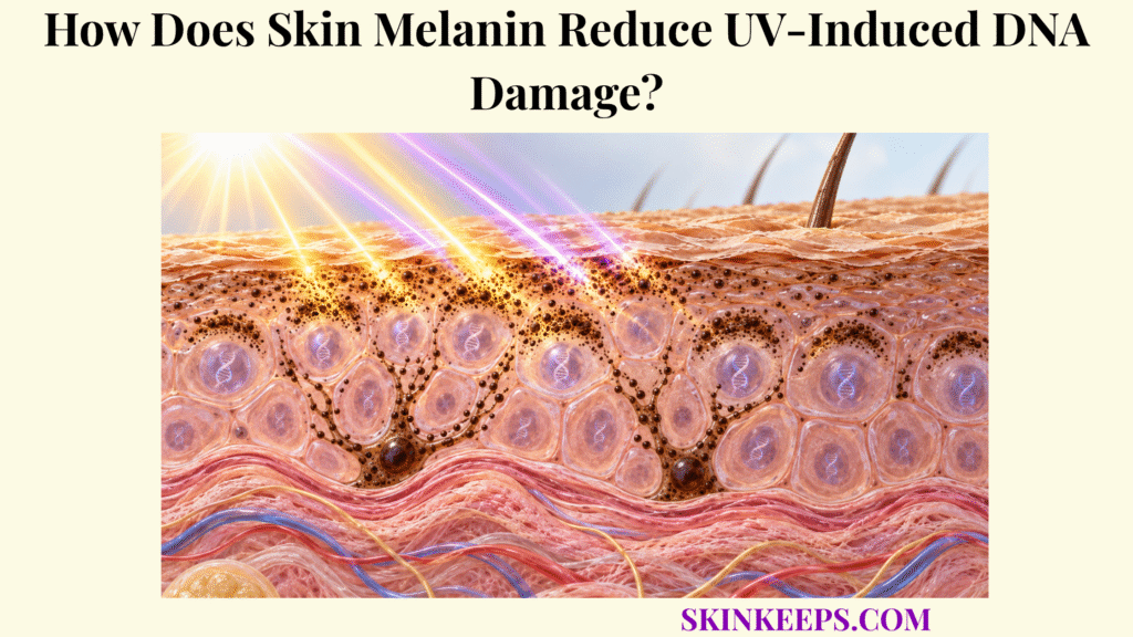
Skin melanin reduces UV-induced DNA damage by absorbing part of incoming UVA and UVB, dissipating absorbed excitation, and positioning pigment-containing melanosomes around keratinocyte nuclei so less radiation reaches genomic DNA. Melanin therefore lowers the photon and oxidative burden that can generate DNA lesions, but it does not make epidermal cells UV-proof.
This page follows that prevention pathway from melanosome transfer and supranuclear positioning to direct photolesions, oxidative DNA injury, adaptive melanogenesis, and the finite limits of natural pigmentation. Melanin reduces the amount of damage that forms; dedicated DNA-repair systems remain responsible for lesions that escape pigment-based prevention.
Where Does Skin Melanin Protect Cellular DNA?
Skin melanin protects cellular DNA mainly within the epidermis, where basal melanocytes produce pigment inside melanosomes and transfer those organelles to keratinocytes that contain most of the epidermal nuclei exposed to solar UV. This pigment-based genome protection operates within the broader network of skin UV defense, not as an isolated mechanism.
Where Is Skin Melanin Produced?
Epidermal melanocytes located mainly along the basal layer synthesize melanin inside specialized membrane-bound organelles called melanosomes. Tyrosinase and related melanogenic machinery generate eumelanin and pheomelanin within these organelles before pigment is distributed through the epidermis.
Why Must Skin Melanin Be Transferred to Keratinocytes?
Melanosome transfer expands photoprotection beyond melanocytes by positioning melanin inside the numerous keratinocytes whose nuclei make up most of the UV-exposed epidermal genome. The precise transfer process is biologically complex, but the functional result is widespread intracellular pigment deployment rather than pigment remaining confined to melanocyte cell bodies.
Melanocyte pigment-producing epidermal cell.
Melanosome organelle in which melanin is synthesized, matured, transported, and transferred.
DNA protection reduced lesion formation before downstream repair becomes necessary.
How Does Melanosome Distribution Affect DNA Protection?
DNA protection depends on where pigment is positioned inside keratinocytes, because perinuclear and supranuclear melanosomes intercept more UV travelling toward the nucleus than poorly positioned or extracellular pigment. Detailed spatial organization is the Better-Owner topic of skin melanin distribution and photoprotection.
| Melanin Mechanism | Cellular Location | DNA-Protective Effect |
|---|---|---|
| Melanin synthesis | Melanocyte melanosomes | Produces UV-absorbing pigment |
| Melanosome transfer | Melanocyte → keratinocyte | Extends pigment defense through the epidermis |
| UV absorption | Pigment throughout epidermis | Reduces photons continuing toward nuclei |
| Energy dissipation | Melanin molecular system | Reduces damaging photochemical excitation |
| Supranuclear positioning | Around keratinocyte nuclei | Reduces direct nuclear UV exposure |
| Eumelanin redox behavior | Pigmented cells | Can reduce part of the UV-driven oxidative burden |
| UV-induced melanogenesis | Melanocyte–keratinocyte signaling unit | Increases later pigmentation and partial future protection |
Melanin primarily prevents or reduces DNA injury; DNA-repair enzymes handle lesions that still form.
How Does Skin Melanin Absorb UV Before It Reaches DNA?
Skin melanin absorbs part of incoming UVA and UVB before those photons reach nuclear DNA, thereby lowering the effective radiation dose capable of producing direct photoproducts or initiating oxidative reactions inside skin cells. The broader optical physics are covered in skin melanin UV absorption; here the focus is the resulting reduction in DNA-lesion pressure.
How Does Skin Melanin Intercept UVA and UVB?
Melanin is a broad-spectrum absorber that intercepts wavelengths across UVA and UVB, although absorption strength varies with wavelength and pigment chemistry. Absorption lowers the number of photons continuing toward DNA, while pigment-containing organelles can also redirect some light through scattering.
What Happens to UV Energy After Skin Melanin Absorbs It?
Eumelanin can dissipate absorbed excitation through rapid non-radiative molecular relaxation, reducing the chance that the absorbed energy will trigger damaging photochemical reactions. The excitation is redistributed through molecular motions and ultimately thermalized; describing the mechanism simply as “UV becomes heat” would erase the ultrafast electronic steps that make this photophysics protective.
How Does Lower UV Transmission Reduce Direct DNA Damage?
When melanin absorbs photons before they reach the nucleus, fewer UV photons are available to excite DNA bases and generate cyclobutane pyrimidine dimers (CPDs) and 6-4 pyrimidine-pyrimidone photoproducts (6-4PPs). UVB is especially efficient at producing these direct photolesions, but UVA can also contribute to CPD formation, so the two wavebands should not be placed into an absolute direct-versus-indirect binary.
CPD UV-generated covalent linkage between adjacent pyrimidine bases.
6-4PP another pyrimidine photoproduct formed especially efficiently by shorter UV wavelengths.
Prevention reduces lesion formation before repair pathways act.
How Does Skin Melanin Shield Keratinocyte Nuclei?
Skin melanin shields keratinocyte nuclei when transferred melanosomes accumulate in perinuclear and supranuclear positions that place UV-absorbing pigment directly between incoming radiation and genomic DNA. Spatial deployment therefore adds a second layer of protection beyond total pigment quantity.
What Is a Supranuclear Melanin Cap?
A supranuclear melanin cap is a pigment-rich arrangement of transferred melanosomes positioned above or around the keratinocyte nucleus relative to the direction of incident UV. This perinuclear “microparasol” concentrates optical attenuation where the genome is located rather than distributing every melanosome randomly through the cytoplasm.
How Does Supranuclear Skin Melanin Reduce DNA Photolesions?
Supranuclear pigment absorbs and redirects part of the radiation that would otherwise reach DNA, lowering the effective ultraviolet dose delivered to nuclear chromophores. Experimental and imaging work supports reduced photodamage when pigment organelles are maintained near the nucleus, including effects on CPD and 6-4PP burden.
Why Does Skin Melanin Distribution Matter as Much as Pigment Amount?
Pigment must be positioned where it can intercept radiation effectively, so total melanin content alone does not fully predict nuclear photoprotection. Reconstructed-epidermis and cell-model findings show that abnormal or extracellular pigment distribution may protect less effectively than physiologic intracellular and perinuclear organization, reinforcing the importance of location as well as amount.
How Does Skin Melanin Limit UV-Driven Oxidative Damage?
Skin melanin limits part of UV-driven oxidative DNA damage by absorbing UVA before it excites cellular photosensitizers and, particularly through eumelanin, contributing radical-scavenging and redox-buffering activity. This is an important secondary DNA-protection mechanism, but it should not be expanded into the whole cutaneous antioxidant system.
How Does UVA Generate Oxidative DNA Damage?
UVA strongly promotes reactive oxygen species through interactions with endogenous cellular photosensitizers, producing oxidative stress that can modify DNA bases as well as proteins and membrane lipids. One commonly discussed oxidative DNA lesion is 8-oxo-7,8-dihydroguanine (8-oxoG), but UVA genotoxicity is not limited to oxidative lesions because UVA can also contribute to CPDs.
How Can Eumelanin Reduce UV-Driven Oxidative Stress?
Eumelanin can reduce oxidative stress by intercepting some incident UV and participating in radical-scavenging and electron-transfer reactions that lower parts of the reactive-species burden. Its broad absorption and relative photostability complement these redox effects, but eumelanin is not a perfect antioxidant and cannot neutralize all ROS generated during exposure.
Why Does Pheomelanin Provide Weaker Oxidative Protection?
Pheomelanin provides weaker photoprotection and can participate in pro-oxidant chemistry under UV or oxidative conditions, making pigment chemistry—not only pigment quantity—important for DNA protection. The appropriate boundary is greater pro-oxidant potential under particular conditions, not the claim that pheomelanin is intrinsically a “dangerous pigment.”
Is Skin Melanin the Main Antioxidant Defense System?
No. Melanin contributes to photochemical and redox protection, but enzymatic and small-molecule skin antioxidant systems provide additional control of reactive oxygen species throughout epidermal and dermal cells. Those systems own the deeper biology of SOD, catalase, glutathione, GPX, and related defenses.
How Does Skin Melanin Respond to Repeated UV Exposure?
Skin melanin responds to repeated UV exposure by increasing melanogenic signaling, pigment synthesis, and melanosome transfer, which can modestly reduce damage from later exposures but only after the initiating UV has already produced cellular stress. Delayed tanning is therefore a damage-triggered adaptation rather than a safe preparatory strategy.
How Does UV Damage Trigger Increased Skin Melanin Production?
UV-induced DNA damage in keratinocytes can stabilize p53, increase POMC and α-MSH signaling, activate melanocyte MC1R, and drive cAMP–MITF-dependent melanogenesis. This pathway sits within the broader skin epidermal UV response, while p53, checkpoints, apoptosis, and related stress biology are Better-Owner mechanisms of the keratinocyte response to UV-induced cellular damage.
How Does Increased Melanosome Transfer Improve Later Shielding?
Additional melanosome production and transfer increase the amount of pigment available within keratinocytes, allowing more UV to be intercepted before reaching cellular DNA during subsequent exposure. The effect varies with pigment chemistry, melanosome distribution, constitutive pigmentation, dose, wavelength, and exposure history; it should not be converted into a universal SPF-equivalent number.
Why Is Tanning Not a Safe DNA-Protection Strategy?
Tanning is not a safe DNA-protection strategy because the signaling that drives delayed pigmentation is initiated by UV exposure that can already have produced DNA photolesions and oxidative stress. Additional pigment may reduce some later lesion burden, but cumulative UV exposure continues to add genotoxic pressure rather than becoming harmless once the skin darkens.
What Limits Skin Melanin Protection From DNA Damage?
Skin melanin protection from DNA damage is limited because pigment cannot absorb every UV photon, oxidative reactions can still occur, and sufficiently intense or cumulative exposure can exceed natural photoprotection even in deeply pigmented skin. Protection is also chemically complex: eumelanin is generally more photoprotective than pheomelanin, and some melanin-related reactions can become damaging under particular experimental conditions.
Why Can UVB Still Damage DNA Despite Skin Melanin?
UVB photons that escape pigment attenuation can be absorbed directly by DNA bases and produce CPDs and 6-4 photoproducts. Greater constitutive pigmentation generally lowers this burden and can reduce deeper epidermal photodamage, but meaningful lesions still form when exposure exceeds the attenuating capacity of pigment.
Why Can UVA Still Damage DNA Despite Skin Melanin?
UVA that reaches living cells can drive reactive oxygen species and oxidative DNA injury and can also contribute to CPD formation through mechanisms that overlap with direct and photosensitized photochemistry. Lack of obvious erythema therefore does not demonstrate that genomic injury did not occur.
Can Skin Melanin Ever Contribute to DNA Damage?
Although melanin is primarily photoprotective in normal epidermal organization, experimental melanocyte research shows that UV-generated reactive chemistry can chemically excite melanin fragments after exposure and produce delayed CPDs through a process called chemiexcitation. These “dark CPDs” have been especially prominent in pheomelanin-rich experimental contexts; the finding adds a mechanistic limit without overturning the dominant protective role of properly organized epidermal melanin.
Evidence boundary: chemiexcitation is a mechanistic experimental phenomenon. It should not be generalized into the claim that normal epidermal melanin is primarily DNA-damaging.
Why Does Skin Still Need DNA Repair After Melanin Protection?
Residual photolesions remain after melanin attenuation, so nucleotide-excision, base-excision, checkpoint, and cell-death systems are required to prevent unrepaired DNA damage from becoming permanent mutations. Those downstream mechanisms are owned by skin DNA repair; melanin itself is best understood as upstream load reduction rather than a repair enzyme.
MC1R signaling also influences antioxidant and DNA-damage-response pathways beyond pigment production, but those pigment-independent effects should not be misattributed to melanin molecules themselves.
Why Is Skin Melanin Only One Part of Solar Defense?
Melanin functions alongside antioxidant systems, DNA repair, cell-cycle control, apoptosis, immune responses, and broader epidermal adaptations. These layers operate together within skin defense against solar injury, which is why natural pigmentation should never be treated as complete protection from cumulative UV exposure.
What Are the Key Takeaways About Skin Melanin and DNA Protection?
The key fact about skin melanin and DNA protection is that pigment lowers the amount of ultraviolet energy reaching epidermal nuclei and therefore reduces—but cannot eliminate—the direct and oxidative DNA damage produced by solar UV exposure.
Protection depends on pigment chemistry, total amount, melanosome transfer, and intracellular positioning. Eumelanin provides stronger absorption and energy dissipation, supranuclear pigment reduces nuclear exposure, and adaptive melanogenesis can modestly increase later attenuation; lesions that still form require downstream antioxidant, repair, checkpoint, and cell-death systems.
- Skin melanin is synthesized inside melanocyte melanosomes.
- Melanosomes must be transferred to keratinocytes for broad epidermal DNA protection.
- Melanin absorbs part of incoming UVA and UVB before those photons reach nuclear DNA.
- Eumelanin dissipates absorbed excitation efficiently through non-radiative pathways.
- Supranuclear melanin caps position pigment around keratinocyte nuclei for additional DNA shielding.
- Melanin reduces formation of UV-induced DNA photolesions, including CPDs.
- Greater constitutive pigmentation generally reduces DNA damage, especially in deeper epidermal layers.
- Pigment distribution matters in addition to pigment amount.
- UVA strongly contributes to oxidative DNA damage and can also contribute to photoproduct formation.
- UVB strongly produces direct DNA lesions such as CPDs and 6-4PPs.
- Eumelanin generally provides stronger DNA photoprotection than pheomelanin.
- Pheomelanin has greater pro-oxidant potential under some conditions.
- UV-induced tanning begins with UV stress and DNA-damage signaling.
- Additional pigmentation can reduce some later UV damage but does not make tanning safe.
- Melanin primarily prevents damage; it does not repair DNA lesions itself.
- Residual DNA injury requires DNA-repair and other cellular defense systems.
- Natural pigmentation reduces UV-induced DNA damage but cannot eliminate it.
What Common Questions Do People Ask About Skin Melanin and DNA Protection?
Common questions about skin melanin and DNA protection focus on whether pigment prevents DNA damage completely, how melanosome caps work, whether eumelanin protects better than pheomelanin, and whether tanning prevents future UV damage.
Can Skin Melanin Completely Prevent UV-Induced DNA Damage?
No. Melanin absorbs and attenuates part of incoming ultraviolet radiation, so more pigmentation generally lowers DNA-damage burden, but CPDs, oxidative DNA lesions, and other UV effects can still occur in all pigmentation phenotypes.
How Do Melanin Caps Protect Keratinocyte DNA?
Transferred melanosomes can organize above and around keratinocyte nuclei, placing UV-absorbing pigment between incident radiation and DNA so fewer photons reach nuclear chromophores.
Does Eumelanin Protect DNA Better Than Pheomelanin?
Generally yes. Eumelanin provides stronger broadband absorption and more efficient energy dissipation, while pheomelanin provides weaker photoprotection and has greater pro-oxidant potential under some UV conditions.
Does Tanning Prevent Future DNA Damage?
Tanning can modestly reduce some damage from later exposure by increasing epidermal pigmentation, but the UV exposure that creates a tan already produces DNA and cellular stress, so tanning should not be treated as a safe photoprotective strategy.
Does Skin Melanin Repair DNA After UV Damage?
No. Melanin primarily reduces how much damage forms in the first place; dedicated DNA-repair pathways must recognize and remove photolesions that still occur after UV exposure.
Sources & Evidence
Mechanisms Regulating Melanogenesis — Melanin synthesis in melanosomes, melanocyte–keratinocyte interaction, UV-regulated melanogenesis, and α-MSH/MC1R signaling.
Melanin’s Journey from Melanocytes to Keratinocytes — Melanosome transfer, keratinocyte uptake and processing, and the evidence that multiple transfer models may contribute.
Keratin Intermediate Filaments Mechanically Position Melanin Pigments for Genome Photoprotection — Modern evidence that K5/K14-dependent three-dimensional perinuclear pigment positioning contributes directly to DNA photoprotection.
The Impact of Skin Colour on Human Photobiological Responses — Human evidence linking constitutive pigmentation with reduced but persistent DNA photodamage and differences in epidermal depth distribution.
The Protective Role of Melanin Against UV Damage in Human Skin — Broadband UV absorption, eumelanin–pheomelanin differences, radical handling, and finite natural photoprotection.
Significance of Melanin Distribution in the Epidermis for Protection Against UV Light — Reconstructed-epidermis evidence showing that pigment distribution and intracellular localization affect photoprotection independently of total melanin content.
MC1R: Front and Center in the Bright Side of Dark Eumelanin and DNA Repair — Eumelanin biology and the distinction between pigment-dependent photoprotection and MC1R-associated repair/antioxidant responses.
UV-Induced Melanin Chemiexcitation: A New Mode of Melanoma Pathogenesis — Mechanistic experimental evidence for delayed “dark CPDs” through reactive chemistry and excited melanin fragments after UV exposure.
Medical note: This page is educational and does not diagnose melanoma, actinic keratosis, photosensitivity, DNA-repair disorders, pigmentation disorders, or sunburn severity. Seek professional assessment for a new or changing pigmented lesion, a bleeding or non-healing lesion, unusual photosensitivity, or severe/recurrent sun reactions; urgent care may be appropriate for extensive blistering burns, dehydration, or systemic illness after major UV exposure.



