
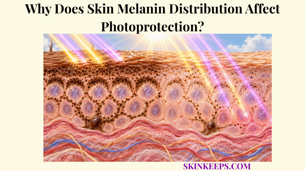
Skin melanin distribution affects photoprotection because UV-absorbing pigment protects most efficiently when melanocyte-derived melanin is transferred broadly into epidermal keratinocytes and positioned close to vulnerable cellular targets, especially their nuclei. Melanin quantity matters, but location determines whether that pigment lies along the optical pathways where ultraviolet radiation is actually travelling.
This page follows pigment from melanocyte production and keratinocyte transfer to perinuclear and supranuclear positioning, coverage across the epidermis, differences in pigment-unit patterns, dynamic UV-driven redistribution, and the finite limits of natural photoprotection. Even well-distributed melanin attenuates rather than completely excludes UVA and UVB.
Where Does Skin Melanin Distribution Occur in the Epidermis?
Skin melanin distribution begins when basal epidermal melanocytes synthesize pigment inside melanosomes and transfer melanin-containing material to surrounding keratinocytes, which then determine much of the pigment’s final intracellular and vertical epidermal location. This spatial deployment is one component of the broader skin UV defense network.
Where Is Skin Melanin Produced Before Distribution?
Melanin is synthesized inside melanosomes within melanocytes located mainly along the stratum basale. These dendritic cells manufacture eumelanin and pheomelanin but remain a relatively small epidermal population; they do not migrate through the epidermis to cover every UV-exposed cell.
How Is Skin Melanin Transferred Into Keratinocytes?
Mature pigment is transported toward melanocyte dendrites and transferred into neighboring keratinocytes through specialized intercellular mechanisms that remain incompletely resolved. Proposed models include coupled exocytosis and uptake, shed pigment-containing vesicles, and cytophagocytosis-like transfer, so no single route should be presented as universally exclusive.
Melanosome the melanocyte organelle in which melanin is synthesized and matured.
Transferred melanin unit a cautious term for pigment after it enters a keratinocyte, where its compartmental organization may differ from the original melanocyte organelle.
Epidermal melanin unit the functional relationship between one melanocyte and multiple pigment-receiving keratinocytes.
Why Does Keratinocyte Skin Melanin Distribution Matter?
Keratinocyte distribution matters because keratinocytes constitute most of the epidermis, so transferring pigment into these cells expands photoprotection far beyond melanocyte cell bodies. Intercellular coverage determines how many UV-exposed keratinocytes contain pigment capable of intercepting radiation.
How Far Through the Epidermis Can Skin Melanin Distribution Extend?
Transferred pigment can persist as keratinocytes differentiate and move toward upper epidermal layers, although persistence varies among pigmentation phenotypes and pigment-processing patterns. More persistent pigment can extend optical attenuation vertically, but persistence alone does not determine the final level of protection.
How Does Skin Melanin Distribution Shield Keratinocyte Nuclei?
Skin melanin distribution shields keratinocyte nuclei most effectively when pigment-containing organelles concentrate close to and above the nucleus, creating a supranuclear microparasol along the path of incoming ultraviolet radiation. This is a spatial extension of skin melanin and UV-induced DNA protection.
What Is a Supranuclear Skin Melanin Cap?
A supranuclear melanin cap is a polarized accumulation of transferred pigment on the UV-facing side of a keratinocyte nucleus. Perinuclear pigment lies close to or around the nucleus more generally; supranuclear pigment is preferentially positioned above it relative to incoming light, so the two terms describe related but not identical geometries.
How Does Skin Melanin Distribution Intercept UV Before It Reaches DNA?
By placing pigment directly along the optical path to the nucleus, supranuclear distribution allows melanin to absorb and scatter part of the incident radiation before it reaches DNA chromophores. A cap reduces nuclear exposure; it does not make the nucleus optically opaque.
Does the Three-Dimensional Position of Skin Melanin Matter?
Yes. Recent peer-reviewed mechanistic work shows that the three-dimensional proximity of pigment organelles to keratinocyte nuclei materially influences UVB genome photoprotection in experimental human keratinocytes. K5/K14 intermediate filaments, microtubules and plectin-mediated coupling participate in maintaining that geometry; these findings identify spatialization machinery rather than a single clinical determinant of human photoprotection.
Why Is Skin Melanin Amount Alone Insufficient for Nuclear Protection?
A large pigment quantity cannot provide optimal nuclear shielding when substantial pigment is extracellular, confined to too few keratinocytes, or poorly positioned relative to nuclei. Reconstructed-human-epidermis experiments illustrate this principle, but those model-specific findings—including nonphysiological distribution and some far-UV-C exposure—should not be generalized to normal deeply pigmented human skin.
How Does Skin Melanin Distribution Improve UV Absorption?
Skin melanin distribution improves functional UV absorption by placing pigment across more UV-exposed keratinocytes and positioning it along relevant optical pathways, increasing the probability that incoming radiation encounters melanin before vulnerable cellular targets. Detailed pigment photophysics belong to the Better-Owner page on skin melanin UV absorption.
Why Does Broader Skin Melanin Distribution Increase Protective Coverage?
When more keratinocytes contain pigment, fewer epidermal regions remain relatively unpigmented, so incoming UV has more opportunities to encounter a broadband absorber. This is an intercellular coverage effect: it concerns how widely pigment is deployed across cells rather than where pigment sits within any one cell.
Why Is Random Skin Melanin Dispersion Not Necessarily Optimal?
Photoprotection depends on strategic organization rather than random pigment spread. Pigment positioned near nuclei can protect genomic DNA more efficiently than the same pigment placed extracellularly or far from sensitive targets, so broad coverage and supranuclear targeting solve different spatial tasks.
How Does Distributed Skin Melanin Absorb and Dissipate UV Energy?
Melanin distributed throughout keratinocytes absorbs part of incoming UVA and UVB, while eumelanin can dissipate much of its electronic excitation through rapid non-radiative relaxation. Distribution changes where this interception can occur; it does not change the fundamental fact that absorption varies with wavelength, pigment chemistry and exposure intensity.
Does Wider Skin Melanin Distribution Automatically Mean More Photoprotection?
Not automatically. Protective effectiveness still depends on total melanin amount, eumelanin–pheomelanin composition, intracellular positioning, pigment persistence, UV wavelength and exposure intensity. Wider coverage is useful only within this larger optical and cellular context.
How Does Skin Melanin Distribution Differ Between Pigmentation Patterns?
Skin melanin distribution differs across constitutive pigmentation patterns through continuous variation in pigment amount, pigment-unit size, clustering, intracellular dispersion and persistence through epidermal layers rather than through sharply separate biological categories.
How Does Melanosome Size Influence Skin Melanin Distribution?
Larger and more heavily melanized pigment units can carry more pigment per structure and are more commonly described in more deeply pigmented epidermal phenotypes. Size alone does not determine protection because pigment chemistry, cell coverage, localization and persistence remain important.
How Does Clustered Skin Melanin Differ From Individually Dispersed Pigment?
Classic microscopy describes smaller pigment units more commonly grouped in clusters in lighter constitutive pigmentation, whereas more deeply pigmented epidermis more often shows larger pigment units distributed individually through keratinocyte cytoplasm. These are tendencies across pigmentation phenotypes, not fixed ethnic or racial categories.
Why Does Skin Melanin Persistence Through Epidermal Layers Matter?
Pigment that remains within differentiating keratinocytes can extend UV attenuation through a greater vertical portion of the epidermis and reduce radiation reaching deeper viable cells. More persistent pigment is one contributor to photoprotection, but it works together with quantity, chemistry and strategic localization.
Why Should Visible Skin Color Not Be Treated as an Exact UV-Protection Measure?
Visible pigmentation reflects several optical and biological variables and cannot precisely reveal eumelanin content, pheomelanin content, supranuclear positioning, pigment persistence, antioxidant capacity, DNA-repair capacity or an individual’s UV-response threshold. Skin tone, phototype, ancestry and exact pigment biology are related but not interchangeable variables.
| Pigment Feature | Typical Distribution Effect | Photoprotective Meaning |
|---|---|---|
| Greater pigment content | More melanin available across epidermis | Increases overall UV attenuation potential |
| Larger pigment units | Greater melanin content per pigment structure | Can increase local optical absorption |
| Smaller pigment units | Less pigment per individual unit | Protection depends strongly on total number and placement |
| Individually dispersed pigment | Broader cytoplasmic coverage | Can increase intracellular optical coverage |
| Clustered pigment | Pigment concentrated into groups | Coverage may be less spatially uniform |
| Supranuclear localization | Pigment concentrated near nucleus | Strong genome-directed photoprotection |
| Greater epidermal persistence | Pigment retained into upper layers | Extends vertical UV attenuation |
| Rapid pigment degradation | Less pigment retained in upper layers | Reduces persistence of optical protection |
| Eumelanin-rich pigment | Stronger absorption and safer energy dissipation | Generally stronger photoprotection |
| Pheomelanin-rich pigment | Weaker absorption and greater pro-oxidant potential | Generally weaker photoprotection |
These are population-level tendencies and mechanistic principles, not fixed categories that can be assigned reliably from race or visual skin color alone.
How Does Skin Melanin Distribution Respond to UV Exposure?
Skin melanin distribution responds to ultraviolet exposure through both increased melanocyte pigment production and active changes in how existing and newly transferred pigment is positioned within keratinocytes. This adaptation sits within the broader skin epidermal UV response.
How Does UV Exposure Increase Skin Melanin Production?
UV-induced cellular stress can activate keratinocyte–melanocyte signaling that increases melanogenesis and expands the pigment available for later distribution. At overview depth, UV-damaged keratinocytes can engage p53–POMC–α-MSH signaling, while melanocyte MC1R and MITF pathways increase pigment production. The cellular stress context is owned more fully by the page on keratinocyte response to UV-induced cellular damage.
How Does Increased Melanin Production Change Skin Melanin Distribution?
Additional melanogenesis can increase the number and pigment content of melanin units delivered to keratinocytes, broadening epidermal pigmentation when transfer and intracellular processing occur normally. Production and distribution are related, but they are not synonymous: new pigment still must reach the correct cells and positions.
Can Existing Skin Melanin Be Repositioned After UV Exposure?
Yes. Experimental human-cell and skin-explant evidence shows that UVA can stimulate keratinocytes to move pigment toward supranuclear regions. OPN3-associated calcium signaling, CaMKII, CREB, Akt and dynein/dynactin-linked transport have been implicated, supporting the broader point that distribution can change without waiting solely for new melanin synthesis.
Does UV-Induced Skin Melanin Redistribution Make Tanning Safe?
No. Increased pigment production and redistribution are adaptive responses to UV exposure that can provide limited additional shielding, but the exposure triggering those responses can already produce DNA photolesions and oxidative stress. A biological adaptation to UV injury is not a reason to deliberately seek the injury that induces it.
What Limits Skin Melanin Distribution as Photoprotection?
Skin melanin distribution has finite photoprotective capacity because even optimally positioned pigment absorbs only part of incoming radiation, while sufficient UVA and UVB can still reach cellular DNA and generate oxidative or photochemical injury.
Why Can UVA Still Cause Injury Despite Skin Melanin Distribution?
UVA can still penetrate pigment-containing epidermis and promote reactive oxygen species, oxidative molecular damage and some DNA photolesions despite natural melanin attenuation. Pigment therefore works alongside skin antioxidant systems, which manage reactive chemistry that optical interception does not prevent.
Why Can UVB Still Reach DNA Despite Skin Melanin Distribution?
UVB photons that escape pigment absorption can reach nuclear DNA and produce photoproducts such as cyclobutane pyrimidine dimers. Lesions that still form require downstream skin DNA repair; pigment distribution reduces the upstream load but does not perform lesion excision itself.
Why Does Skin Melanin Type Matter in Addition to Distribution?
Well-positioned pigment cannot be evaluated independently of pigment chemistry because eumelanin generally provides stronger broadband absorption and photochemical protection than pheomelanin. Two epidermal regions with similar spatial pigment coverage can therefore differ in functional protection if the pigment chemistry differs.
Can High or Prolonged UV Exposure Overwhelm Skin Melanin Distribution?
Yes. As UV intensity, duration or cumulative exposure increases, the photon and oxidative burden can exceed the protection provided by pigment quantity and spatial organization. No universal exposure threshold can be inferred from skin tone or melanin pattern alone.
Why Is Skin Melanin Distribution Only One Part of Photoprotection?
Residual ultraviolet injury requires epidermal stress responses, antioxidant control, DNA repair, checkpoint signaling, apoptosis and immune responses. Melanin distribution is therefore one layer of skin defense against solar injury, not a standalone guarantee against photodamage.
What Are the Key Takeaways About Skin Melanin Distribution?
The key fact about skin melanin distribution is that pigment protects most effectively when it is transferred broadly to epidermal keratinocytes and strategically positioned near vulnerable targets, especially keratinocyte nuclei, rather than being considered only as a total quantity of skin pigment.
- Melanocytes synthesize melanin, but keratinocytes provide much of its final protective distribution.
- Melanin must be transferred into keratinocytes to produce broad epidermal photoprotection.
- Pigment distribution has two levels: coverage across keratinocytes and localization inside each keratinocyte.
- Perinuclear pigment places melanin close to genomic DNA.
- Supranuclear melanin caps place pigment along the path of incoming UV.
- Three-dimensional pigment positioning influences UVB genome photoprotection in mechanistic human-keratinocyte models.
- K5/K14 intermediate filaments, microtubules and plectin participate in maintaining pigment position in experimental systems.
- Broad pigment coverage increases opportunities for UV interception, but random distribution is not automatically optimal.
- Melanin quantity alone does not determine photoprotection.
- Pigment-unit size, clustering, individual dispersion and persistence also influence protection.
- More deeply pigmented epidermal phenotypes tend to retain larger and more individually dispersed pigment units through more epidermal layers.
- Pigmentation patterns exist on a continuum and should not be reduced to rigid racial categories.
- UV can increase both melanogenesis and intracellular pigment redistribution.
- UVA-induced supranuclear cap formation demonstrates that distribution can be dynamic.
- Tanning is a limited damage-triggered adaptation, not a safe photoprotective strategy.
- Even optimally distributed melanin cannot absorb every UVA or UVB photon.
- Melanin distribution works with pigment amount, pigment type, antioxidants, DNA repair and other UV defenses.
What Common Questions Do People Ask About Skin Melanin Distribution?
Common questions about skin melanin distribution focus on why pigment surrounds keratinocyte nuclei, whether dispersed melanin protects better than clustered pigment, how distribution differs among pigmentation patterns, and whether UV exposure can change pigment placement.
Why Does Skin Melanin Distribution Form Caps Around Keratinocyte Nuclei?
Pigment-containing organelles can accumulate around and above keratinocyte nuclei, positioning UV-absorbing melanin between incoming radiation and DNA and thereby reducing nuclear UV exposure.
Is Dispersed Skin Melanin More Protective Than Clustered Melanin?
Broader intracellular dispersion can increase pigment coverage, but photoprotection depends on more than dispersion alone; total melanin, pigment type, supranuclear positioning, persistence, and UV wavelength all influence the final protective effect.
Does Darker Skin Have More Melanocytes to Improve Skin Melanin Distribution?
Not primarily. Epidermal melanocyte density is broadly comparable across constitutive pigmentation phenotypes at equivalent sites, while differences in melanin production, pigment-unit size, clustering, distribution, and persistence contribute more strongly to visible pigmentation.
Can UV Exposure Change Skin Melanin Distribution?
Yes. UV exposure can increase new melanogenesis and pigment transfer, and experimental evidence also shows that UVA can stimulate redistribution of existing pigment toward supranuclear keratinocyte regions.
Does Optimal Skin Melanin Distribution Completely Protect Against UV?
No. Strategic pigment distribution reduces UV exposure and DNA damage, but some UVA and UVB still penetrate pigmented epidermis, so oxidative stress and DNA photolesions can occur despite natural pigmentation.
Sources & Evidence
1. Melanin Transfer and Fate within Keratinocytes in Human Skin Pigmentation
2. Melanin Transfer in the Epidermis: The Pursuit of Skin Pigmentation Control Mechanisms
3. Significance of Melanin Distribution in the Epidermis for the Protective Effect Against UV Light
4. Keratin Intermediate Filaments Mechanically Position Melanin Pigments for Genome Photoprotection
5. The Impact of Skin Colour on Human Photobiological Responses
6. The Protective Role of Melanin Against UV Damage in Human Skin
7. Opsin 3 Mediates UVA-Induced Keratinocyte Supranuclear Melanin Cap Formation
8. MITF and UV Responses in Skin: From Pigmentation to Addiction
Medical note: This page explains normal pigmentation biology and natural photoprotection; it does not diagnose pigment disorders, photosensitivity, or skin cancer. Seek dermatologic evaluation for a changing or bleeding pigmented lesion, persistent unexplained pigment change, or unusual photosensitivity. Severe widespread blistering or systemic illness after major UV exposure warrants urgent medical assessment.



