
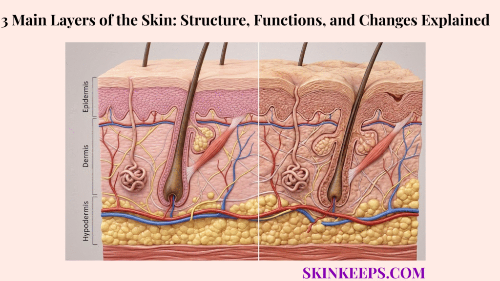
The essential distinction: Skin proper means epidermis + dermis. The hypodermis lies below it but is often shown as the third layer in illustrations because these tissues work closely together. This distinction matters when discussing protection, aging and visible skin changes.
This guide compares the three anatomical levels, shows how their functions interact, and explains why aging, UV exposure, and daily care affect different tissues. The diagrams are educational and not to scale.
What Are the Most Common Questions About the Three Skin Layers?
What are the three main layers of skin?
From the outside inward, the common teaching model shows the epidermis, dermis, and hypodermis. Anatomically, only the first two are skin proper; the hypodermis is subcutaneous tissue beneath them.
Which layer is thickest?
The dermis is typically thicker than the epidermis, although thickness varies by location. Subcutaneous tissue can be even thicker, but it is not part of skin proper.
Which skin layer contains blood vessels and nerves?
The dermis contains blood vessels and many sensory structures. The hypodermis also contains vessels and nerves; the normal epidermis is avascular.
Why Does the Three-Layer Skin Model Include the Hypodermis?
The epidermis and dermis together constitute skin proper, whereas the hypodermis is underlying subcutaneous tissue. Because the hypodermis supports and insulates the structures above, it is commonly included in a three-layer educational model of the skin region. [1]
Why Do the Epidermis and Dermis Form the Skin Proper?
The epidermis is a superficial layer of keratinized stratified squamous epithelium; the dermis is the connective tissue beneath it. Their interface connects the protective epithelial surface with the vascular and mechanically supportive tissue below. These two layers constitute the skin proper. [1]
Why Is the Hypodermis Shown Beneath the Skin Proper?
The hypodermis contains varying amounts of fat and loose connective tissue and helps connect skin to underlying fascia. Its location and functions make it useful to show alongside epidermis and dermis, even though it is not classified as skin proper. [1] [5]
| Model | Anatomical structures | Meaning |
|---|---|---|
| Skin proper | Epidermis + dermis | The two layers comprising the skin anatomically |
| Common three-layer model | Epidermis + dermis + hypodermis | Skin plus supportive subcutaneous tissue |
How Does the Epidermis Form the Skin’s Protective Barrier?
The epidermis limits excessive water loss and exposure to many external irritants through a renewing layer of keratinocytes and an organized surface lipid barrier. It also contains melanocytes involved in pigmentation. The epidermis is avascular, meaning it normally contains no blood vessels of its own. [1] [5]
How Do Keratinocytes and Lipids Limit Transepidermal Water Loss?
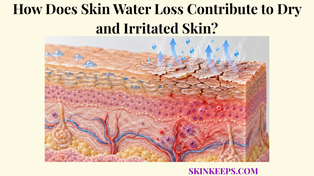
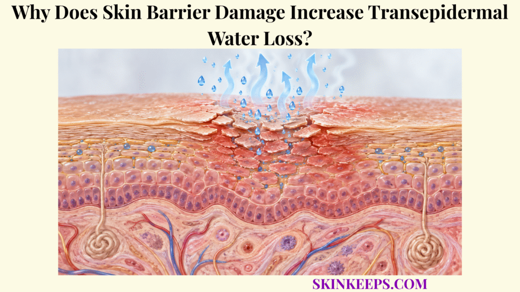
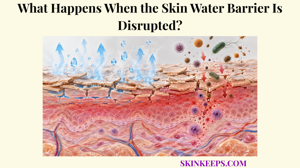
Keratinocytes mature into flattened corneocytes in the stratum corneum. Corneocytes and surrounding extracellular lipids form a “brick-and-mortar” arrangement that helps restrict excessive transepidermal water loss (TEWL). Healthy skin remains permeable to some water; the barrier regulates rather than eliminates its movement. [1] [5]
For more detail, see how the epidermis limits transepidermal water loss.
How Does Epidermal Renewal Maintain the Surface?
Keratinocytes arise in the deeper basal epidermis, differentiate, move toward the surface, and eventually shed. This continuous process renews the surface. Turnover varies with age, body site, and biological conditions, so a single fixed rate is not universal. [1] [3]
How Do Melanocytes and Melanin Influence Pigmentation?
Melanocytes produce melanin and transfer pigment to surrounding epidermal cells. Skin color reflects melanin production, type, distribution, and other individual factors. Melanin contributes to the response to ultraviolet (UV) exposure, but it does not prevent all sun-related injury. [1] [7]
How Do Langerhans Cells Contribute to Epidermal Immune Defense?
The skin barrier is not only physical. Langerhans cells are specialized immune cells found in the epidermis, particularly in the stratum spinosum. They help recognize and process foreign material and support communication with the immune system. They work alongside keratinocytes and other immune defenses; they do not make the epidermis completely resistant to infection. [1]
What Gives the Dermis Strength, Sensation, and Vascular Support?
The dermis is a living connective-tissue layer that provides the skin with mechanical strength, stretch and recoil, sensation, and circulation. It contains blood vessels, nerves, sweat glands and hair follicles, as well as collagen-rich extracellular matrix. [1] [2]
How Do the Papillary and Reticular Dermis Differ?
The superficial papillary dermis consists of relatively loose connective tissue near the epidermis. The deeper reticular dermis contains denser connective tissue that helps resist mechanical stress. These are related regions of one dermal layer. [1]
How Do Collagen and Elastic Fibers Influence Skin Mechanics?
Collagen fibers contribute tensile strength, while elastic fibers support stretch and recoil. Fibroblasts make and maintain components of the extracellular matrix. The organization of these structures influences tissue resilience; collagen and elastic fibers do not perform identical functions. [1] [8]
For a closer examination, explore how collagen and elastin are organized within the dermis.
How Do Dermal Blood Vessels Support the Avascular Epidermis?
Dermal capillaries supply oxygen and nutrients to surrounding living tissues. The epidermis itself does not normally contain vessels, so nutrients and oxygen reach it largely by diffusion from dermal circulation. This makes vascular support and surface barrier protection complementary roles. [1]
How Do Dermal Nerves, Sweat Glands, and Hair Follicles Function?
Sensory nerve structures detect stimuli including touch, pain, and temperature. Sweat glands participate in evaporative cooling, and hair follicles produce hairs as part of the skin appendage system. The density and function of these structures vary by body region. [1] [2] Sebaceous glands commonly open into hair follicles and produce sebum, an oily secretion that helps lubricate skin and hair. They are absent from some specialized regions, including the palms and soles. [1]
How Does the Hypodermis Provide Cushioning, Insulation, and Attachment?
The hypodermis is the subcutaneous region of adipose and loose connective tissue. It cushions against pressure, contributes to insulation and energy storage, and helps attach the skin to deeper fascia. Its thickness varies with body site and individual biology. [1] [5]
How Does Subcutaneous Fat Store Energy and Cushion Tissues?
Adipocytes store energy as lipids. Adipose lobules can deform under pressure, helping cushion underlying structures. Cushioning is not uniform: fat distribution and volume differ across the body and between individuals. [5]
How Does Subcutaneous Fat Contribute to Thermal Insulation?
Subcutaneous fat slows passive heat transfer between deeper tissues and the environment. This insulation is different from active temperature control through sweating or changes in skin blood flow. [2] [5]
For the insulation mechanism, read how hypodermal fat regulates body temperature.
How Does Fibrous Connective Tissue Attach Skin to Fascia?
Connective-tissue partitions and bands link subcutaneous tissue with dermis and underlying fascia. Their arrangement affects both attachment and how freely the skin can move over deeper structures. [1]
How Do the Skin Layers Work Together to Protect and Regulate the Body?
The epidermis chiefly supplies a protective surface barrier; the dermis supports sensory function, circulation and sweating; and the hypodermis cushions and insulates. These mechanisms act together but are not interchangeable. [1] [2]
How Do the Epidermal Barrier and Dermal Circulation Complement One Another?
The surface barrier restricts excessive moisture loss and the entry of many environmental substances. Beneath it, dermal blood vessels support living skin tissues. Epidermal protection and dermal circulation are distinct skin functions that together contribute to tissue maintenance. [1]
How Do Blood Flow, Sweating, and Fat Insulation Control Temperature?
Dermal vessel dilation or constriction influences heat exchange. Sweat evaporation removes heat when environmental conditions permit, while subcutaneous fat slows passive heat transfer. Each mechanism contributes differently to thermoregulation. [2] [5]
Why Does Wound Depth Affect Which Tissues Participate in Repair?
A superficial wound may involve epidermal structures, while deeper wounds can reach vascular dermis or subcutaneous tissues. Depth changes the tissue types and repair processes involved. A wound’s surface appearance does not reliably reveal its depth or determine its recovery time. [1]
Why Does Skin Layer Thickness Vary Across Body Areas?
Skin structure is specialized by location. Palms and soles have a particularly thick protective stratum corneum; eyelids have thin skin; and both dermal tissue and subcutaneous fat vary independently across body sites. [1] [5]
Why Is the Epidermis Thicker on Palms and Soles Than on Eyelids?
Palms and soles experience repeated friction and pressure, and their specialized epidermis includes a thick stratum corneum. Eyelid skin is comparatively thin. A thicker epidermis does not mean that the dermis or hypodermis must also be thick at that site. [1] [5]
How Does Dermal Thickness Vary Between Body Regions?
The dermis varies in thickness and arrangement of connective tissue according to local mechanical demands. These differences are normal forms of specialization, not a straightforward measurement of which region is healthier. [5]
Why Does Subcutaneous Fat Distribution Differ Across the Body?
Subcutaneous fat varies by anatomical region and individual characteristics. Its local distribution affects contour, cushioning, and mobility, but visible contour alone cannot establish the amount or health of the deeper tissues. [5]
| Region or tissue | Main variation | Important distinction |
|---|---|---|
| Palms and soles | Relatively thick outer epidermal surface | Other layers need not be proportionally thick |
| Eyelids | Relatively thin skin | Specify which layer is being compared |
| Dermis | Site-specific connective-tissue structure | Normal anatomical specialization |
| Hypodermis | Variable distribution of adipose tissue | Contour differences do not prove disease |
What Changes Occur in Each Skin Layer With Age?
Aging can affect epidermal renewal and recovery, dermal connective tissue and vasculature, and the distribution of subcutaneous fat. The pattern differs by person and body location and is also influenced by long-term environmental exposure. [3] [6]
How Can Epidermal Renewal and Barrier Recovery Change With Age?
Epidermal turnover, surface characteristics, and recovery following barrier disruption may change with aging. Effects vary between people and body sites; older age alone does not mean that the barrier is necessarily impaired in every area. [3]
How Do Dermal Collagen and Elastic Fiber Changes Affect Skin Resilience?
Alterations in the dermal extracellular matrix can contribute to differences in strength, recoil and wrinkle formation. Intrinsic aging and accumulated UV exposure contribute differently, so visible changes should not automatically be attributed to age alone. [3] [6]
How Can Subcutaneous Fat Redistribution Affect Skin Contour?
Subcutaneous fat may redistribute or diminish in some regions with age, changing tissue padding and external contours. Connective tissue, underlying bone and other structures also influence appearance; a visible contour change cannot identify a single cause. [3]
| Layer | Age-associated process | Potential appearance or function |
|---|---|---|
| Epidermis | Changes in renewal and recovery | Altered surface maintenance |
| Dermis | Changes in connective tissue and vessels | Differences in resilience and elasticity |
| Hypodermis | Regional changes in adipose distribution | Changes in cushioning and contour |
How Do UV Radiation and Environmental Stress Affect the Skin Layers?
Ultraviolet radiation, friction, pressure, and injury affect skin in different ways and to different depths. UV exposure can alter epidermal pigmentation and damage dermal connective tissue over time; repeated mechanical stress can thicken the stratum corneum or produce injury. [7] [9]
How Does UV Exposure Affect Epidermal Pigment and Dermal Connective Tissue?
UV exposure can stimulate pigment changes in the epidermis and contribute to long-term deterioration of dermal collagen and elastic fibers. This helps explain photoaging. Effects vary with wavelength, cumulative exposure, skin biology and tissue depth. [6] [7]
How Do Repeated Friction and Pressure Alter the Epidermis?
Repeated mechanical stress can produce localized thickening of the stratum corneum, sometimes forming a callus. Such thickening is often adaptive, but persistent friction or pressure may also result in pain or tissue injury. [9]
How Can Physical Injury Involve Multiple Skin Layers?
Injuries may be limited to the epidermis or extend into dermis and subcutaneous tissue. Inflammation and damage need not remain limited to the visible surface. Their depth cannot always be determined simply from appearance. [1]
| Stressor | Possible tissue involvement | Typical concern |
|---|---|---|
| UV radiation | Epidermal pigmentation and dermal connective tissue | Pigment changes and photoaging |
| Friction or pressure | Outer epidermis; deeper tissue if severe | Callus formation or injury |
| Trauma | Variable skin and subcutaneous depth | Repair depends on severity and depth |
How Can Daily Care Protect the Skin Barrier and Reduce UV Damage?
Practical care can help protect the outer barrier and reduce avoidable ultraviolet injury, although it cannot stop natural aging or prevent every skin condition. Sun protection, gentle cleansing and regular moisturizing serve different purposes and complement—not replace—medical evaluation when changes are concerning. [11] [12]
How Does Broad-Spectrum Sunscreen Reduce UV-Related Skin Damage?
Choose a broad-spectrum sunscreen of SPF 30 or higher, ideally water-resistant when outdoors or sweating, and use it on exposed skin. Broad-spectrum means it covers both UVA and UVB rays. Reapply about every two hours while outdoors and after swimming or heavy sweating according to the product instructions. Shade and protective clothing are also important: sunscreen does not block all UV exposure. [11]
Why Does Gentle Cleansing Help Preserve the Epidermal Barrier?
Gentle cleansing removes dirt, excess oil and residues without needing harsh scrubbing. Use a mild cleanser suited to your skin and lukewarm rather than very hot water. Frequent abrasive washing and irritating products can leave the surface dry or uncomfortable. The aim is a clean surface, not removal of the skin’s natural protective barrier. [12]
How Do Moisturizers Support Water Retention in the Stratum Corneum?
Moisturizers can help the outer skin retain water, reduce the feeling of dryness and support comfort when applied regularly, especially after washing while skin is still slightly damp. Suitable formulations may include humectants that attract water, emollients that soften the surface, and occlusives that reduce water escape. Moisturizers support routine barrier care but do not replace sunscreen or treat every cause of irritation. [12]
Which Daily Habits Help Limit Preventable Skin Irritation?
A straightforward routine is often enough: avoid unnecessarily harsh exfoliation, introduce potentially irritating products cautiously, and protect skin from excessive sun and friction. Seek advice rather than repeatedly experimenting with products if irritation persists. These steps can help reduce avoidable stress without promising to reverse deep tissue aging. [12]
Which Visible Skin Changes May Involve Different Layers?
Visible changes can suggest anatomical mechanisms but are not a reliable diagnosis of the affected layer. Dryness and flaking commonly involve epidermal processes; wrinkles and reduced elasticity may involve dermal tissue; contour changes may reflect deeper tissue and other factors. [3] [10]
Why Do Dryness and Flaking Often Involve Epidermal Surface Processes?
Dryness and flaking commonly relate to water retention, shedding, and surface barrier behavior. Environment, irritants, and multiple conditions may cause similar appearances; these symptoms do not identify one specific defect. [10]
How Can Dermal Connective-Tissue Changes Contribute to Wrinkles?
Dermal collagen and elastic fibers influence strength and recoil. Their alteration may contribute to wrinkles and reduced elasticity, alongside muscle movement, cumulative exposure and other factors. A wrinkle alone does not measure collagen content. [3] [6]
Why Can Subcutaneous Tissue Changes Affect Surface Contours?
Regional changes in fat volume or distribution can influence the visible shape of the overlying skin. Yet connective tissues and deeper structures also affect contour, so appearance alone does not reveal the precise anatomical cause. [3]
Why Can Appearance Alone Not Identify the Affected Layer?
A visible spot, bump, patch or line may involve overlapping structures beneath the skin surface. Medical assessment can require clinical history, examination and sometimes additional testing. Observation describes what is visible; diagnosis explains its cause. [4]
| Observation | Possible contributors | Limitation |
|---|---|---|
| Dryness or flaking | Epidermal surface processes | Many causes can look similar |
| Wrinkles | Dermal matrix and other structures | Cannot quantify collagen by appearance |
| Reduced elasticity | Mechanical tissue properties | Does not isolate one underlying change |
| Contour change | Subcutaneous tissue and deeper supports | Cannot establish the cause visually |
When Should Skin Changes Be Evaluated by a Clinician?
New, evolving, persistent or otherwise concerning skin changes should not be automatically dismissed as normal anatomy or aging. Changing spots, bleeding or nonhealing lesions, unexplained pain, and wounds with delayed healing deserve assessment appropriate to the situation. [4]
When Do Persistent Irritation or Delayed Healing Warrant Medical Advice?
Unexplained or worsening irritation, pain or wounds that do not heal as expected can have many causes. A clinician can assess timing, associated symptoms and relevant exposures. No single fixed timeframe diagnoses a particular condition by itself. [10]
Why Should Changing, Bleeding, or Nonhealing Lesions Be Examined?
Dermatology guidance recommends attention to new, changing, bleeding or nonhealing lesions. These features do not automatically mean skin cancer; an evaluation helps distinguish among potential causes. [4]
Why Should Sudden Changes Not Automatically Be Attributed to Aging?
Age-related changes often develop gradually, but sudden or unexplained symptoms can have other explanations. A qualified clinician can evaluate the medical history, medications and findings instead of assuming that a specific skin layer is responsible. [3] [4]
How Do the Three Skin Layers Compare in Structure, Function, and Change?
For an at-a-glance comparison, the epidermis chiefly provides barrier, renewal and pigmentation functions; the dermis contributes connective-tissue, vascular and sensory support; and the hypodermis cushions, insulates and connects skin to deeper structures. [1] [2]
| Layer / status | Tissue type and defining cells | Blood vessels | Main functions | Examples of changes |
|---|---|---|---|---|
| Epidermis (skin proper) | Keratinized stratified squamous epithelium; keratinocytes, melanocytes and immune cells | No vessels in normal epidermis; supported by dermal diffusion | Barrier, cell renewal, pigmentation, immune surveillance | Surface dryness, thickening and pigment variation |
| Dermis (skin proper) | Fibrous connective tissue; fibroblasts, collagen, elastic fibers and appendages | Contains blood vessels and lymphatic vessels | Strength, elasticity, sensation, circulation and thermoregulation | Connective-tissue and vascular changes |
| Hypodermis (below skin proper) | Adipose lobules and loose connective tissue; adipocytes and fibrous septa | Contains larger vessels and nerves | Padding, energy storage, insulation and attachment | Fat distribution and tissue-volume changes |
These interconnected tissues explain why skin structure, function and visible changes need to be interpreted at the correct anatomical depth.
Which Medical References Support This Guide?
These references support the anatomical, physiological and clinical concepts discussed above.
- OpenStax. 5.1 Layers of the Skin.
- Cleveland Clinic. Skin: Layers, Structure and Function.
- MedlinePlus. Aging changes in skin.
- American Academy of Dermatology. Skin cancer: Symptoms, diagnosis, and causes.
- MSD Manual. Structure and Function of the Skin.
- DermNet. Skin ageing.
- DermNet. Ultraviolet radiation and human health.
- DermNet. Collagen.
- DermNet. Corns and calluses.
- MedlinePlus. Dry skin.
- American Academy of Dermatology. How to prevent skin cancer (sun protection).
- American Academy of Dermatology. Skin care on a budget (gentle cleansing and moisturizing).
Medical note: This article provides general educational information. It does not diagnose or treat an individual skin condition. Seek advice from a qualified clinician for persistent, unexplained or changing symptoms. Figures are conceptual illustrations and are not to scale. This guide credits the named author and references; an independent medical review is not claimed unless one has actually occurred.
7 Responses